2270
Feasibility of Accelerated MRI in Temporomandibular Joints Using AI-assisted Compressed Sensing Technique1Department of Radiology, West China Hospital, Sichuan University, Chengdu, China, 2Department of Orthodontics, West China Hospital of Stomatology, Chengdu, China
Synopsis
Keywords: Other Musculoskeletal, MSK, Temporomandibular Joints
Motivation: Patients with temporomandibular joint (TMJ) disorders often cannot endure long MRI examination due to facial pain, thus necessitating accelerated MRI.
Goal(s): To investigative the feasibility of AI-assisted compressed sensing (ACS) accelerated MRI technique in TMJ, and compare its performance with parallel imaging (PI) protocol and standard protocol.
Approach: Forty-four participants with 88 TMJs were qualitatively and quantitatively evaluated. Diagnostic agreement of joint effusion and disc displacement were analyzed.
Results: Overall image quality, SNR, and most structures visibility of ACS protocol were significantly higher than standard protocol, and similar to PI protocol. Diagnostic agreement was excellent with kappa values ranging from 0.81 to 1.00.
Impact: This study demonstrated that ACS accelerated MRI is feasible in TMJ with reduced acquisition times, good image quality, and excellent diagnostic precision, which holds great promise in clinical practice and is especially helpful for patients with TMJ disorders.
Introduction
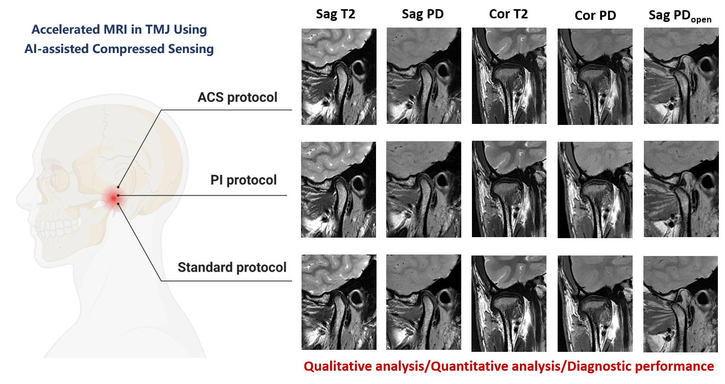
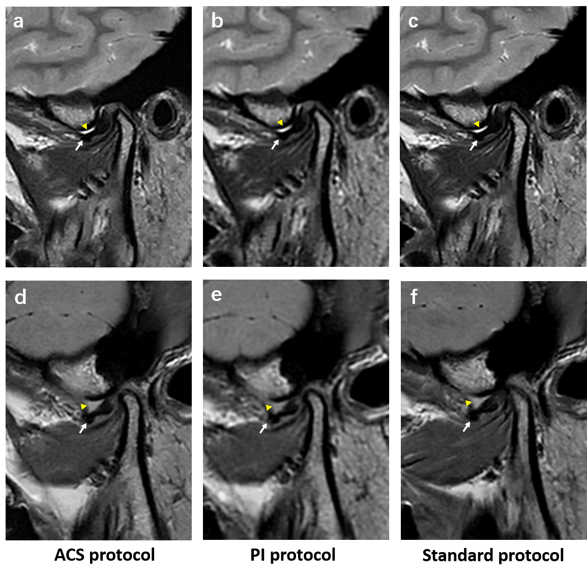
MRI is regarded as the reference standard for diagnosing temporomandibular joint (TMJ) disc displacement and joint effusion [1]. For comprehensive assessment, patients with TMJ disorders (TMD) are often instructed to maintain close-mouth position or open-mouth position for imaging during examination. However, the relatively long acquisition time of MRI can decrease patient comfort and cause motion artifact, especially in the open-mouth position, which may reduce image quality and diagnostic confidence, thus necessitating accelerated MRI of the TMJ.The recently proposed AI-assisted compressed sensing (ACS) technique combines deep learning neural network, compressed sensing, parallel imaging (PI), and partial Fourier [2]. Previous studies showed that ACS technique can facilitate noise suppression and greatly accelerate MRI examination [3,4]. Therefore, this study aimed to investigate the feasibility of accelerated MRI protocols with ACS technique in TMJ, and compare its performance with PI protocol and standard protocol (Figure 1).
Materials and Methods
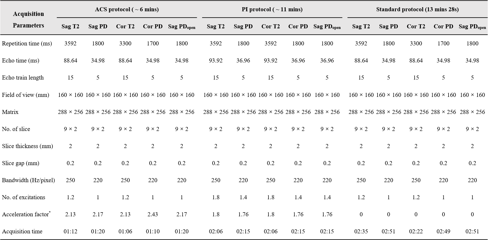
This prospective study was approved by the local Institutional Review Board. Participants with symptomatic TMJ disorders were enrolled from March 2023 to October 2023. MRI examinations were performed on the 3T MRI scanner (uMR 790, United Imaging Healthcare) with 32-channel head coil using ACS protocol, PI protocol and standard protocol. Each protocol included sagittal/coronal T2 weighted imaging and sagittal/coronal proton density weighted imaging in the closed-mouth position, as well as sagittal proton density weighted imaging in the open-mouth position (Figure 2).Qualitative analysis, quantitative analysis and diagnostic performance evaluation were conducted by two independent radiologists in a blind manner. For qualitative analysis, a 4-point Likert scale (4-excellent, 3-good, 2-moderate, 1-poor) was used to rate overall image quality and visibility of clinically relevant structures, including TMJ disc, mandibular fossa, mandibular condyle, lateral pterygoid muscle condyle (LPM). For quantitative analysis, the signal-to-noise ratio (SNR) of TMJ disc, condyle and LPM were measured. The free-hand region of interests (ROI) was draw at the largest cross-section of the disc, and ellipse ROIs were placed on the condylar head, inferior head of LPM, and background air, respectively. Diagnostic agreement of ACS protocol with the other two protocols was evaluated for joint effusion, sideways disc displacement, and anterior disc displacement. Statistical difference among three protocols was assessed by using Friedman rank sum test with Dunn-Bonferroni method or Kruskal-Wallis ANOVA test with Bonferroni correction.
Results
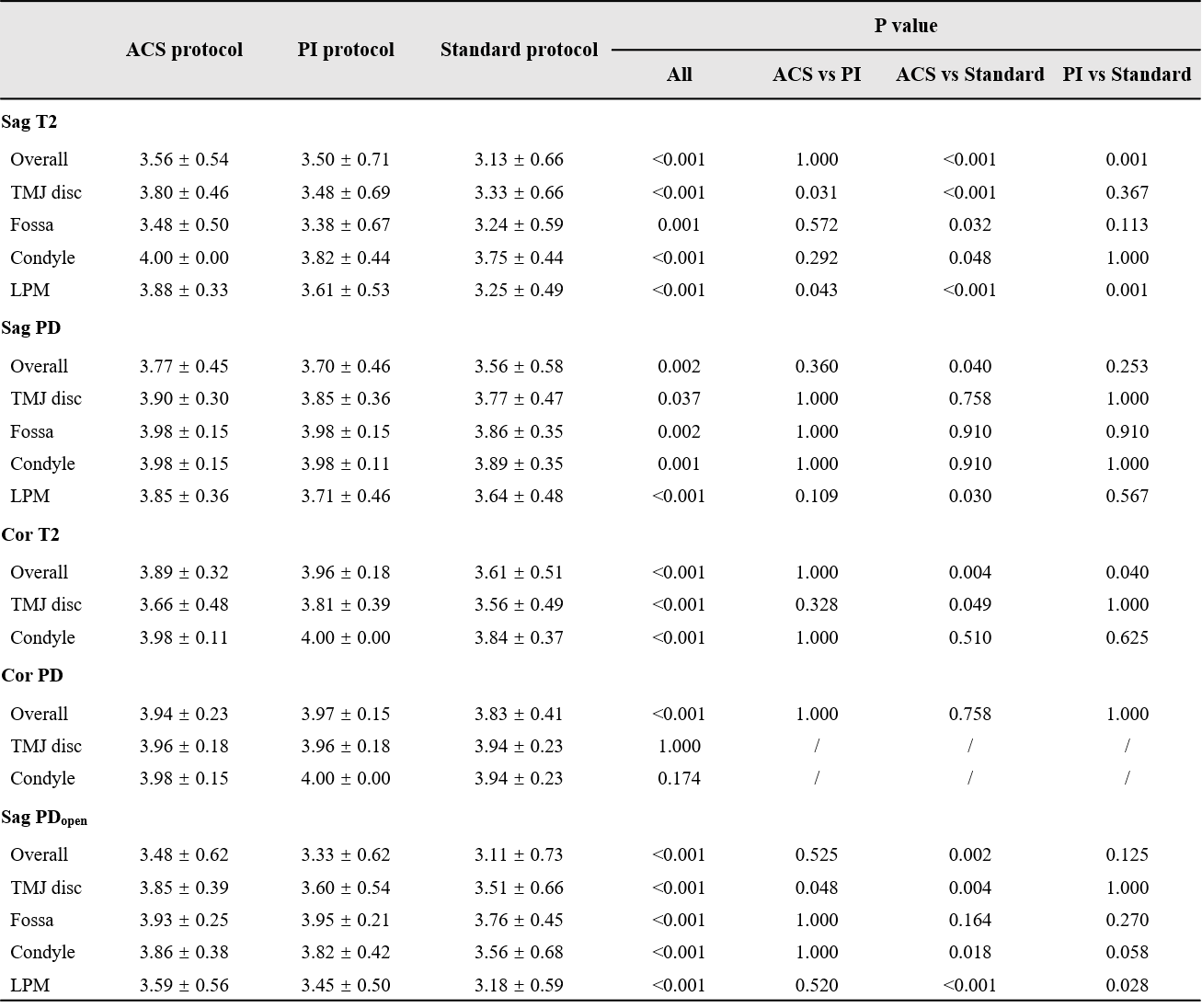
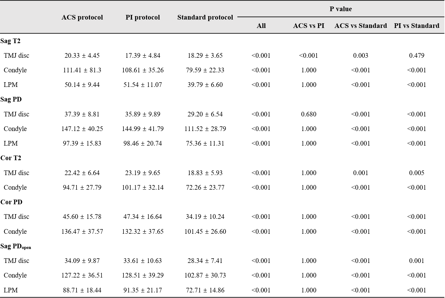
A total of 44 participants (10 male and 34 female, mean age ± standard deviation: 23.32 years ± 3.24) with 88 TMJs were included in this study. Among the analyzed TMJs, joint effusion, sideways disc displacement and anterior disc displacement were diagnosed in 61, 53 and 43 joints, respectively. For qualitative analysis, the overall image quality and most structures visibility of ACS protocol were significantly higher than standard protocol, and similar to PI protocol (Figure 3). For quantitative analysis, ACS protocol demonstrated significantly higher SNR than standard protocol in the TMJ disc, condyle and LPM (all P < 0.05), and ACS protocol showed similar SNR to PI protocol except for the TMJ disc in sagittal T2 weighted imaging (Figure 4). The diagnostic agreement of ACS protocol with the other two protocols was excellent (Figure 5). In specific, the kappa values between ACS protocol and PI protocol, ACS protocol and standard protocol were 0.97 (95% CI: 0.92-1.00) and 0.89 (95% CI: 0.79-0.99) for or joint effusion, and 1.00 (95% CI: 1.00-1.00) and 0.81 (95% CI: 0.69-0.93) for sideways disc displacement, and 0.93 (95% CI: 0.85-1.00) and 0.98 (95% CI: 0.93-1.00) for anterior disc displacement, respectively.Discussion
The novel ACS technique is developed to balance speed and image quality. It integrates the output of the trained AI module as an additional constraint into the compressed sensing framework by adding a regularization term based on the difference between the reconstructed image and the predicted image of the AI module [5]. Previous studies have shown that ACS technique has great potential in abdominal and musculoskeletal imaging, with faster acquisition speed, better image quality and higher lesion detectability [2-4, 6]. TMD patients with facial pain and restricted mandibular movement often have difficulties in holding still in close-mouth position and maintaining open-mouth position for a long time. In this study, the acquisition time of ACS protocol was approximately 45% faster than PI protocol and nearly 55% faster than standard protocol. Therefore, TMJ imaging with accelerated ACS technique is promise to be a great alternative to PI and standard protocols.Conclusion
Accelerated MRI with ACS technique can greatly reduce acquisition time of TMJ, while providing superior or equivalent image quality and excellent diagnostic agreement with PI and standard protocolsAcknowledgements
This work was supported by the Technology Innovation Project of Science and Technology Bureau of Chengdu (2022-YF05 -01691-SN) and Clinical Research Project of West China Hospital of Stomatology, Sichuan University (LCYJ-2023-YY-2).References
[1] Xiong X, Ye Z, Tang H, et al. MRI of temporomandibular joint disorders: Recent advances and future directions[J]. Journal of Magnetic Resonance Imaging, 2021, 54(4): 1039-1052.
[2] Liu J, Li W, Li Z, et al. Magnetic resonance shoulder imaging using deep learning–based algorithm. European Radiology, 2023: 1-11.
[3] Li H, Hu C, Yang Y, et al. Single-breath-hold T2WI MRI with artificial intelligence-assisted technique in liver imaging: As compared with conventional respiratory-triggered T2WI. Magnetic Resonance Imaging, 2022, 93: 175-180. [4] Wang Q, Zhao W, Xing X, et al. Feasibility of AI-assisted compressed sensing protocols in knee MR imaging: a prospective multi-reader study. European Radiology, 2023: 1-12.
[5] Zhai R, Huang X, Zhao Y et al. Intelligent incorporation of AI with model constraints for MRI acceleration. Proceedings of the 29th Annual Meeting of ISMRM, 2021.
[6] Zhao Q, Xu J, Yang Y X, et al. AI-assisted accelerated MRI of the ankle: clinical practice assessment. European Radiology Experimental, 2023, 7(1): 62.
Figures