2267
Accelerated 3D isotropic multi-echo UTE using CG-SENSE and Deep Learning Reconstruction for CT-like Imaging & Ultrashort T2* Mapping1Canon Medical Systems USA, Inc., Tustin, CA, United States, 2Canon Medical Systems Corporation, Otawara, Japan
Synopsis
Keywords: Whole Joint, Bone
Motivation: MRI provides superior soft-tissue contrast, however, cortical bone and ultrashort-T2* tissues are invisible in routine clinical MRI. Multi-echo Ultrashort TE (mecho-UTE) may allow visualization and quantification of bone and ultrashort-T2* tissues, however, its acquisition time is often long.
Goal(s): We used CG-SENSE and Deep Learning Reconstruction (CG-SENSE+DLR) to accelerate mecho-UTE, making it clinically feasible.
Approach: 5-min, 3-min, and 2-min mecho-UTE scans were prospectively acquired and reconstructed with Gridding and CG-SENSE+DLR. Resolution, sharpness, and T2* were measured and compared.
Results: CG-SENSE+DLR allows 2- and 3-min mecho-UTE with improved resolution and sharpness compared to 5-min mecho-UTE with Gridding reconstruction.
Impact: Routine MRI and mecho-UTE enable comprehensive MSK imaging, providing soft-tissue contrast and visualization-and-quantification of bone and ultrashort-T2* tissues. CG-SENSE+DLR allows accelerated 2- and 3-minunte mecho-UTE, which would enable widespread clinical utilization to demonstrate the value of mecho-UTE compared to CT.
Introduction
MRI provides superior soft-tissue contrast compared to CT, however, in musculoskeletal (MSK) conditions, where bone assessment is needed, a CT scan is often required. Single-echo Zero TE (ZTE) and Ultrashort TE (UTE) have the potential for radiation-free imaging of cortical bone, however, poor resolution, long scan time, and fundamental tradeoffs between scan time, SNR, and resolution[1] need to be addressed before wider adoption for clinical evaluation.In addition to CT-like contrast, dual-echo and multi-echo UTE can provide visualization of ultrashort-T2* tissues (tendon, ligament, meniscus, etc.) and quantification of bone porosity index[2] and quantitative T2* of ultrashort T2* tissues[3]–[7], which correlated well with CT bone density measurements and ultrashort-T2* tissue health, respectively. While providing more information compared to single-echo counterpart, multi-echo UTE is even more demanding in terms of scan time requirements.
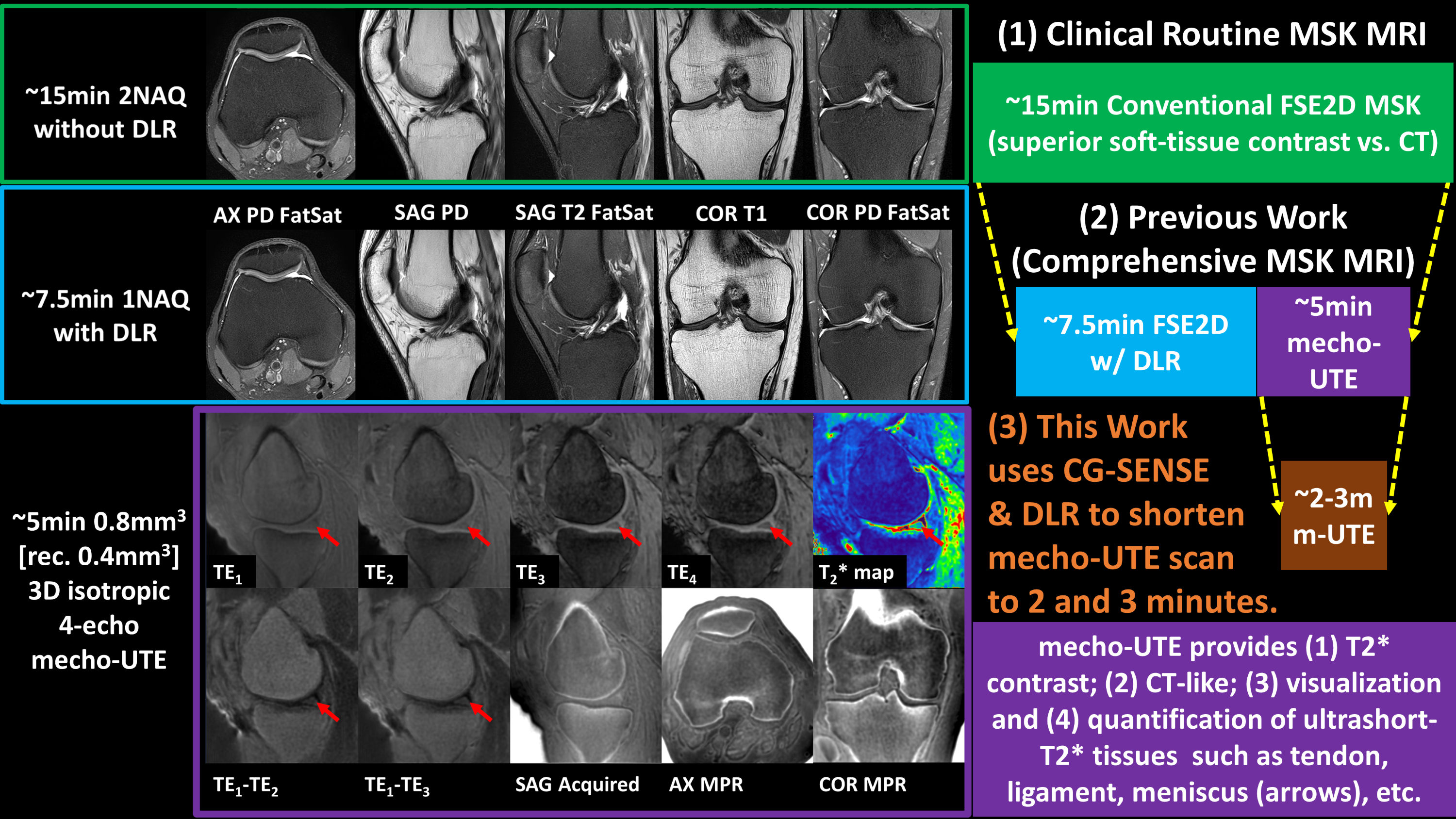
As seen in Figure 1, routine FSE2D and mecho-UTE would allow comprehensive MSK imaging by providing excellent soft-tissue contrast and visualization/quantification ultrashort-T2* tissues (cortical bone, tendon, meniscus, ligament, etc.), respectively. This work aimed to use CG-SENSE[8] and Deep Learning Denoising Reconstruction[9], [10] (CG-SENSE+DLR) to accelerate and improve the resolution and sharpness of multi-echo UTE (mecho-UTE), making it viable for clinical adoption. Our goal is not to replace CT, but rather to enable a radiation-free bone imaging option for patients who are CT-contraindicated and need radiation-exposure minimization (for example, repeated CT exams, pediatric patients, etc.).
Methods
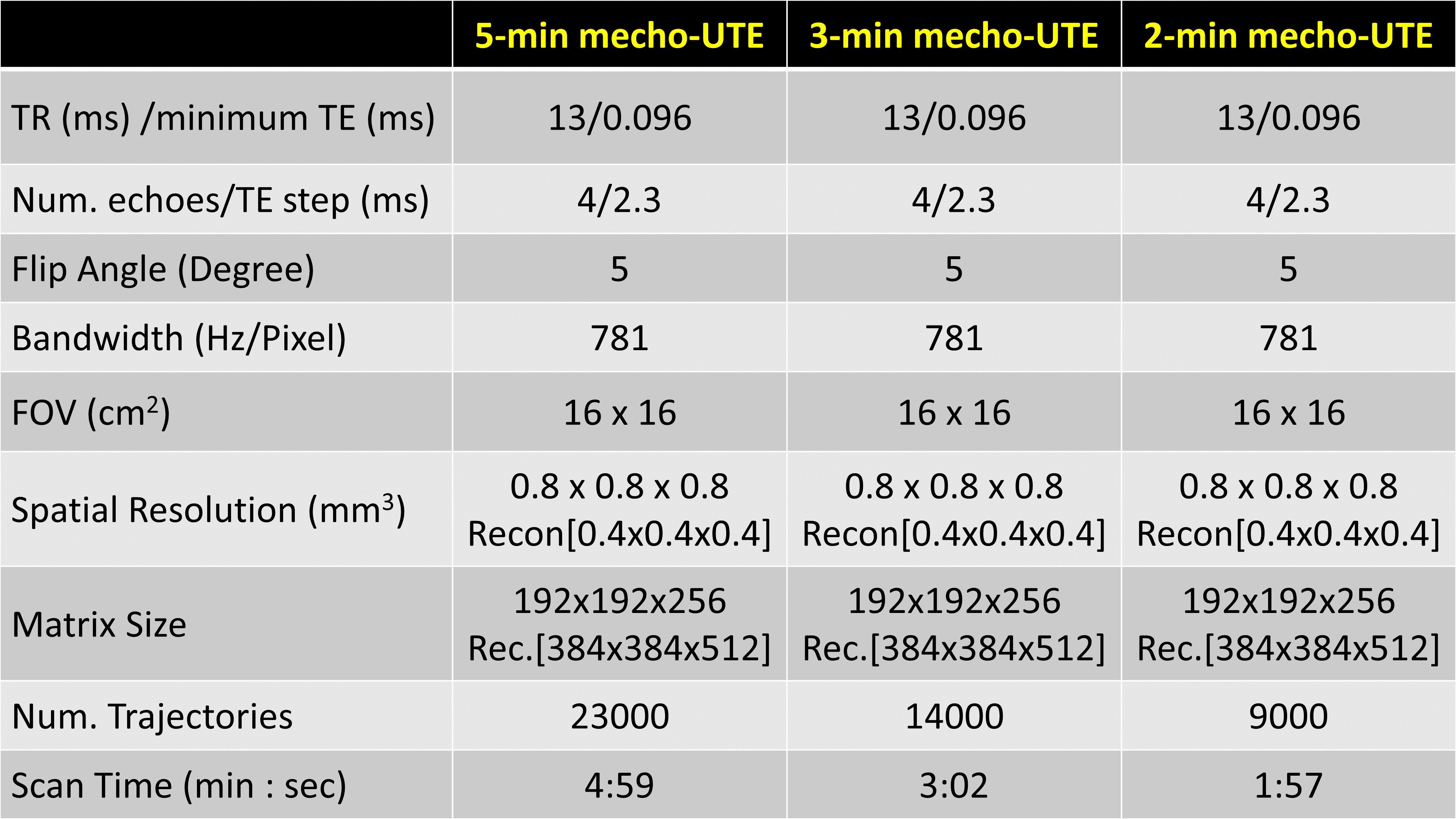
Data Collection:Experiments were performed on four subjects (four knees and one shoulder) using a 3T scanner with approved IRB. 5-min, 3-min, and 2-min 3D-isotropic 0.8mm3 [reconstructed as 0.4mm3] resolution 4-echo mecho-UTE scans were prospectively acquired using the protocols in Figure 2. Three scans were reconstructed with both gridding and CG-SENSE+DLR.
Data Analysis:
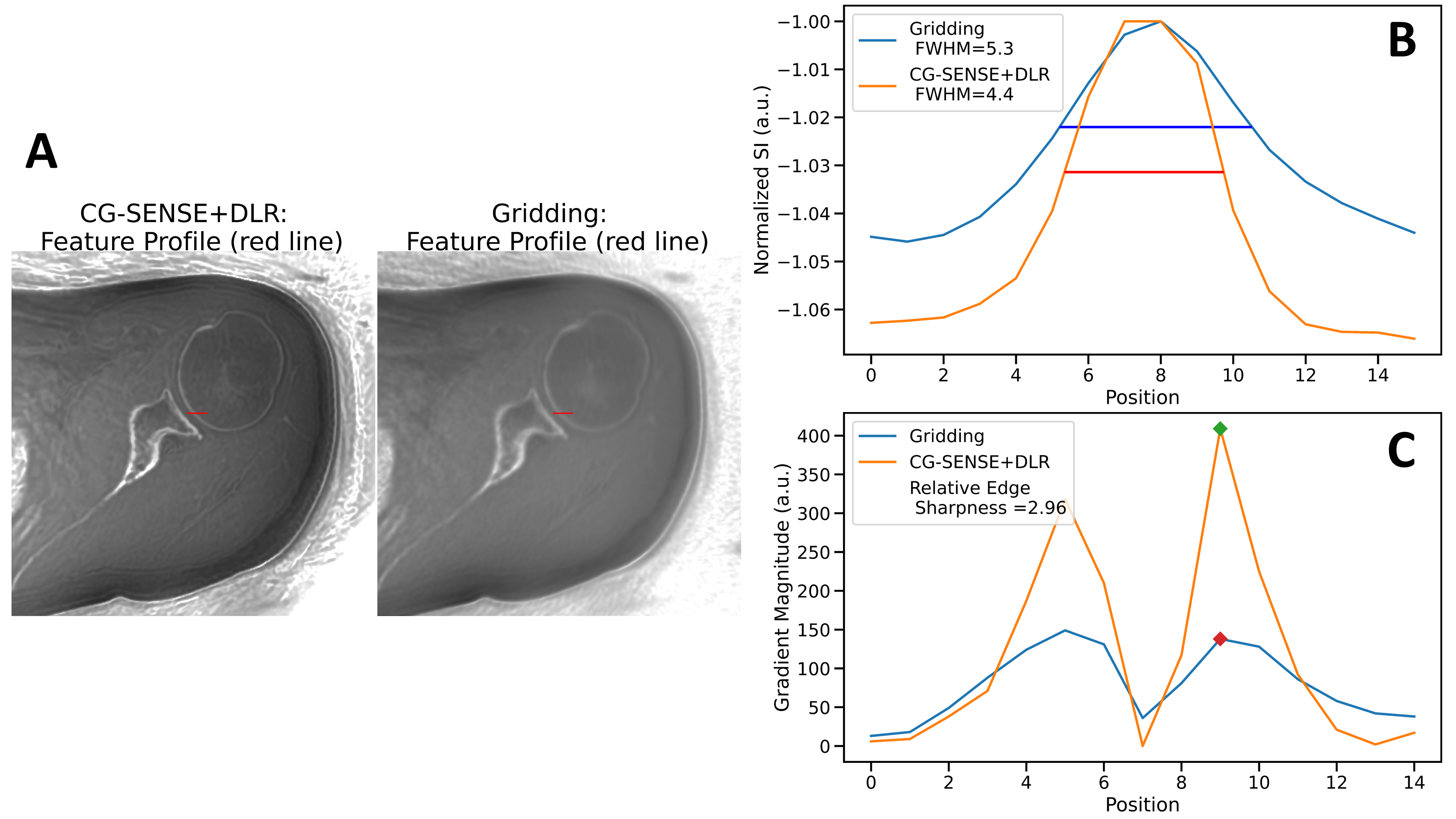
Logarithmic inversion[11] was applied to the first echo (TE1 = 0.096ms) image to generate CT-like images. The second echo was subtracted from the first echo (TE1-TE2) for better delineation/visualization of ultrashort-T2* tissues (tendons, ligaments, etc.). Mono-exponential fitting was used to generate T2* map. Region of interests on tendons were drawn for T2* measurements. Full-Width at Half-Maximum (FWHM) and Relative Edge SHarpness (RESH)[9] were measured to assess resolution and sharpness, as seen in Figure 3.
Results and Discussion
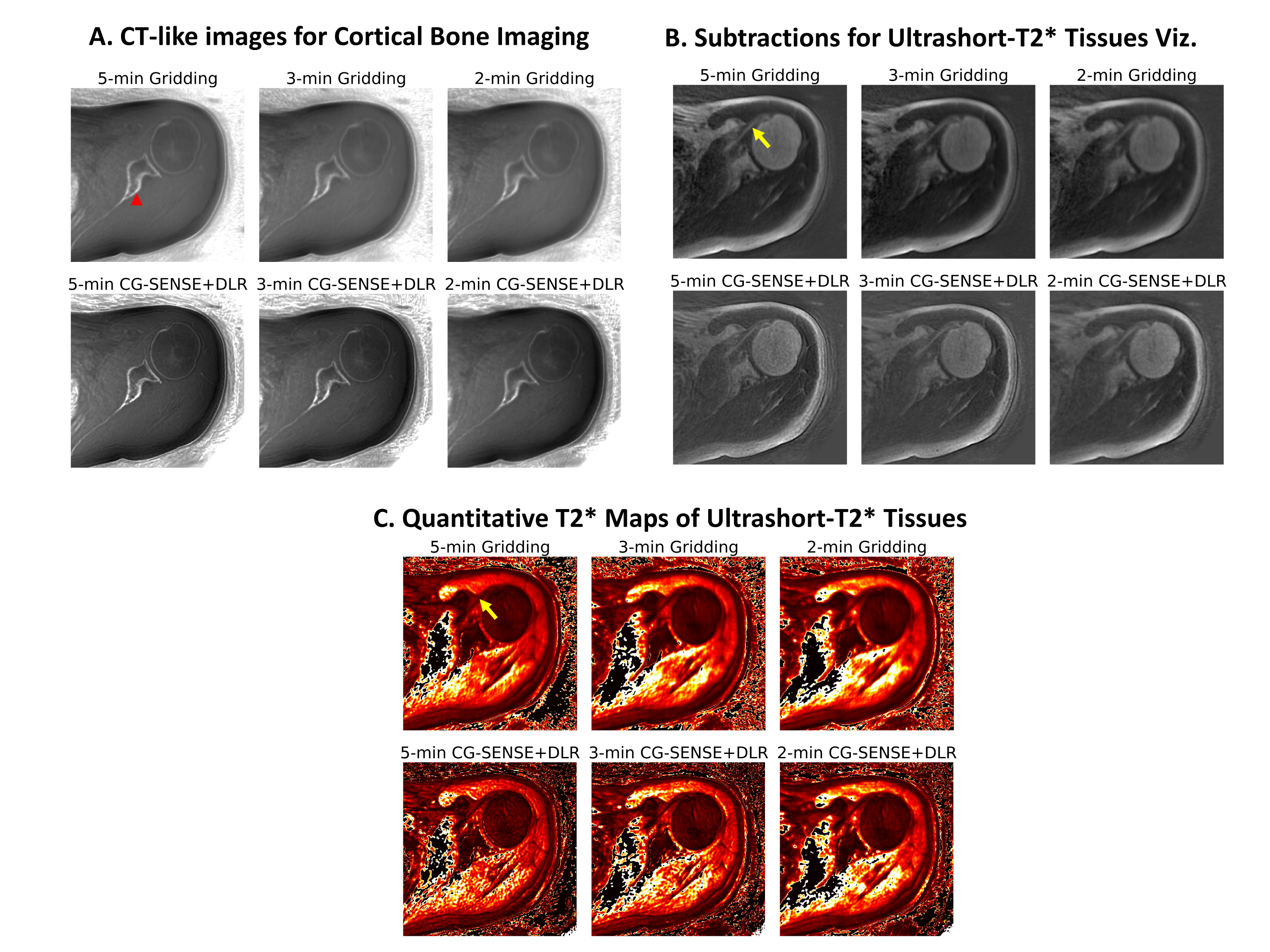
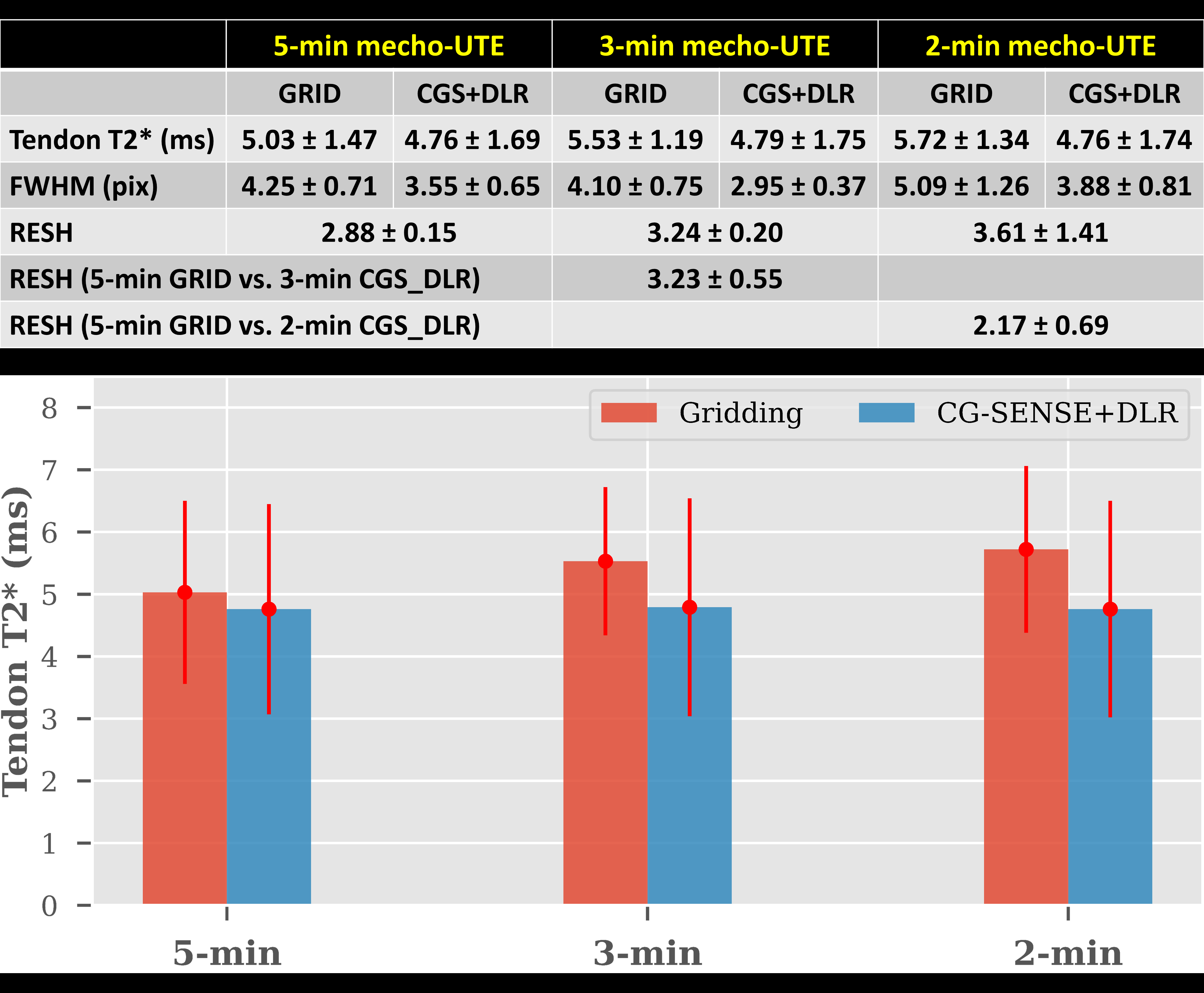
Figure 4 shows CT-like, subtraction, and T2* map images. Figure 5 shows quantitative comparisons of T2*, FWHM, and RESH.Qualitatively, as shown in Figure 4, CG-SENSE+DLR images show improved image quality and sharpness compared to Gridding counterparts. Furthermore, 3-min and 2-min with CG-SENSE+DLR show improved image quality and sharpness compared to the 5-min with Gridding.
Quantitatively, as shown in Figure 5, tendon T2* values were similar between scans and reconstructions and in the range from literature[12], [13]. As seen in the bar plot, mean T2* values were increased in Gridding reconstruction with shorter acquisition times (likely due to streaking and blurring artifacts) while those in CG-SENSE+DLR stayed relatively consistent around 4.76-4.79ms.
Compared to Gridding counterparts, CG-SENSE+DLR images have an average of 22.76% smaller FWHM and 3.24 times higher RESH. Compared to 5-min Gridding, 3-min and 2-min CG-SENSE+DLR have an average of 30.59% and 8.71% smaller FWHM, respectively and 3.23 and 2.17 times higher RESH, respectively.
Conclusion
This work demonstrated that CG-SENSE+DLR allows accelerated 2- and 3-minute 3D-isotropic 0.8mm3 [reconstructed as 0.4mm3] 4-echo mecho-UTE acquisition with improved resolution and sharpness compared to 5-min mecho-UTE with Gridding reconstruction. The accelerated mecho-UTE may enable widespread clinical adoption to evaluate its diagnostic performance and economic value of comprehensive MSK MRI (routine FSE2D and mecho-UTE) compared to CT or to CT & MRI.Acknowledgements
No acknowledgement found.References
Figures