2261
Quantitative Ultrashort Echo Time Double Echo Steady State (qUTE-DESS) for T1, T2, and Diffusivity Mapping of Human Cartilage1Radiology, University of California, San Diego, San Diego, CA, United States, 2Research Service, Veterans Affairs San Diego Healthcare System, San Diego, CA, United States, 3Bioengineering, University of California, San Diego, San Diego, CA, United States, 4Radiology Service, Veterans Affairs San Diego Healthcare System, San Diego, CA, United States
Synopsis
Keywords: Cartilage, Cartilage, UTE, DESS, UTE-DESS, Diffusion, T1, T2
Motivation: A comprehensive validation of parameter mapping for T1, T2, and ADC based on qUTE-DESS has not been conducted yet.
Goal(s): To investigate the feasibility and accuracy of qUTE-DESS for estimating T1, T2, and ADC parameters in human patellar cartilage and to compare these results with conventional MR techniques.
Approach: The study used a qUTE-DESS sequence with variable flip angles and gradient moment adjustments. The cartilage sample was imaged using this approach and conventional imaging sequences, and the acquired data were processed using signal fitting.
Results: Significant correlations exist between the parameters estimated by qUTE-DESS and those from conventional sequences.
Impact: This study pioneers the comprehensive validation of T1, T2, and ADC parameter mapping using qUTE-DESS for knee joint imaging. Its findings offer a promising avenue to enhance the assessment of short T2 tissues and advance clinical applications in musculoskeletal diagnostics.
INTRODUCTION
The Double Echo Steady State (DESS) has been employed in the imaging of the human knee joint due to its ability to achieve flexible image contrast, including T1, T2, and diffusion weighting1–3. One of the major applications of DESS in the musculoskeletal system is the assessment of cartilage, which can benefit from fluid suppression achieved through diffusion weighting. Recently, quantitative Ultrashort Echo Time DESS (qUTE-DESS) has been investigated as a promising imaging technique to assess T2 parameters for short T2 tissues in the knee4–6. More notably, qUTE-DESS can be applied to estimate the apparent diffusion coefficient (ADC) from short T2 tissues such as tendon, ligament, meniscus, and deep cartilage. Given that conventional diffusion imaging is challenging for short T2 tissues due to its long TE, it is of high importance to investigate qUTE-DESS-based diffusion imaging. The feasibility of qUTE-DESS has been demonstrated to estimate T1, T2, and diffusivity simultaneously7. However, the accuracy of these parameters has not been validated yet. In this study, we investigated the association of quantitative parameters from qUTE-DESS, for the first time, with those obtained from conventional MR techniques in a human patellar cartilage sample.METHODS
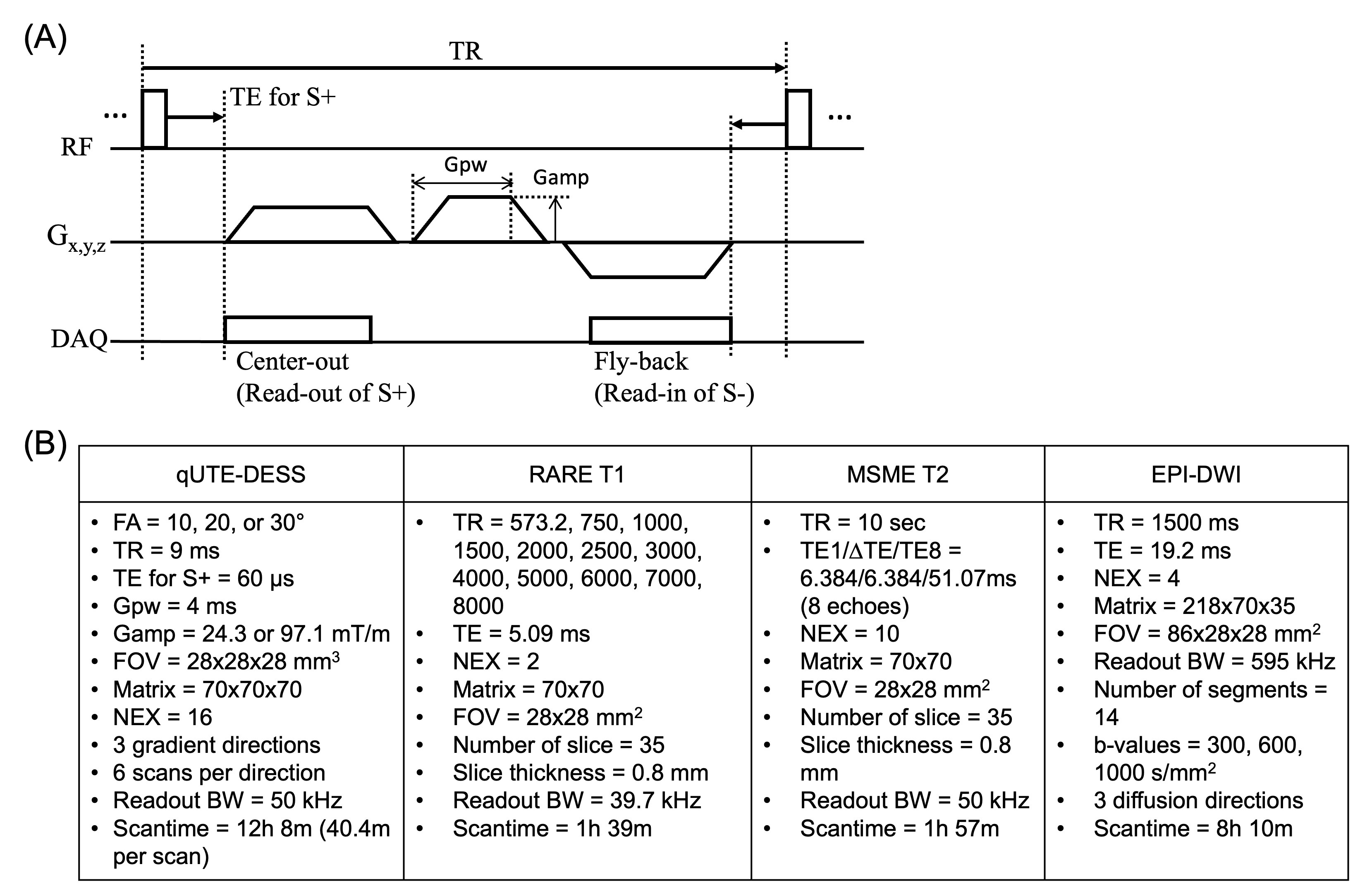
qUTE-DESS: Figure 1A shows the pulse sequence diagram. A variable flip angle (FA) scheme is utilized to achieve different T1 weighting, where image acquisition is repeated with different FAs. Different diffusion weighting is achieved by using the variable gradient moment of the spoiling gradients, controlled by adjusting the gradient amplitude (Gamp) with a fixed gradient pulse width (Gpw) in each scan. Variable image T1, T2, and diffusion weighting are present, and T1, T2, and ADC parameters can be estimated through signal fitting.Sample Preparation: A human patellar cartilage sample was harvested from a 73-year-old donor. The specimen was inserted into a 20cc syringe with fomblin. A rubber piece was used to hold the specimen and prevent its movement during the MRI scan.
MR Imaging: The qUTE-DESS sequence was implemented in a 3T preclinical MR scanner (Biospec-3T, Bruker). The prepared cartilage sample was imaged using a 23mm birdcage coil. MRI was conducted using qUTE-DESS, as well as conventional quantitative imaging sequences, including Rapid Imaging with Refocused Echoes (RARE) for T1 mapping, Multi-Slice Multi-Echo (MSME) for T2 mapping, and Echo Planar Imaging-based Diffusion Weighted Imaging (EPI-DWI) for ADC mapping. qUTE-DESS comprised a total of 18 acquisitions (3 FAs x 2 Gamps x 3 gradient-orientations). Additionally, standard B1 mapping was performed. The imaging parameters are shown in Figure 1B.
Data Processing: All images were reconstructed using online reconstruction software by Bruker. Data fitting and analysis were conducted using Matlab. For qUTE-DESS, T1, T2, and ADC were fitted utilizing the steady-state free precession (SSFP) signal model8,9. The parameter fitting was performed in each gradient direction and averaged to yield the final parameter maps.
RESULTS
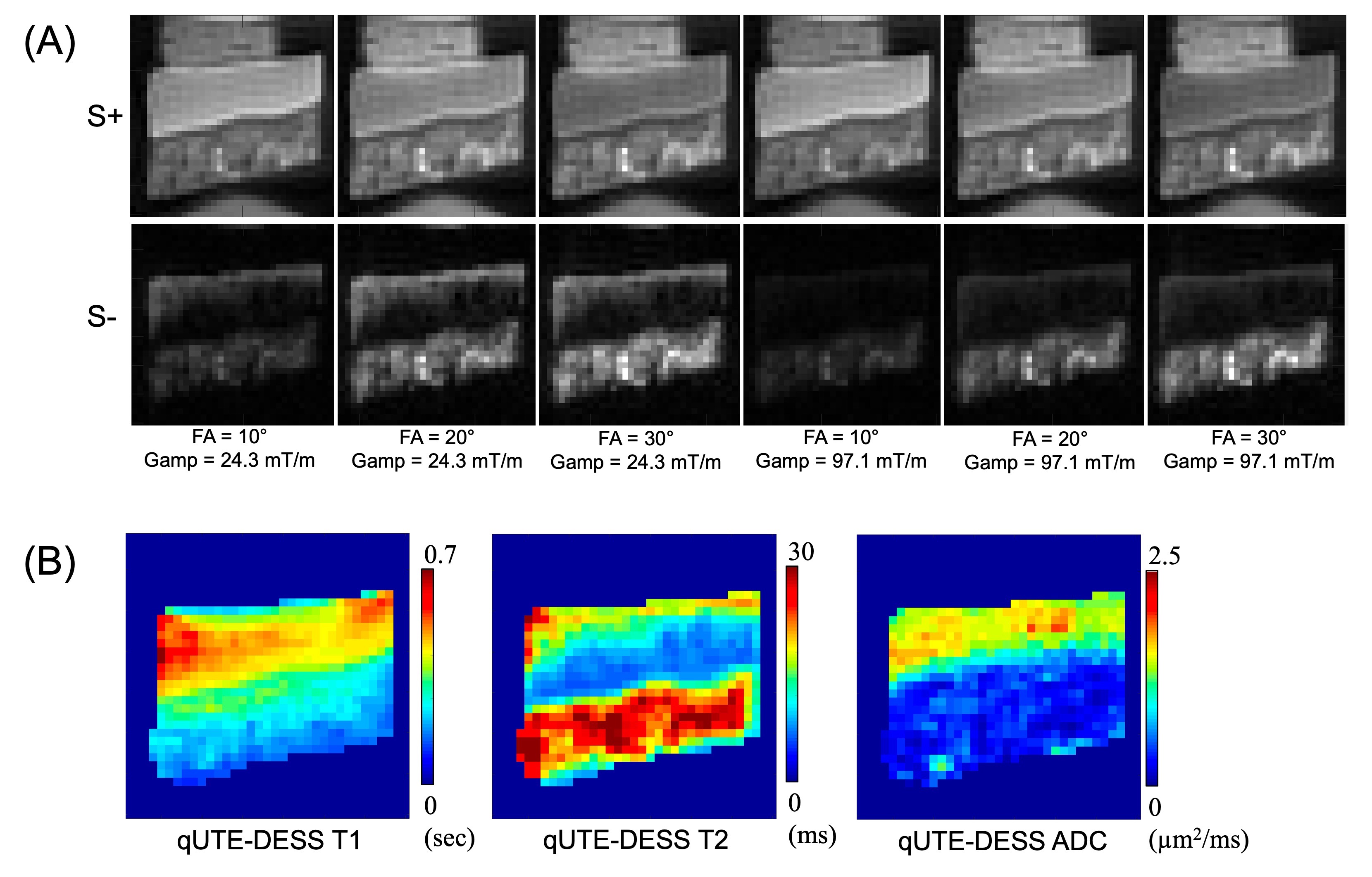
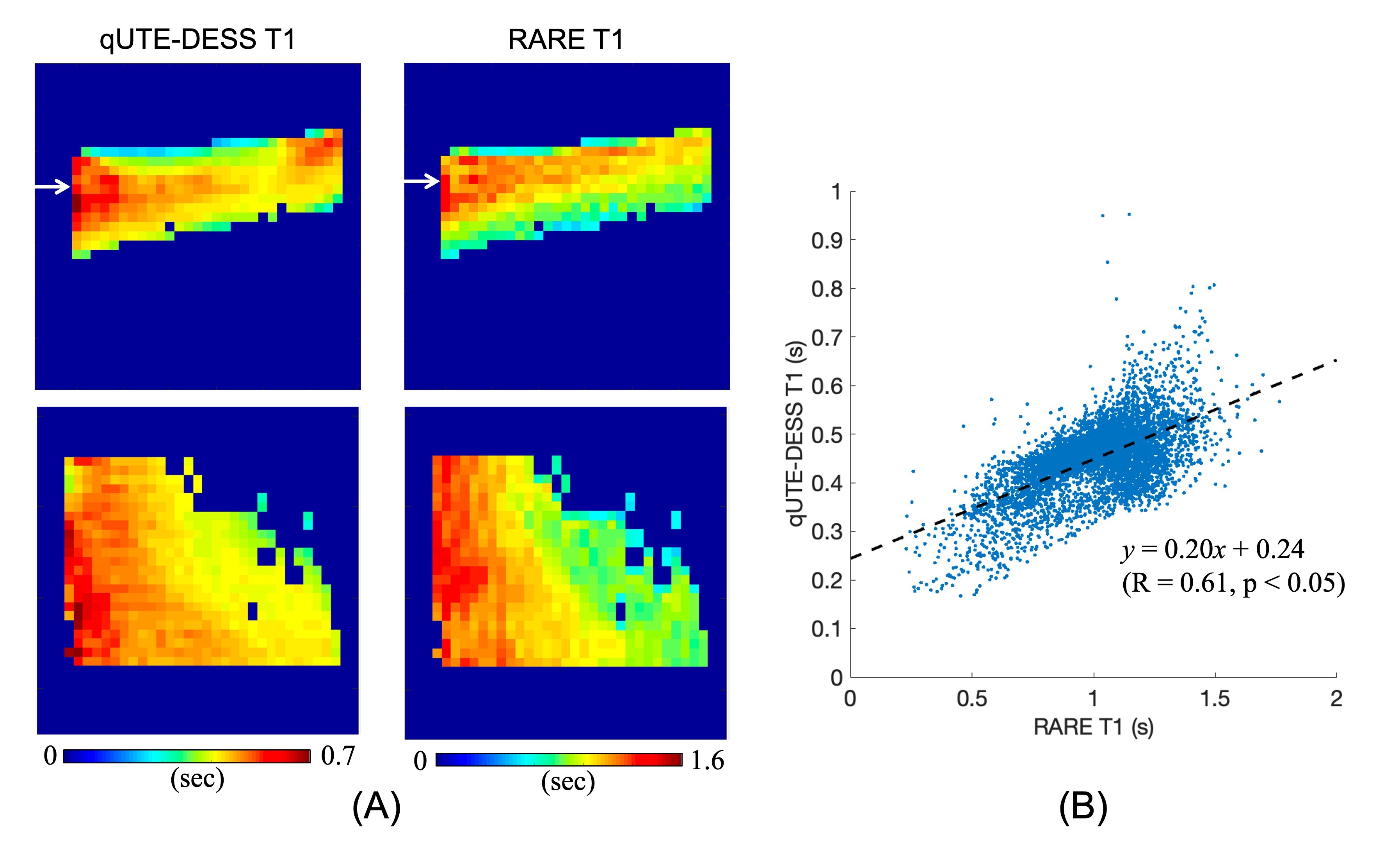
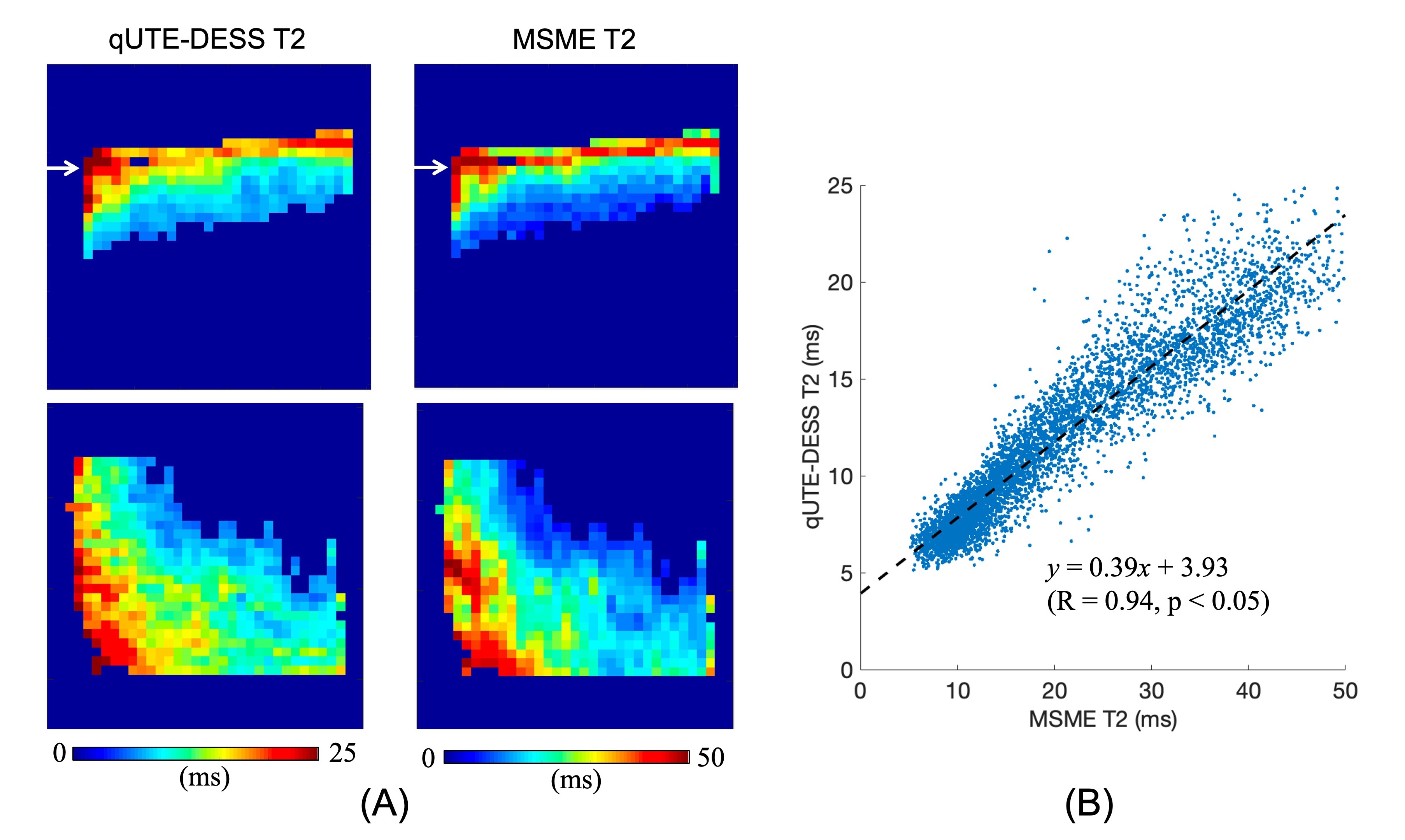
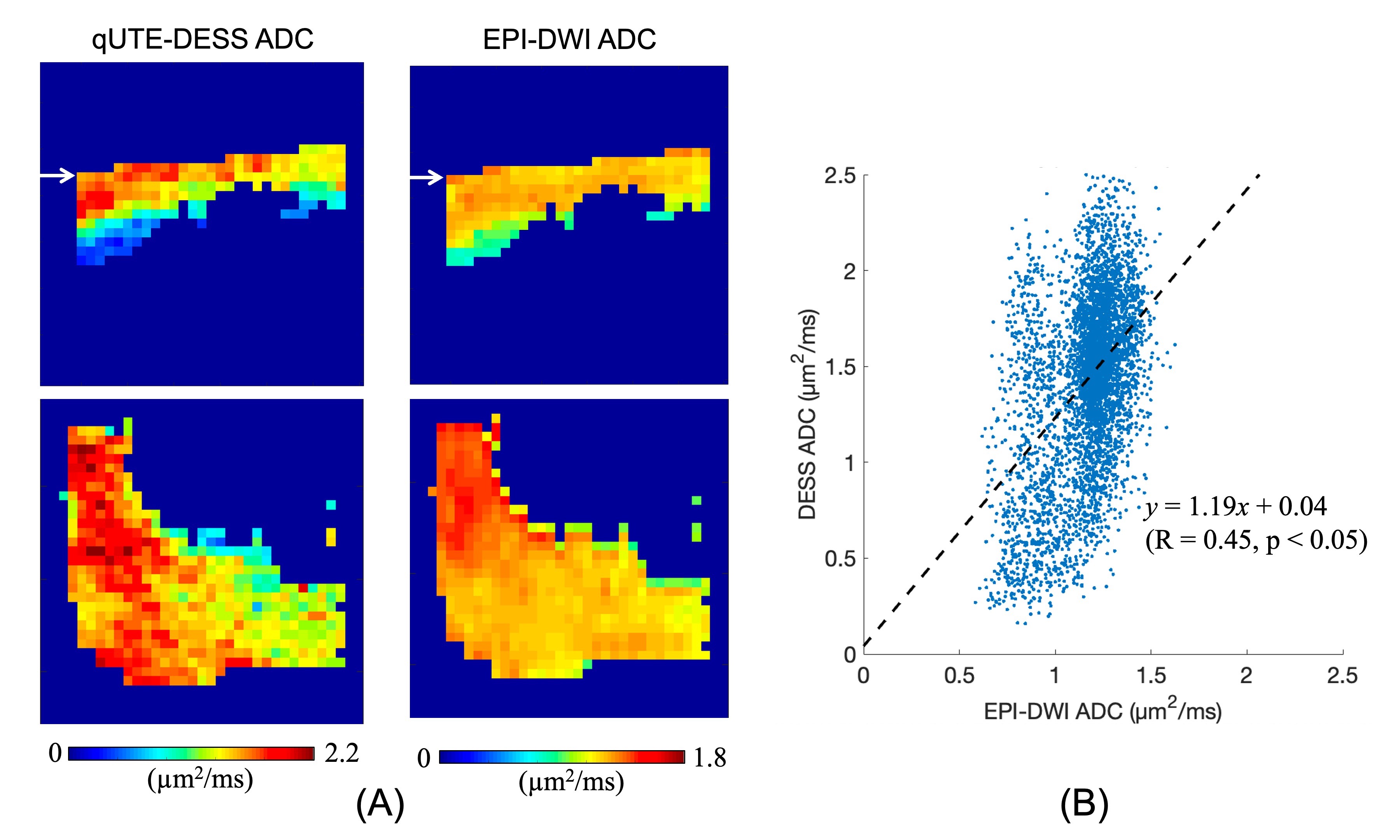
Figure 2A shows an example of the acquired qUTE-DESS S+/S- images acquired with different FAs and Gamps activated in a logical x-direction. Figure 2B shows the resultant parameter maps from qUTE-DESS fitting. Figures 3-5 show a comparison between qUTE-DESS and corresponding clinical sequence parameters, demonstrated in the coronal (top images in Figures 3A-5A) and axial (bottom images in Figures 3A-5A) planes at the location indicated by a white arrow. The T1, T2, and ADC estimated by qUTE-DESS showed a significant correlation with those from RARE, MSME, and EPI-DWI (Pearson correlation R = 0.61, 0.94, and 0.45, respectively).DISCUSSION AND CONCLUSION
Despite the strong correlation, T1 and T2 parameters from qUTE-DESS were underestimated compared to those from RARE and MSME. ADC was slightly overestimated in qUTE-DESS. Since those sequences utilize different schemes for parameter mapping and fitting, it may be difficult to directly compare without any bias. The ADC from qUTE-DESS was found to be weaker in correlation than other parameters. Notably, the qUTE-DESS demonstrated a wider dynamic range of values and was able to estimate parameters in the deep layer of the cartilage, whereas EPI-DWI failed due to low SNR at the long TE. However, the estimated ADC map showed high visual similarity between the two techniques, which is promising, and it remains to be seen whether the greater dynamic range may be more useful for diagnoses.The scan time for qUTE-DESS was approximately 40 minutes per scan, with NEX of 16 to secure enough SNR for 0.4mm isotropic resolution, which is not clinically feasible. However, by matching the spatial resolution to that commonly used for in vivo knee imaging, such as 0.7x0.7x2mm³, the scan time can be dramatically reduced. In our future studies, we will investigate qUTE-DESS in the knee joint of human subjects.
Acknowledgements
The authors acknowledge grant support from the NIH (R01AR078877, R01AR062581, R01AR068987, R01AR075825, R01AR079484, RF1AG075717, and R21AR075851), Veterans Affairs (I01CX001388, I01BX005952, I01CX002211), and GE Healthcare.References
1. Hardy PA, Recht MP, Piraino D, Thomasson D. Optimization of a Dual Echo in the Steady State (DESS) free-precession sequence for imaging cartilage. J. Magn. Reson. Imaging 1996;6:329–335.
2. Welsch GH, Scheffler K, Mamisch TC, et al. Rapid estimation of cartilage T2 based on double echo at steady state (DESS) with 3 Tesla. Magn. Reson. Med. 2009;62:544–549.
3. Moriya S, Miki Y, Yokobayashi T, Ishikawa M. Three-Dimensional Double-Echo Steady-State (3D-DESS) magnetic resonance imaging of the knee: Contrast optimization by adjusting flip angle. Acta radiol. 2009;50:507–511.
4. Chaudhari AS, Sveinsson B, Moran CJ, et al. Imaging and T2 relaxometry of short-T2 connective tissues in the knee using ultrashort echo-time double-echo steady-state (UTEDESS). Magn. Reson. Med. 2017;78:2136–2148.
5. Jang H, Athertya JS, Jerban S, et al. Correction of B0 and linear eddy currents: Impact on morphological and quantitative ultrashort echo time double echo steady state (UTE‐DESS) imaging. NMR Biomed. 2023:1–15.
6. Jang H, Ma Y, Carl M, Jerban S, Chang EY, Du J. Ultrashort echo time Cones double echo steady state (UTE‐Cones‐DESS) for rapid morphological imaging of short T 2 tissues. Magn. Reson. Med. 2021;86:881–892.
7. Jang H, Ma Y, Masoud-Afsahi A, et al. Quantitative UTE Double Echo Steady State (qUTE-DESS) for Simultaneous Mapping of T1, T2, and Diffusivity of Short T2 Tissues – Ex Vivo study. In: Joint Annual Meeting ISMRM-ESMRMB & ISMRT 31st Annual Meeting. London, UK; 2022. p. 1411.
8. Freed DE, Scheven UM, Zielinski LJ, Sen PN, Hürlimann MD. Steady-state free precession experiments and exact treatment of diffusion in a uniform gradient. J. Chem. Phys. 2001;115:4249–4258.
9. Bieri O, Ganter C, Scheffler K. Quantitative in vivo diffusion imaging of cartilage using double echo steady-state free precession. Magn. Reson. Med. 2012;68:720–729.
Figures