2260
ROI based Multi-parameter Quantitative Network(RMQ-Net) with Uncertainty-awareness for Quantitative UTE MRI Study of Cartilage1Department of Radiology, University of California, San Diego, San Diego, CA, United States, 2Radiology Service, Veterans Affairs San Diego Healthcare System, San Diego, CA, United States, 3Department of Orthopaedic Surgery, University of California San Diego, San Diego, CA, United States
Synopsis
Keywords: Cartilage, MSK
Motivation: Quantitative MRI (qMRI) studies of cartilage regions need both regional segmentation and pixel-wise fitting analysis, which can be time-consuming and subject to inter-individual variability.
Goal(s): To design a deep neural network for simultaneous qMRI mapping and accurate tissue segmentation.
Approach: By leveraging different scan sequences, we proposed a RMQ-net with Uncertainty-awareness(UA) module, or UA-QMR-net. A majority-voting strategy was applied for robust cartilage segmentation and accelerated qMRI analysis.
Results: The results demonstrated that the UA-RMQ-net achieved higher performance than the original RMQ-net for both UTE-T1 and UTE-T1r analyses of articular cartilage.
Impact: By leveraging information from different scan sequences, the proposed UA-RMQ-net could obtain higher performance for accelerated qMRI analysis.
Introduction
While conventional clinical MRI sequences show little or no signal for many short-T2 tissues such as the deep cartilage, menisci, and bone, ultrashort echo time (UTE) MRI techniques allow high-resolution morphological and quantitative imaging of both short- and long-T2 tissues in the joint[1]. It would be of great clinical value to simultaneously segment and quantify tissues accurately and effectively. Deep learning models with multi-task design, generally with one as the major task and others as auxiliary tasks with weaker constraints, have been widely used to finish several tasks simultaneously[2]. It also has been testified that multi-task DCNN with reduced scan time could achieve similar qMRI results compared with the traditional methods in our previous studies[3,4]. In this study, by leveraging different scan sequences that are used for parameter mappings, we proposed a Regions-of-interest Based Multi-parameter Quantitative Network (RMQ-net) with Uncertainty-awareness(UA) module or UA-QMR-net. A majority-voting strategy was applied in the UA-QMR-net to generate more confident segmentation results for the cartilage area with a more robust accelerated qMRI analysis.Method
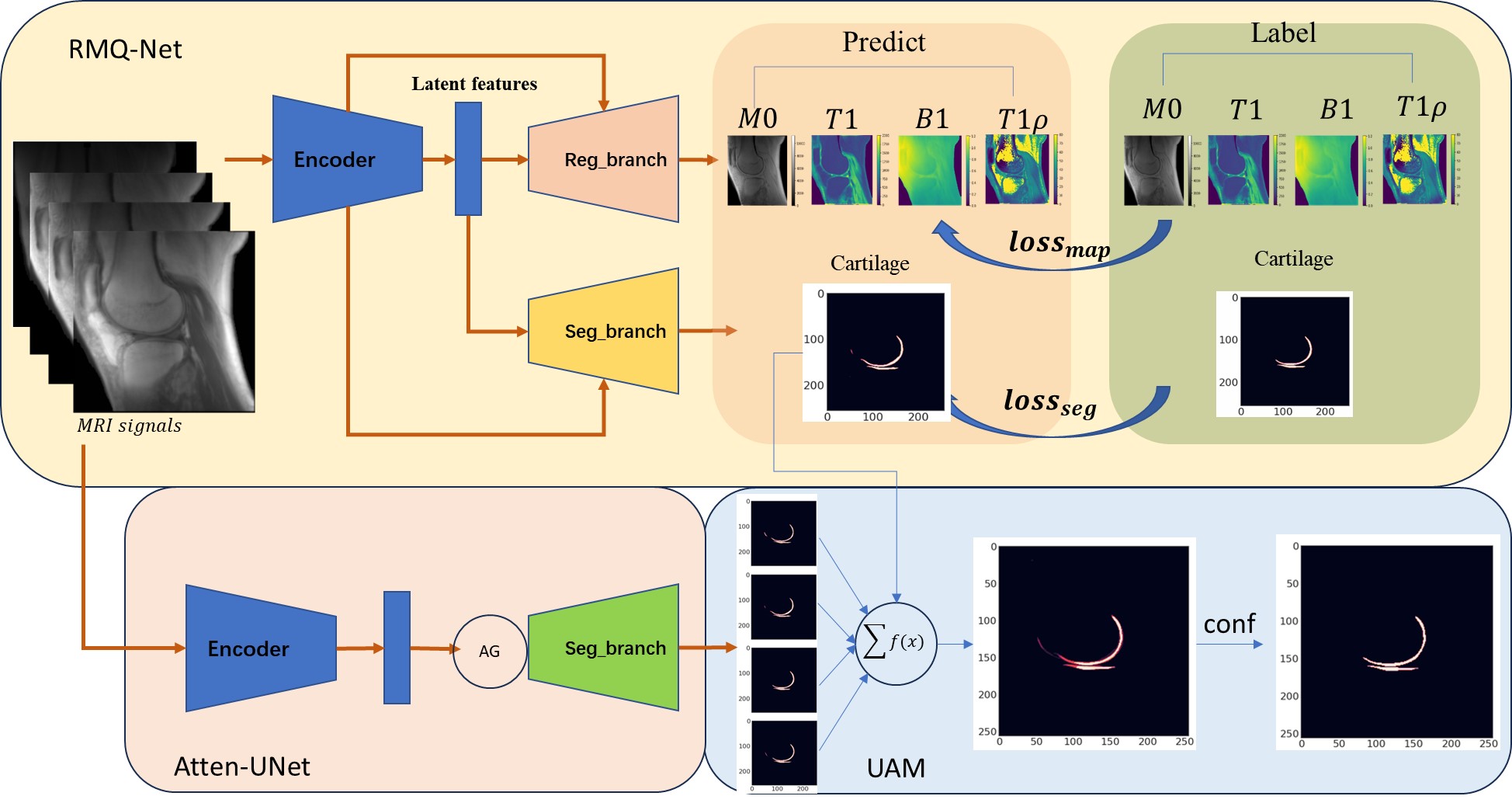
As shown in Figure 1, the UA-RMQ-Net mainly comprises two parts: RMQ-net and UA module. RMQ-net is based on a U-Net style DCNN with two branches for outputs, namely Reg_branch and Seg_branch, as introduced before[3,4]. UA module includes two steps. First, Attention-Unet is applied as a segmentation task for each of the input MRI images. Then, the predicted segmentations (with predicted segmentation from the RMQ-Net) were combined according to a weighting function to generate a soft segmentation prediction. Finally, a majority-voting strategy is applied to generate the final segmentation area, which could be used to combine with the predicted parameter mapping (i.e., T1 and T1ρ) to generate the final ROI analysis of the target tissue (e.g., articular cartilage).Three-dimensional UTE cones sampling was performed using an echo time (TE) of 32 μs, repetition time (TR) of 20 ms, and flip angles (FAs) of 5°, 10°, 20°, and 30°, respectively, for UTE-T1 mapping. For UTE-T1ρ mapping, the scan parameters were TR=500ms, flip angle=10°, and 7 spin-lock times (TSLs)=0, 12, 24, 36, 48, 72, and 96 ms. Subsequently, image registration was performed to minimize inter-scan motion. M0, UTE-T1, UTE-T1ρ, and B1 maps were derived via non-linear fitting using the Levenberg‐Marquardt algorithm. ROIs for the cartilage area were labeled with homemade Matlab code by three experienced radiologists[5,6].
A total of 1056 slice images from 44 subjects (including healthy volunteers and patients with different degrees of OA) were used for model training, and 144 images of six additional subjects were used for model validation. The DCNNs were implemented with pytorch 1.1.0 on a workstation with an Nvidia GTX 1080 Ti (11 GB GPU memory). Several experiments with two different MRI scan parameter combinations were studied, including UTE-T1 data with one FA and UTE-T1ρ data with two TSLs (1FA+2TLS), and UTE-T1 data with two FAs and UTE-T1ρ data with three TSLs (2FAs+3TSLs).
The threshold rate for selecting the final segmentation prediction area is a key parameter for generating better segmentation results. According to the majority-voting strategy, 0.5, 0.7, and 0.9 were tested, respectively. We also tested whether including the segmentation prediction of the RMQ-net had any influence. Pearson correlation was performed to evaluate the relationship between the predicted values from the UA-RMQ-net and the ground truth for all regions of interest (ROIs).
Results and Discussion
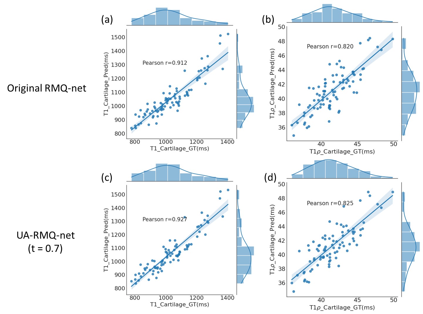
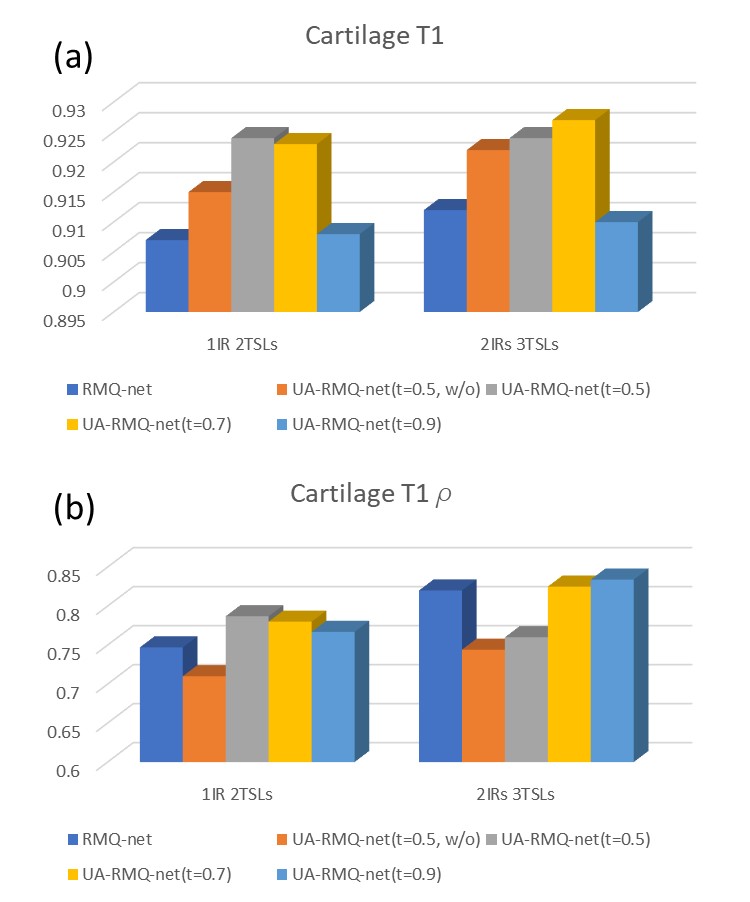
As shown in Figure 2, for the 2FAs+3TSLs combination, Pearson correlation studies were performed between the predicted UTE-T1 and UTE-T1ρ ROIs analysis from DCNNs and the ground truth. For the original RMQ-Net, the R-values are 0.912 and 0.820 for UTE-T1 and UTE-T1ρ, respectively. For the UA-RMQ-Net, the R-values are 0.927 and 0.825. The R-values of UA-RMQ-Net are higher than the results of the original RMQ-Net.In Figure 3 (a), it could be found that for UTE-T1 analysis, UA-RMQ-Net would normally have a better correlation r-value than the RMQ-Net, except when t =0.9. For UTE-T1ρ analysis with 1FA+2TSLs, UA-RMQ-net has a higher r-value than RMQ-net, except without the input of the segmentation prediction from RMQ-net. For UTE-T1ρ analysis with 2FAs+3TSLs, UA-RMQ-net with a threshold(t) =0.7 and 0.9 higher than the original RMQ-net.
Conclusion
To fully leverage multi-sequences of MRI scans for UTE-T1 and UTE-T1ρ mapping, we proposed an Uncertainty-awareness module to further improve the performance of the RMQ-net. By a series of hyper-parameter tuning for the majority-voting strategy, we found that UA-RMQ-net with the output of the RMQ-net and threshold(t) value of 0.7 would normally obtain better performance for the correlation studies for the cartilage area for both UTE-T1 and UTE-T1ρ analyses.Acknowledgements
The authors acknowledge grant support from the National Institutes of Health (R01AR062581, R01AR068987, R01AR075825, and R01AR079484), VA Clinical Science and Rehabilitation Research and Development Services (Merit Awards I01CX001388, I01CX002211, and I01BX005952), and GE Healthcare.References
1. Chang EY, Du J, Chung CB. UTE imaging in the musculoskeletal system. J Magn Reson Imaging 2015; 41(4):870-883.
2. Alexander Selvikvåg Lundervold, Arvid Lundervold.An overview of deep learning in medical imaging focusing on MRI, Zeitschrift für Medizinische Physik,Volume 29, Issue 2, 2019,Pages 102-127.
3. Lu X, Ma YJ, Saeed J, Jang H, Xue YP, Zhang XD, Wu M, Gentili A, Hsu CN, Chang EY, Du J. Deep CNNs with Physical Constraints for simultaneous Multi-tissue Segmentation and Quantification (MSQ-Net) of Knee from UTE MRIs. ISMRM 2021.
4. Lu X, Ma YJ, et al. Deep CNNs with Physical Constraints for simultaneous Multi-tissue Segmentation and Multi-parameter Quantification (MSMQ-Net) of Knee. International Society for Magnetic Resonance in Medicine(ISMRM) 2022, 07-15 May, London, England, United Kingdom
5. Ma YJ, Lu X, Carl M, Zhu Y, Szeverenyi NM, Bydder GM, Chang EY, Du J. Accurate T1 mapping of short T2 tissues using a three-dimensional ultrashort echo time cones actual flip angle imaging-variable repetition time (3D UTE-Cones AFI-VTR) method. Magn Reson Med. 2018 Aug;80(2):598-608.
6. Ma YJ, Carl M, Searleman A, Lu X, Chang EY, Du J.3D adiabatic T1ρ prepared ultrashort echo time cones sequence for whole knee imaging. Magnetic resonance in medicine 80 (4), 1429-1439. 2018
Figures