2252
Machine Learning Assisted Prediction of Cartilage Proteoglycan Content Using MR Fingerprinting1Research Unit of Health Sciences and Technology, University of Oulu, Oulu, Finland, 2University of Eastern Finland, Kuopio, Finland, 3Oulu University Hospital, Oulu, Finland, 4Department of Diagnostic Radiology, Oulu University Hospital, Oulu, Finland, 5Medical Research Center, University of Oulu and Oulu University Hospital, Oulu, Finland
Synopsis
Keywords: Cartilage, Cartilage, MRF
Motivation: Early signs of cartilage degeneration include changes in proteoglycan content, which cannot be diagnosed using standard clinical imaging tools.
Goal(s):
Prediction of cartilage proteoglycan content from quantitative MR fingerprinting data at 3T.
Approach:
Gaussian process regression (GPR) models were trained to predict optical density of safranin-O stained cartilage sections, representing proteoglycan content, from MRF data on a voxel-by-voxel basis.
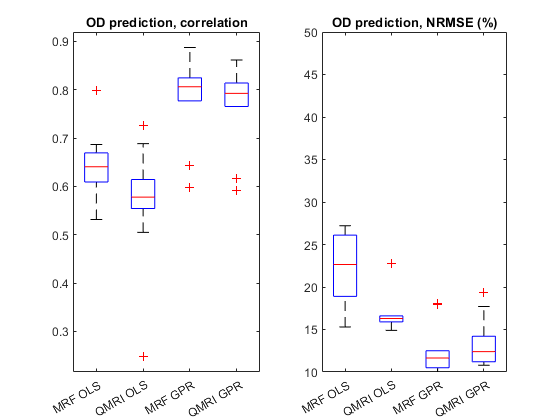
Results: The trained GPR models reached very high accuracy (mean correlation of 0.81 with a respective NRMSE of 11.7%) and had clearly enhanced performance when compared to linear models.
Impact: Non-invasive prediction of
proteoglycan content in cartilage using MR fingerprinting at clinical field
strength is feasible, holding promise for direct clinical imaging of cartilage
composition in the future.
Introduction
Early signs of degenerative cartilage pathologies (such as osteoarthritis) include changes in cartilage proteoglycan content and collagen organization, which cannot be currently diagnosed using standard clinical imaging tools. Quantitative magnetic resonance imaging (qMRI) parameters have been shown to be sensitive to macromolecular structure of cartilage [1,2], and a combination of qMRI and machine learning has previously been used to predict cartilage composition at preclinical and clinical field strengths [3,4]. These approaches have relied on the measurement of multiple qMRI parameters with tailored sequences, which are often too time-consuming for clinical application. The aim of this study was to use the previously proven machine learning techniques to predict the compositional properties of cartilage from data gathered using magnetic resonance fingerprinting (MRF), which allows simultaneous measurement of multiple qMRI parameters, thus reducing the imaging time significantly.Methods
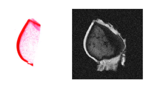
The studied samples consisted of three bovine patellae, which were imaged at 3 T (four 2-mm slices for each patella, 0.6x0.6 mm2 in-plane resolution) using an MRF sequence dedicated for qMRI of articular cartilage [5]. The utilized sequence allows simultaneous acquisition of T1, T2, proton density (PD) and B1 maps. After MR imaging, tissue corresponding to each MRI slice was cut into three 3.0 μm thick histological sections that underwent safranin-O staining of proteoglycans (Fig. 1). Digital densitometry measurements were carried out on the sections using 492-nm wavelength monochromatic light to estimate the proteoglycan content [6]. The high-resolution histological images were downsampled to match the resolution of the MR images (256x256 px), followed by co-registraiton to the proton density (PD) maps of their corresponding slices using the ELASTIX registration toolbox [7,8]. The averaged voxel values of these sets were then used in training of Gaussian process regression (GPR) models to predict voxel-by-voxel optical density separately from the parameter maps and raw MRF data. The model was validated using leave-one-out cross-validation, where a single slice was utilized for testing of a model, which was trained using the data from the other slices. This testing was repeated individually for each slice to test the generalizability of the model. The performance of the trained model was evaluated with visual inspection of the predicted optical density as well as with voxel-wise normalized root-mean-squared error (NRMSE) and Pearson correlation coefficients for each test iteration. The performance of GPR models was compared to a linear regression model to study the benefits of using machine learning for the different predictions.Results
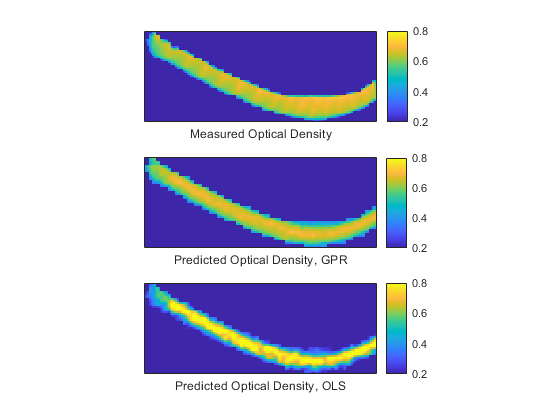
In visual evaluation (Figure 2), optical density could be predicted by the model, although the quality of the predicted virtual histology images was somewhat limited by the low resolution of the MRI data. The GPR models trained using raw MRF data (Figure 3) reached very high median accuracy (r = 0.81 and NRMSE = 11.7%) and demonstrated enhanced performance when compared to the linear model (r = 0.64 and NRMSE = 22.7%). Similarly, the GPR models trained using qMRI maps reached high accuracy (r = 0.79 and NRMSE = 12.4%) superior to the linear models (r = 0.58 and NRMSE = 16.3%).Discussion & Conclusions
The results of this study demonstrate that the spatial proteoglycan content of articular cartilage can be predicted from clinical MRF data with high accuracy. Furthermore, the results underline the need to utilize non-linear methods in constructing the prediction models, as using GPR approximately halved the prediction error as compared to the linear model. The results also indicate a benefit from using the raw MRF data as a predictor instead of the parameter maps obtained from the same data. However, the difference is quite small, indicating that to improve the predictions there may be a need to sensitize the utilized MRF sequence to other processes such as diffusion or T1ρ relaxation. Similarly to previous studies, the trained GPR model was able to accurately predict the optical density of the studied cartilage (r = 0.80, RMSE = 1.28) [3] (r = 0.81, RMSE = 2.55) [4] despite differences in MRI acquisition protocols. The study is limited by the small set of measurement data, necessitating the use of voxel-by-voxel training approach instead of more powerful image-to-image deep-learning approaches. A further limitation is the homogeneity of the dataset as it did not include samples suffering from cartilage degeneration e.g. reduced optical density and vertical fissures. Finally, a limitation arises from comparison of thin histological sections to thick MRI slices. These findings suggest that non-invasive prediction of PG content in cartilage from MRF measurements using a 3 T clinical scanner is feasible, holding promise for future clinical applications.Acknowledgements
Academy of Finland, grant number: 354692References
[1] M.T. Nieminen, J. Rieppo, J. Silvennoinen, J. Töyräs, J.M. Hakumäki, M.M. Hyttinen, H.J. Helminen, J.S. Jurvelin, Spatial assessment of articular cartilage proteoglycans with Gd-DTPA-enhanced T1 imaging, Magn. Reson. Med. 48 (2002) 640–648. https://doi.org/10.1002/mrm.10273.
[2] M.J. Nissi MJ, E.-N. Salo, V. Tiitu, T. Liimatainen, S. Michaeli, S. Mangia, J. Ellermann, M.T. Nieminen, Multi-Parametric MRI Characterization of Enzymatically Degraded Articular Cartilage, J. Orthop. Res. Off. Publ. Orthop. Res. Soc. 34 (2016) 1111–1120. https://doi.org/10.1002/jor.23127.
[3] K. Linka, J. Thüring, L. Rieppo, R.C. Aydin, C.J. Cyron, C. Kuhl, D. Merhof, D. Truhn, S. Nebelung, Machine learning-augmented and microspectroscopy-informed multiparametric MRI for the non-invasive prediction of articular cartilage composition, Osteoarthritis Cartilage. 29 (2021) 592–602. https://doi.org/10.1016/j.joca.2020.12.022.
[4] S.A. Mirmojarabian, A.W. Kajabi, J.H.J. Ketola, O. Nykänen, T. Liimatainen, M.T. Nieminen, M.J. Nissi, V. Casula, Machine Learning Prediction of Collagen Fiber Orientation and Proteoglycan Content From Multiparametric Quantitative MRI in Articular Cartilage, J. Magn. Reson. Imaging. 57 (2023) 1056–1068. https://doi.org/10.1002/jmri.28353.
[5] M.A. Cloos, J. Assländer, B. Abbas, J. Fishbaugh, J.S. Babb, G. Gerig, R. Lattanzi, Rapid Radial T1 and T2 Mapping of the Hip Articular Cartilage With Magnetic Resonance Fingerprinting, J. Magn. Reson. Imaging. 50 (2019) 810–815. https://doi.org/10.1002/jmri.26615.
[6] I. Kiviranta, J. Jurvelin, M. Tammi, A.M. Säämänen, H.J. Helminen, Microspectrophotometric quantitation of glycosaminoglycans in articular cartilage sections stained with Safranin O, Histochemistry. 82 (1985) 249–255. https://doi.org/10.1007/BF00501401.
[7] S. Klein, M. Staring, K. Murphy, M.A. Viergever, J.P.W. Pluim, elastix: A Toolbox for Intensity-Based Medical Image Registration, IEEE Trans. Med. Imaging. 29 (2010) 196–205. https://doi.org/10.1109/TMI.2009.2035616.
[8] D. Shamonin, Fast parallel image registration on CPU and GPU for diagnostic classification of Alzheimer’s disease, Front. Neuroinformatics. 7 (2013). https://doi.org/10.3389/fninf.2013.00050.
Figures