2248
Assessment of Synthetic 3T Image Generator for Accurate CSF Volume Measurement in T1-w, T2-w, and FLAIR Sequences Compared to Low-Field Imaging1Monash Biomedical Imaging, Monash University, Clayton, Victoria, Australia, 2Australian National Imaging Facility, Queensland, Australia, 3Neuroscience, Monash University, Clayton, Victoria, Australia, 4Radiology, Alfred Hospital, Victoria, Australia, 5Herston Imaging Research Facility, University of Queensland, Queensland, Australia, 6School of Electrical Eng. and Computer Science, University of Queensland, Queensland, Australia, 7School of Clinical Science, Queensland University of Technology, Queensland, Australia, 8David Hartley Chair of Radiology, Royal Perth Hospital, Western Australia, Australia, 9Medical School, University of Western Australia, Western Australia, Australia, 10South Australian Health and Medical Research Institute, South Australia, Australia, 11Data Science and AI, Monash University, Clayton, Victoria, Australia
Synopsis
Keywords: AI/ML Image Reconstruction, Low-Field MRI
Motivation: Addressing limited access to high-field MRI systems, our study investigates whether a Synthetic 3T Image Generator can enhance low-field MRI to match high-field image quality, crucial for accurate cerebrospinal fluid (CSF) volume analysis.
Goal(s): We aimed to validate the efficacy of the Synthetic 3T generator in improving CSF volume measurements on low-field MRI, in comparison to high-field T1-w, T2-w, and FLAIR sequences.
Approach: A cGAN was employed to enhance 64mT MRI data to synthetic 3T images, for evaluation in comparison to high-field MRIs.
Results: The synthetic 3T images demonstrated significant improvements in CSF volume estimation across all sequences when compared to low-field images.
Impact: The synthetic 3T MRI enhancements could advance neuroimaging in resource-limited settings, improve diagnostic precision for brain injuries and potentially broaden the application of neurological and psychiatric patient care worldwide and expand neuroimaging research opportunities.
Background
Magnetic Resonance Imaging (MRI), through T1-weighted, T2-weighted, and Fluid-Attenuated Inversion Recovery (FLAIR) sequences, is essential for evaluating Cerebrospinal Fluid (CSF) volumes in brain anatomy, crucial for diagnosing brain injuries1. Alterations in CSF flow and volume can signal severe issues like hydrocephalus or indicate brain swelling severity2. The Munro-Kellie hypothesis underlines the brain’s compensation for volume shifts, affecting intracranial pressure and compliance, key to managing potential herniation and optimizing medical responses3. Accurate CSF analysis is vital in cases of head trauma, where swift detection of hematomas or leaks is imperative4,5.The motivation for this study stems from the need to enhance low-field MRI data to match the superior resolution of high-field 3T MRIs, which is often limited by availability and cost. Our study focuses on the Synthetic 3T image generation framework, using advanced algorithms to improve low-field MRI quality, aiming to improve CSF volume estimation. The Synthetic 3T's capability to produce images comparable to high-field MRI holds the potential to transform neuroimaging practices, particularly for evaluating and managing intracranial pathologies where accurate CSF assessment is critical.
Methods
Our study leveraged Synthetic 3T, an advanced image generator framework, to enhance low-field MRI data using a conditional generative adversarial network (cGAN) model6. This model consists of a generator designed to translate 64mT MRI data into synthetic 3T images. It operates with a noise vector to guide the translation process, ensuring the output mirrors the HF 3T domain with high fidelity. The discriminator component of the GAN is tasked with distinguishing between genuine 3T images and synthetic outputs, providing iterative feedback that refines the generator's performance.A key feature of Synthetic 3T is its conditional generation capability, which maintains the intricate structural details of the original MRI data during the upscaling process. It further employs L1 loss to enhance pixel accuracy and the structural similarity index measure (SSIM) loss to preserve structural integrity. These elements are instrumental in replicating the quality and precision of higher-field strength MRI images.
For training, Synthetic 3T was trained and validated on a paired dataset from 50 healthy subjects, utilizing both the Hyperfine Swoop (64mT) and Siemens Biograph mMR (3T) systems to collect the necessary MRI data (IRB approved). To assess the practical application and validate the effectiveness of the Synthetic 3T framework, we tested it on an independent set of 50 healthy volunteers. The CSF volumes derived from the synthetic 3T images underwent segmentation using the SynthSeg+ algorithm7. These volumes were then meticulously compared to those obtained from actual HF 3T MRI scans across T1-w, T2-w, and FLAIR sequences, allowing us to evaluate the accuracy of CSF volume estimations facilitated by synthetic image enhancements.
Results
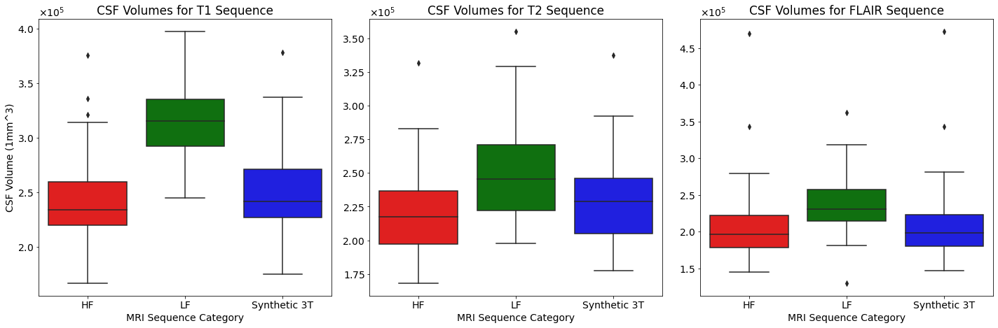
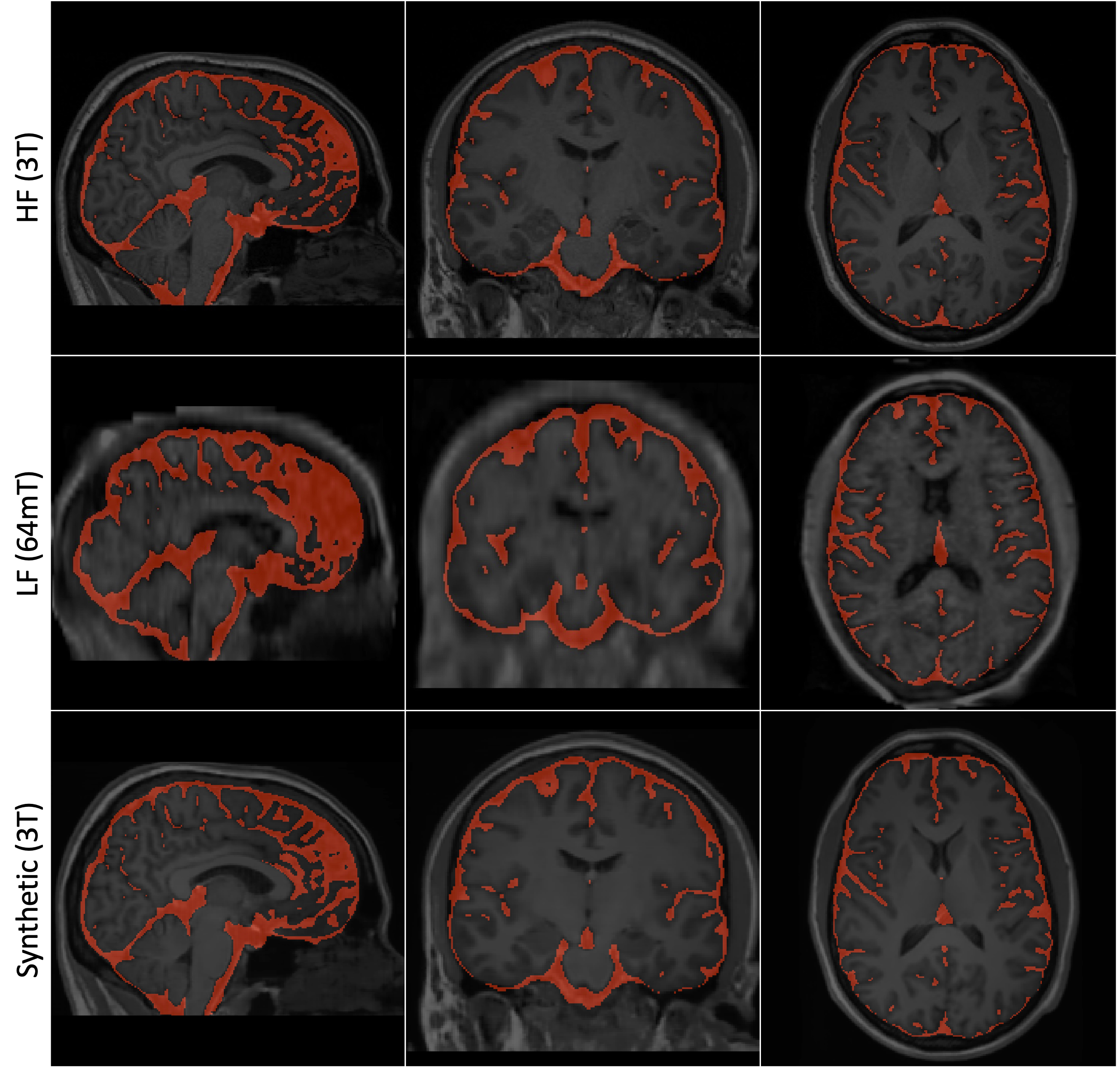
Figure 1 illustrates the variability in CSF volumes measured from different MRI modalities; T1-w sequences are shown in red, T2-w in green, and FLAIR in blue, with each modality further broken down into high-field (HF), low-field (LF), and synthetic 3T imaging, indicating distinct distributions and central tendencies for each category-sequence combination. Shapiro-Wilk tests showed non-Gaussian distributions for most groups, necessitating non-parametric analysis8. The Kruskal-Wallis test identified significant differences in CSF volume estimation across sequences: for T1 (H=65.5, p<0.001), T2 (H=21.1, p<0.001), and FLAIR (H=26.7, p<0.001). Post-hoc analyses using Dunn's test indicated significant enhancements in CSF volume delineation between HF and synthetic images against LF images for all sequences. Specifically, in T1, HF and synthetic images differed significantly from LF images (p<0.001). In T2, significant enhancements were noted between LF images and both HF (p<0.001) and synthetic images (p<0.001). For FLAIR, notable improvements were evident between LF and HF (p<0.001) and between LF and synthetic images (p<0.001).Example results of CSF segmentation on MRI brain scans obtained from three different modalities are showcased in Figure 2: HF on the top row, LF in the middle row, and Synthetic 3T on the bottom row, providing a visual comparison of the segmentation quality and anatomical details captured across field strengths and imaging techniques.
Conclusions
The use of the Synthetic 3T image generator has shown substantial improvement in CSF volume estimations across T1-w, T2-w, and FLAIR sequences, surpassing low-field MRI accuracy. These findings underscore the potential of synthetic imaging as an effective surrogate for high-field MRI, offering a more accessible option for precise neuroimaging. This could significantly impact diagnosing and treating brain injuries, particularly in settings where high-field MRI is not available. While the results are promising, further research is warranted to confirm these benefits in a broader clinical context.Acknowledgements
This study was funded by the National Imaging Facility (NIF) and Hyperfine Inc. We extend our gratitude to the NIF, an NCRIS capability, for their facilities and invaluable support at Monash Biomedical Imaging, Monash University.References
- De Leon, M. J., DeSanti, S., Zinkowski, R., Mehta, P. D., Pratico, D., Segal, S., ... & Rusinek, H. (2004). MRI and CSF studies in the early diagnosis of Alzheimer's disease. Journal of internal medicine, 256(3), 205-223.
- Grimm, F., Edl, F., Kerscher, S. R., Nieselt, K., Gugel, I., & Schuhmann, M. U. (2020). Semantic segmentation of cerebrospinal fluid and brain volume with a convolutional neural network in pediatric hydrocephalus—transfer learning from existing algorithms. Acta Neurochirurgica, 162, 2463-2474.
- Mokri, B. (2001). The Monro–Kellie hypothesis: applications in CSF volume depletion. Neurology, 56(12), 1746-1748.
- Vella, M. A., Crandall, M. L., & Patel, M. B. (2017). Acute management of traumatic brain injury. Surgical Clinics, 97(5), 1015-1030.
- Oh, J. W., Kim, S. H., & Whang, K. (2017). Traumatic cerebrospinal fluid leak: diagnosis and management. Korean journal of neurotrauma, 13(2), 63.
- Isola, P., Zhu, J. Y., Zhou, T., & Efros, A. A. (2017). Image-to-image translation with conditional adversarial networks. In Proceedings of the IEEE conference on computer vision and pattern recognition (pp. 1125-1134).
- Billot, B., Magdamo, C., Cheng, Y., Arnold, S. E., Das, S., & Iglesias, J. E. (2023). Robust machine learning segmentation for large-scale analysis of heterogeneous clinical brain MRI datasets. Proceedings of the National Academy of Sciences, 120(9), e2216399120.
- Luengo, J., García, S., & Herrera, F. (2009). A study on the use of statistical tests for experimentation with neural networks: Analysis of parametric test conditions and non-parametric tests. Expert Systems with Applications, 36(4), 7798-7808.
Figures