2241
Physics-informed Variational Auto-Encoder to generate synthetic multi-echo chemical shift-encoded liver MR images1Biomedical Imaging Center, Pontificia Universidad Catolica de Chile, Santiago, Chile, 2i-Health Millennium Institute for Intelligent Healthcare Engineering, Santiago, Chile, 3Pediatric Gastroenterology and Nutrition Department, Division of Pediatrics, School of Medicine, Pontificia Universidad Catolica de Chile, Santiago, Chile, 4Nutrition & Dietetics. Department of Health Sciences; Faculty of Medicine, Pontificia Universidad Catolica de Chile, Santiago, Chile, 5Department of Nutrition, Diabetes and Metabolism. Faculty of Medicine, Pontificia Universidad Catolica de Chile, Santiago, Chile, 6Department of Electrical Engineering, Pontificia Universidad Catolica de Chile, Santiago, Chile, 7Monash Biomedical Imaging, Monash University, Melbourne, VIC, Australia, 8Department of Data Science and AI, Monash University, Melbourne, VIC, Australia, 9Department of Medical Imaging and Radiation Sciences, Monash University, Melbourne, VIC, Australia
Synopsis
Keywords: Other AI/ML, Machine Learning/Artificial Intelligence, Generative Model
Motivation: Deep Learning (DL)-based methods to quantify liver PDFF have had robustness difficulties due to the lack of large and heterogeneous training datasets with known results.
Goal(s): To create a DL algorithm to synthesize realistic multi-echo liver MR images given a set of arbitrary MR scan parameters.
Approach: To use a physics-driven approach to create a DL-based generative model able to synthesize realistic liver CSE-MR images with different compositions and geometries.
Results: Our framework enabled a reliable customization of MR scan parameters, by directly adjusting them in the physical model. Feasibility of training a DL method purely based on synthetic data was also demonstrated.
Impact: We successfully generated realistic multi-echo liver MR images with diverse geometries and compositions, which can be used to efficiently train DL-based methods for liver PDFF quantification. The physics-driven nature of our model enables the customization of MR scan parameters.
Introduction
MRI Proton Density Fat Fraction (PDFF) is a biomarker that is highly correlated to Metabolic dysfunction-Associated Fatty Liver Disease (MAFLD)1,2. In recent studies, numerous Deep Learning (DL)-based PDFF quantification methods have been proposed to accelerate the required scan times and post-processing stages. However, they have not achieved an optimal robustness to different MR scanners or scan protocols3, mainly due to cost and time limitations associated to the gathering of a heterogeneous enough dataset.To address this issue, we propose a physics-based generative model to artificially synthesize multi-echo Chemical Shift-Encoded (CSE) liver MR images, which correspond to the required data to calculate water and fat compositions.
Theory
The CSE-MR signal at the n-th echo (In), at echo-time TEn, is defined as a function of the water and fat proton densities (ρW, ρF), the R2* signal decay, and the off-resonance field (ϕ):$$I_n=e^{-R_2^*\cdot TE_n}\cdot e^{i2\pi\phi^*\cdot TE_n}\cdot [\rho_W+\rho_F\sum_p\alpha_p\cdot e^{i2\pi f_p\cdot TE_n}]$$
where an a priori known 6-peak fat signal spectrum is considered (fp and αp are each peak's frequency and relative amplitude, respectively)4. If the MR signal is separated into water-only and fat-only components, then the PDFF corresponds to the ratio ρF / (ρW + ρF).
Methods
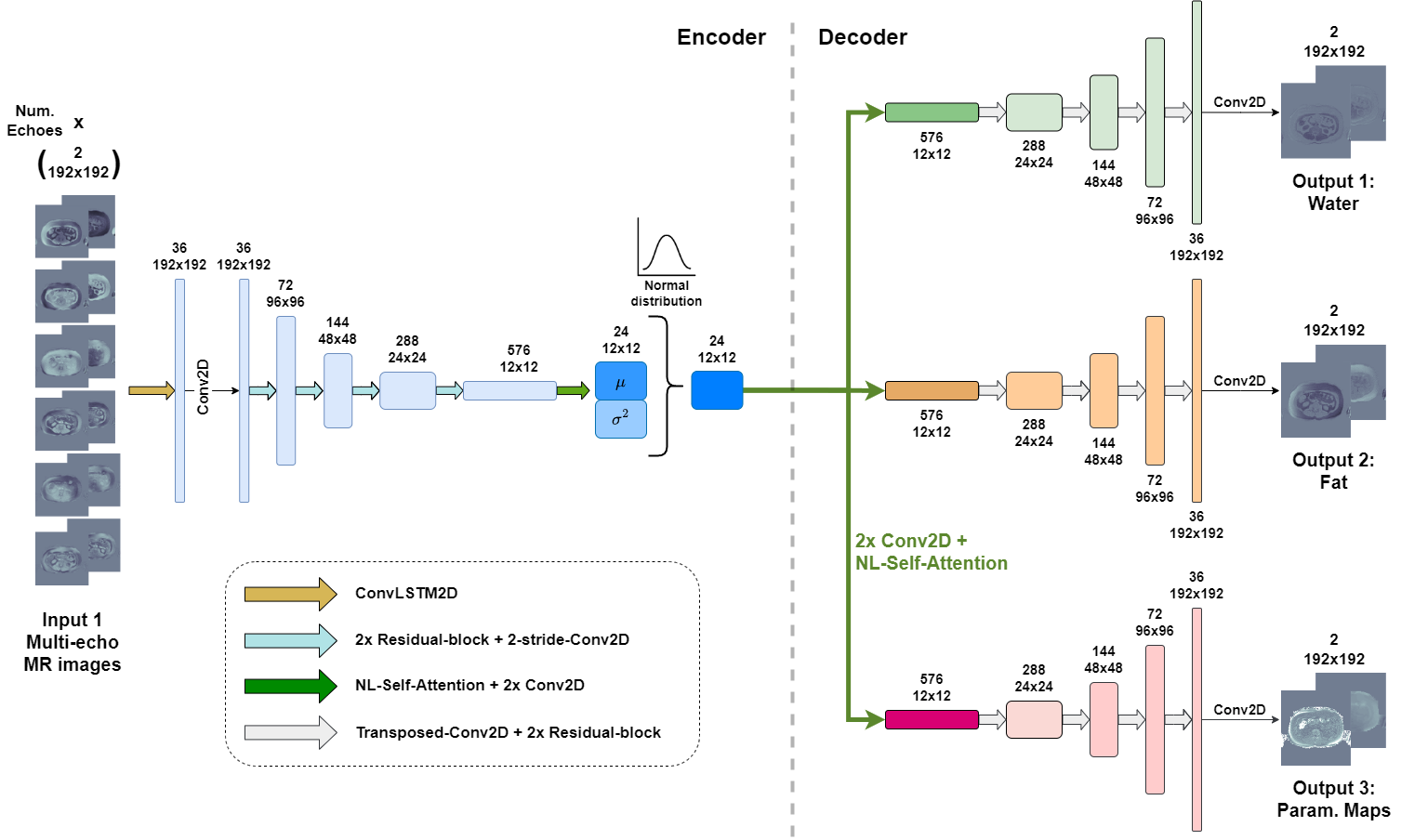
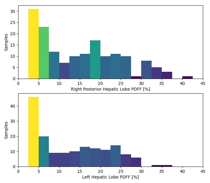
We propose a Physics-Informed Variational Auto-Encoder (PI-VAE), an attention-based convolutional neural network with an encoder-multi-decoder architecture whose outputs are the variables involved in the MR signal model (ρW, ρF, R2*, ϕ), whilst the input are series of multi-echo liver CSE-MR images (Figure 1). Specifically, PI-VAE is composed of three decoders to separately estimate water-only images, fat-only images, and the parametric maps R2* and ϕ, which are merged into a single complex-valued field-map: ξ=ϕ+iR2*/2π.PI-VAE was trained using multi-slice CSE-MR scans from 172 subjects (3842 axial slices) with heterogeneous levels of hepatic PDFF (Figure 2). The chosen loss function measured: 1) the perceptual similarity between original and reconstructed liver MRI ($$$\mathcal{L}_P$$$), 2) the numerical error between reference and reconstructed water/fat images (same for R2* and ϕ) ($$$\mathcal{L}_S$$$), and 3) an adversarial loss that assessed the realism of the generated MR images ($$$\mathcal{L}_D$$$):
$$\mathcal{L}=\alpha_P\cdot\mathcal{L}_P+\alpha_S\cdot\mathcal{L}_S+\alpha_D\cdot\mathcal{L}_D+\alpha_{KL}\cdot\mathcal{L}_{KL}$$
The latent space was regularized to achieve a zero-centered and low-variance feature space ($$$\mathcal{L}_{KL}$$$), which enables the posterior generation from latent space features that are randomly sampled from a standard normal distribution. Then, since the decoders can generate ρW, ρF, R2*, ϕ from the sampled latent space, our framework enables the generation of liver CSE-MR images with customizable echo-times and field-strengths (by varying the fat spectrum).
Results
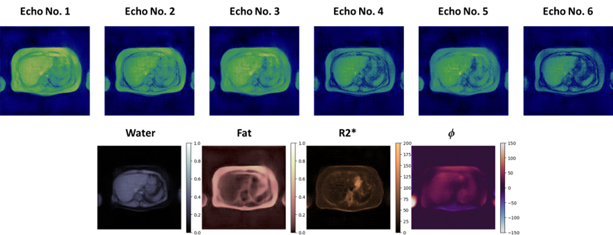
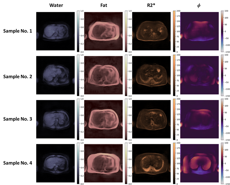
As showed in Figure 3, PI-VAE can generate realistic liver water-fat images and parametric maps (R2*, ϕ) with random geometries, and multi-echo MR images at customized echo times.Compared to a validation dataset (384 slices, unobserved during training), a randomly synthesized dataset showed a similar distribution. Distribution similarity metrics such as Fréchet Inception Distance (FID) and Maximum Mean Discrepancy (MMD) were 9.65% and 0.46%, respectively. Both scores measure the distance between two different distributions, and therefore values closer to zero indicate a greater similarity. Moreover, single and Multi-Scale (MS) Structural Similarity Index Measure (SSIM) scores between pairs of synthetic samples also showed an acceptable variability (SSIM = 78.72%; MS-SSIM = 56.37%), as shown in Figure 4.
Finally, we also trained a previously proposed DL-based method for PDFF quantification5 only using synthetic data, and we compared it to the original version trained using a data-augmentation workflow. Considering measurements at two ROIs on a liver CSE-MRI testing dataset with different echo-times, we observed a bias of 0.81±2.57% and -0.41±0.88% for synthetic-data and original versions, respectively. Reference values were obtained using iterative Graph Cuts6. Nevertheless, reproducibility coefficients (RDC) of 1.78±1.00 and 6.96±13.64% for synthetic-data and original versions, respectively, showed a precision diminishing.
Discussion
PI-VAE can synthesize realistic liver CSE-MR images with various geometries, along with their water/fat compositions. Unlike previously proposed generative models for similar medical imaging applications, PI-VAE allows an easy and reliable adjustment of the scanning parameters by incorporating the signal generation physics into the generation process.Moreover, due to the reduced dimensionality of the latent space, the generative process is highly efficient in terms of computational cost. This represents a great opportunity to train DL-based methods using data synthesized with PI-VAE, since the generation could be performed during training by randomly sampling latent spaces and decoding them at each iteration.
Conclusion
The generation of realistic and diverse liver CSE-MR images using our PI-VAE can be useful to address the lack of large datasets to train DL methods. PI-VAE may also be extended to artificially synthesize other MR modalities related to an analytical physical model (i.e.: MR relaxometry, QSM, etc.).Acknowledgements
This work was funded by Fondecyt 1200839. J.M. was funded by the National Agency for Research and Development (ANID) / Scholarship Program / DOCTORADO BECAS CHILE/2020 – 21210665. CT was funded by Fondecyt 1231535 and Millennium Institute for Intelligent Healthcare Engineering, iHEALTH (ICN2021_004).References
1. Eslam, M. et al. A new definition for metabolic dysfunction-associated fatty liver disease: An international expert consensus statement. J Hepatol 73, 202–209 (2020).
2. Idilman, I. S. et al. Hepatic steatosis: Quantification by proton density fat fraction with MR imaging versus liver biopsy. Radiology 267, 767–775 (2013).
3. Daudé, P. et al. Comparative review of algorithms and methods for chemical‐shift‐encoded quantitative fat‐water imaging. Magn Reson Med (2023) doi:10.1002/mrm.29860.
4. Hamilton, G. et al. In vivo characterization of the liver fat 1H MR spectrum. NMR Biomed 24, 784–790 (2011).
5. Meneses, J. P. et al. Reproducible DL-based approach for liver PDFF quantification. in ISMRM Annual Meeting (2023).
6. Hernando, D., Kellman, P., Haldar, J. P. & Liang, Z. P. Robust water/fat separation in the presence of large field inhomogeneities using a graph cut algorithm. Magn Reson Med 63, 79–90 (2010).
Figures