2239
Three-Dimensional Amyloid-Beta PET Synthesis from Structural MRI with Conditional Generative Adversarial Networks1Biomedical Engineering, University of Calgary, Calgary, AB, Canada, 2Electrical & Software Engineering, University of Calgary, Calgary, AB, Canada, 3Radiology, Cumming School of Medicine, University of Calgary, Calgary, AB, Canada, 4Hotchkiss Brain Institute, University of Calgary, Calgary, AB, Canada
Synopsis
Keywords: AI/ML Image Reconstruction, Alzheimer's Disease, MRI, PET, Image Translation
Motivation: Alzheimer’s Disease hallmarks include amyloid-beta deposits and brain atrophy, detectable via PET and MRI scans, respectively. PET is expensive, invasive and exposes patients to ionizing radiation. MRI is cheaper, non-invasive, and free from ionizing radiation but limited to measuring brain atrophy.
Goal(s): To develop an 3D image translation model that synthesizes amyloid-beta PET images from T1-weighted MRI, exploiting the known relationship between amyloid-beta and brain atrophy.
Approach: The model was trained on 616 PET/MRI pairs and validated with 264 pairs.
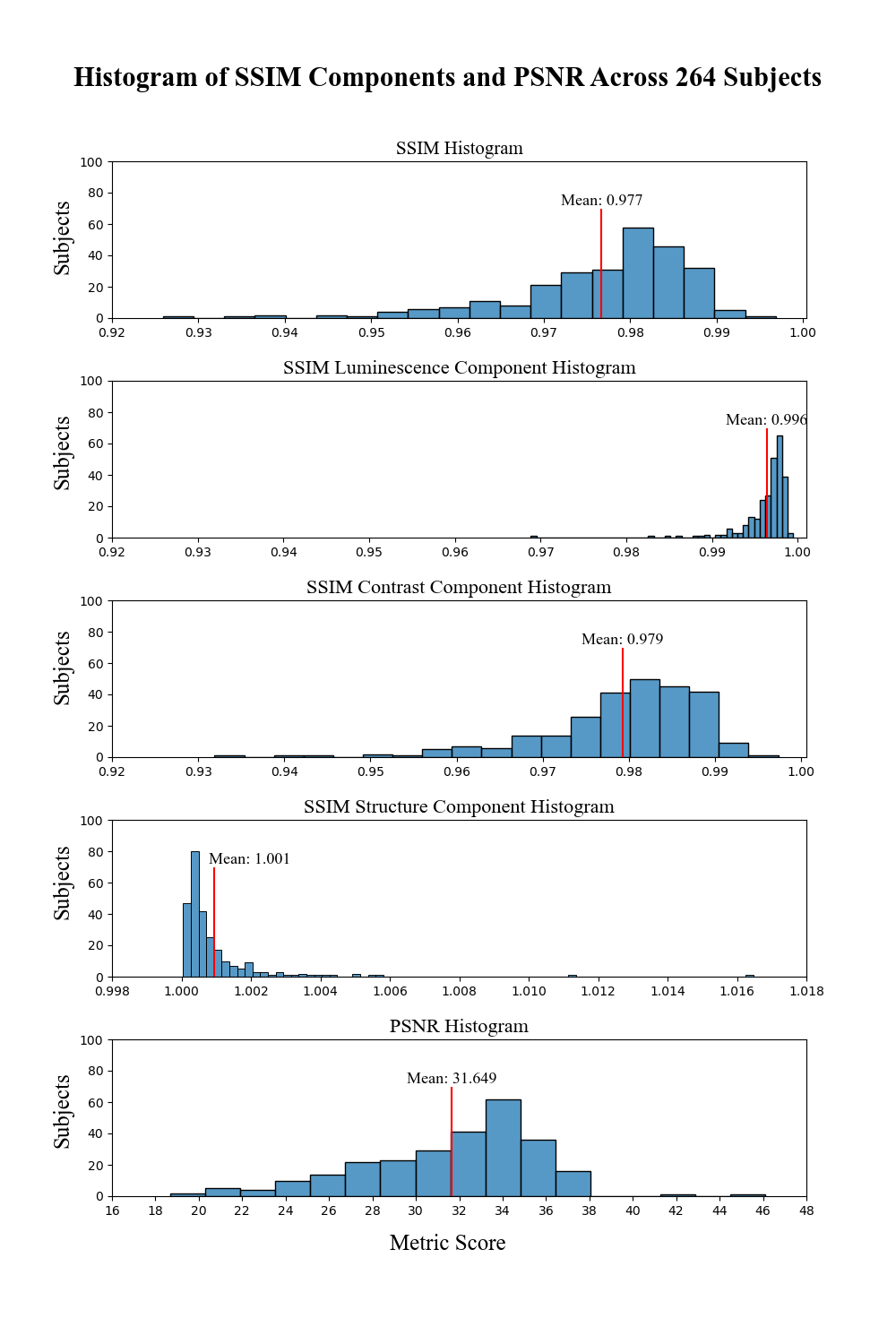
Results: The model synthesized amyloid-beta PET images from T1-weighted MRI with high-degree of similarity showing high SSIM and PSNR metrics (SSIM>0.97&PSNR=34).
Impact: Our model proves the feasibility of synthesizing amyloid-beta PET images from structural MRI ones, significantly enhancing accessibility for large-cohort studies and early dementia detection, while also reducing cost, invasiveness, and radiation exposure.
Introduction
Alzheimer’s Disease (AD) is the predominant form of dementia.1 AD is a progressive neurodegenerative disease characterized by memory impairment and cognitive decline.2 Dementia had an enormous global economic burden of $1.3 trillion USD in 2019.3 Histopathologically, AD is characterized by the deposition of amyloid-beta within the brain, a key molecule associated with AD progression.4 When amyloid-beta accumulates between neurons, it can lead to synaptic failure and neuronal death, manifesting as brain atrophy.5 In recent decades, imaging amyloid-beta has become possible using positron emission tomography (PET) with radiotracers such as Pittsburgh Compound B (PiB)6. However, PET imaging is costly ($5000-$8000 per scan)7, invasive (requiring injection of a radiotracer), and involves exposure to harmful ionizing radiation.8 These factors limit its availability in many jurisdictions and make it challenging to use for early-onset AD detection.Structural MRI is a more affordable, non-invasive, alternative to PET that does not use ionizing radiation.9 While it primarily targets brain atrophy driven by AD, MRI cannot provide amyloid-beta measurements as PET does.10 Addressing this limitation, image translation models have been developed to generate synthetic amyloid-beta PET images from structural MRI11,12, using the known relationship between amyloid-beta burden and brain atrophy.13,14
Image translation models can generate synthetic images from other image types, providing access to images that might be difficult to obtain.15 These models are commonly implemented with Conditional Generative Adversarial Networks (cGAN) due to their ability to generate realistic synthetic images.16 Prior research has demonstrated success in generating 2D axial amyloid-beta PET images from structural MRI.17
This work builds on the previous study by introducing a 3D-cGAN model with significant architectural improvements, enhancing the amyloid-beta PET synthesis shown in the image comparison metrics.
Methods
The Open Access Series of Imaging Studies (OASIS-3)18 dataset included 1098 subjects with both PiB PET and MRI images, comprising 609 cognitively normal (CN) individuals and 489 at different levels of cognitive decline. The MRI images were captured using three scanners: Siemens Biograph mMR 3T, Siemens Trio Tim 3T, and Siemens Sonata 1.5T with a resolution of 1mm´1mm´1mm. The amyloid-beta images were acquired with two scanners: Siemens Biograph 40 PET/CT and Siemens ECAT 962 using PiB radiotracer with an injected dose ranging from 6 to 20 millicuries and a 60-minutes dynamic scan and standard uptake value ratio (SUVR) was obtained using the PET Unified Pipeline.The MRI images were preprocessed with FreeSurfer to produce brain extractions. Dynamic PET images were converted to static by summing frames between 30 and 60 minutes. These images were co-registered to the MRI images using an affine registration with the Advanced Normalization Tools (ANTs), and brain extraction was performed using the MRI as a mask. PET images were SUVR normalized by dividing the mean tracer concentration in the cerebellum cortex, a standard reference region for PiB PET imaging.19,20
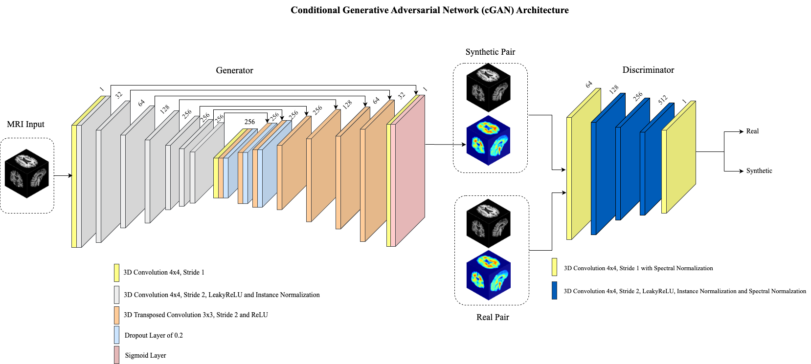
The 3D-cGAN architecture as shown in Figure 1 was implemented using MONAI. A brain area mask loss was incorporated to focus on brain information while disregarding the background. To prevent exploding gradients, spectral normalization was applied in the discriminator. PET images were z-score normalized to a range between 0 and 1, with a mean=0 and standard-deviation=49.72. This normalization facilitates denormalization without compromising tracer quantification and improves the stability of the training process.
The model was trained with 616 PET/MRI pairs and validated with 264 pairs. The training and validation cohorts were stratified with equal proportion of females/males and different levels of cognitive decline.
The validation cohort was evaluated with Structural Similarity Index Measure (SSIM)21 and Peak Signal-to-Noise Ratio (PSNR).22
Results
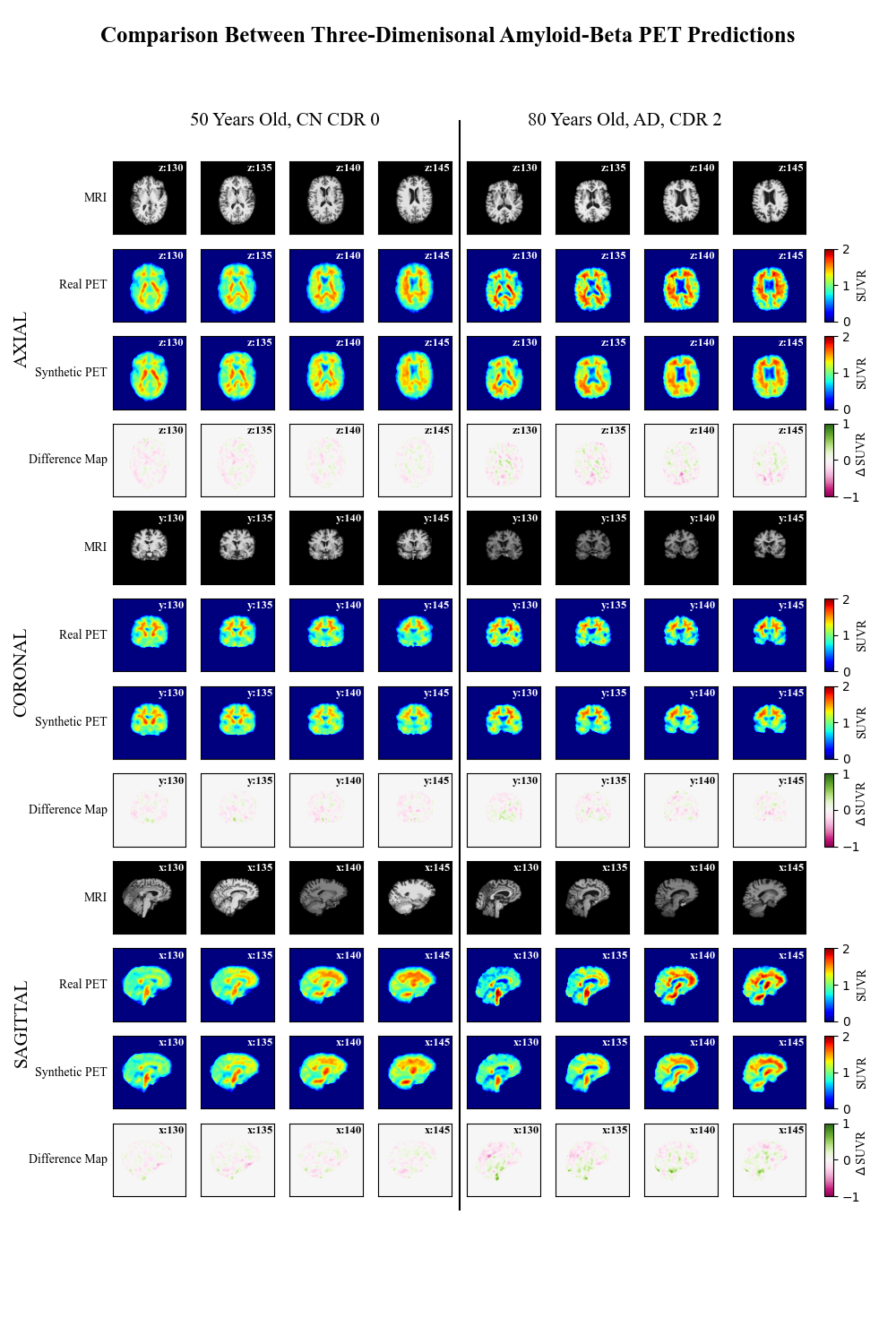
Figure 2 showcases two representative subjects, illustrating the model’s ability to synthesize high-fidelity amyloid-beta PiB PET images from T1-weighted MRI for both CN and AD cases, presented in three anatomical planes. Figure 3 displays the distribution of SSIM in contrast, luminescence, and structural components, alongside the PSNR. The model achieved a mean SSIM=0.977 and PSNR=31.64Discussion
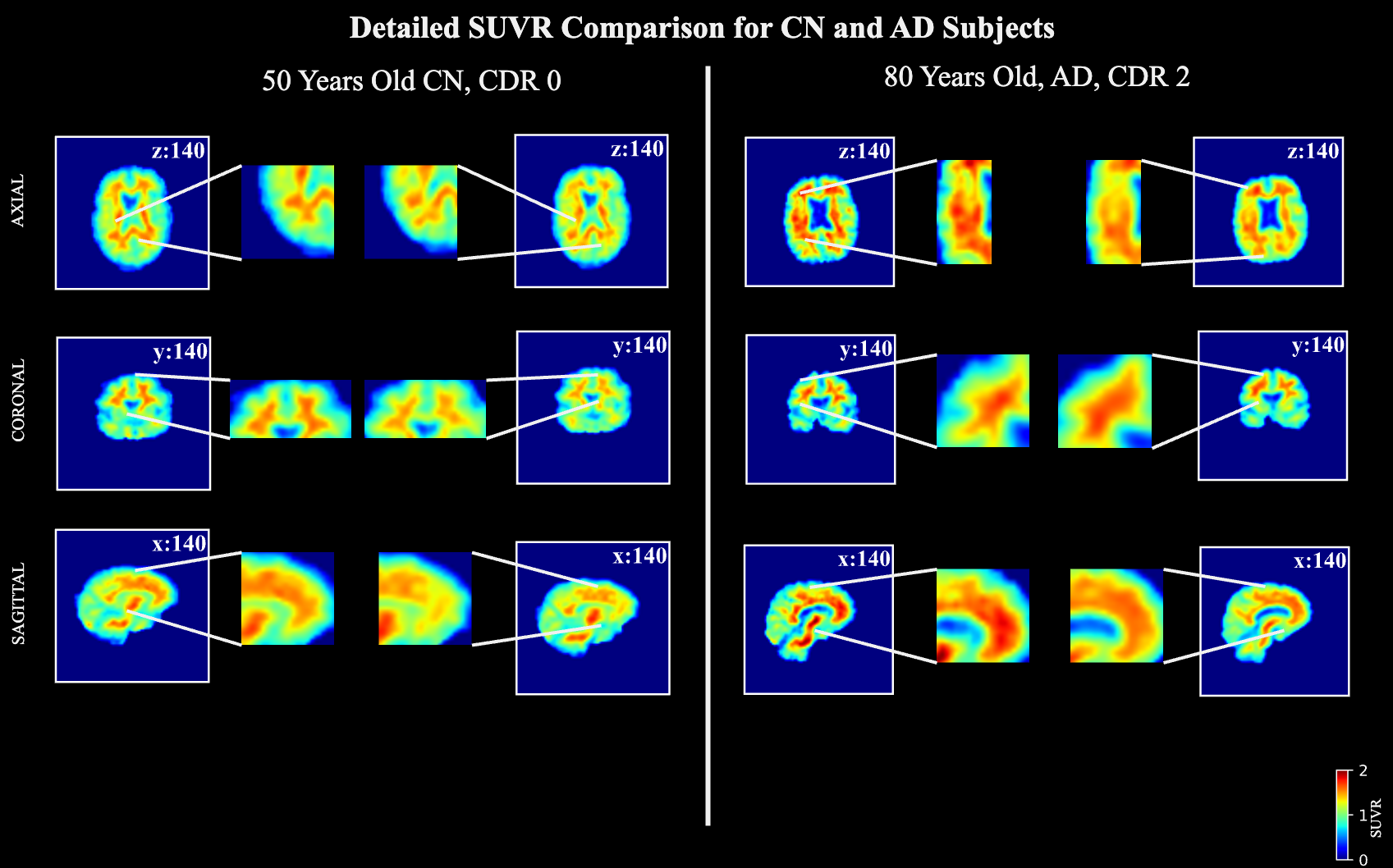
Future work will aim to improve the precision of the synthetic PET images, particularly for subtle discrepancies shown in Figure 4. This can be addressed by exploring model architectures such as Self-Attention Conditional GAN, vision transformers GAN, and stable diffusion models. A prevalent challenge in GAN is the emphasis on relative image quality at the expenses of quantitative contrast accuracy. Future work will include developing metrics that better quantify contrast fidelity.Conclusion
This study demonstrates that a three-dimensional image translation model is capable of synthesizing amyloid-beta PiB PET images from structural MRI with high-degree of similarity, achieving mean SSIM>0.97 and PSNR>31. This approach offers a cost-effective, less-invasive, and radiation-free method for early AD screening.Acknowledgements
The authors would like to thank the University of Calgary, in particular the Schulich School of Engineering and Departments of Biomedical Engineering and Electrical & Software Engineering; the Cumming School of Medicine and the Departments of Radiology and Clinical Neurosciences; as well as the Hotchkiss Brain Institute, Research Computing Services and the Digital Alliance of Canada for providing resources. The authors would like to thank the Open Access of Imaging Studies Team for making the data available. JA – is funded in part from a graduate scholarship from the Natural Sciences and Engineering Research Council Brain Create. MEM acknowledges support from Natural Sciences and Engineering Research Council of Canada Discovery Grant (RGPIN-03552), Early Career Researcher Supplement (DGECR-00124), and Natural Sciences and Engineering Research Council (NSERC) Alliance – Alberta Innovates Advance Program Grant (ALLRP 580342-22).References
1. Wimo, A., et al., The worldwide costs of dementia in 2019. Alzheimer's & Dementia, 2023. 19(7): p. 2865-2873.
2. Kumar, A., et al., Alzheimer Disease, in StatPearls. 2022: Treasure Island (FL).
3. Wong, W., Economic burden of Alzheimer disease and managed care considerations. Am J Manag Care, 2020. 26(8 Suppl): p. S177-S183.
4. Sadigh-Eteghad, S., et al., Amyloid-beta: a crucial factor in Alzheimer's disease. Med Princ Pract, 2015. 24(1): p. 1-10.
5. Sala-Llonch, R., et al., Inflammation, Amyloid, and Atrophy in The Aging Brain: Relationships with Longitudinal Changes in Cognition. J Alzheimers Dis, 2017. 58(3): p. 829-840.
6. Yamin, G. and D.B. Teplow, Pittsburgh Compound-B (PiB) binds amyloid beta-protein protofibrils. J Neurochem, 2017. 140(2): p. 210-215.
7. Wittenberg, R., et al., Economic impacts of introducing diagnostics for mild cognitive impairment Alzheimer's disease patients. Alzheimers Dement (N Y), 2019. 5: p. 382-387.
8. Marti-Climent, J.M., et al., Effective dose estimation for oncological and neurological PET/CT procedures. EJNMMI Res, 2017. 7(1): p. 37.
9. McMahon, K.L., G. Cowin, and G. Galloway, Magnetic Resonance Imaging: The Underlying Principles. Journal of Orthopaedic & Sports Physical Therapy, 2011. 41(11): p. 806-819.
10. Vemuri, P. and C.R. Jack, Jr., Role of structural MRI in Alzheimer's disease. Alzheimers Res Ther, 2010. 2(4): p. 23.
11. Zhang, J., et al., BPGAN: Brain PET synthesis from MRI using generative adversarial network for multi-modal Alzheimer's disease diagnosis. Comput Methods Programs Biomed, 2022. 217: p. 106676.
12. Sikka, A., et al. MRI to PET Cross-Modality Translation using Globally and Locally Aware GAN (GLA-GAN) for Multi-Modal Diagnosis of Alzheimer's Disease. 2021. arXiv:2108.02160.
13. Tosun, D., et al., Spatial patterns of brain amyloid-beta burden and atrophy rate associations in mild cognitive impairment. Brain, 2011. 134(Pt 4): p. 1077-88.
14. Chételat, G., et al., Relationship between atrophy and β-amyloid deposition in Alzheimer disease. Annals of Neurology, 2010. 67(3): p. 317-324.
15. Goodfellow, I.J., et al. Generative Adversarial Networks. 2014. arXiv:1406.2661.
16. Isola, P., et al., Image-to-Image Translation with Conditional Adversarial Networks. 30th Ieee Conference on Computer Vision and Pattern Recognition (Cvpr 2017), 2017: p. 5967-5976.
17. Vega, F., et al., Image Translation for Estimating Two-Dimensional Axial Amyloid-Beta PET From Structural MRI. Journal of Magnetic Resonance Imaging. n/a(n/a).
18. LaMontagne, P.J., et al., OASIS-3: Longitudinal Neuroimaging, Clinical, and Cognitive Dataset for Normal Aging and Alzheimer Disease. medRxiv, 2019.
19. Price, J.C., et al., Kinetic Modeling of Amyloid Binding in Humans using PET Imaging and Pittsburgh Compound-B. Journal of Cerebral Blood Flow & Metabolism, 2005. 25(11): p. 1528-1547.
20. Jack, C.R., Jr., et al., Serial PIB and MRI in normal, mild cognitive impairment and Alzheimer's disease: implications for sequence of pathological events in Alzheimer's disease. Brain, 2009. 132(Pt 5): p. 1355-65.
21. Zhou, W., et al., Image quality assessment: from error visibility to structural similarity. IEEE Transactions on Image Processing, 2004. 13(4): p. 600-612.
22. Horé, A. and D. Ziou. Image Quality Metrics: PSNR vs. SSIM. in 2010 20th International Conference on Pattern Recognition. 2010.
Figures