2233
Synthesizing high-resolution brain MR T1-MPRage-like Images from low-dose CT1Neurology, Washington University School of Medicine, St. Louis, MO, United States, 2Mallinckrodt Institute of Radiology, Washington University School of Medicine, St. Louis, MO, United States
Synopsis
Keywords: Other AI/ML, Machine Learning/Artificial Intelligence, image conversion, MR to CT conversion, image synthesis
Motivation: Using deep learning to improve the soft tissue contrast in low-dose brain CT similar to MRI.
Goal(s): Converting low-dose brain CT to high-resolution T1-MPRAGE-like Images.
Approach: A ResUNet-based deep learning approach is developed to learn the complex transformation from low-dose brain CT to its corresponding T1-MPRAGE.
Results: With the proposed approach, we obtained high-resolution MR T1-MPRAGE-like images with superior soft-tissue contrasts from noisy low-dose brain CT images.
Impact: By transferring brain CT to T1-MPRAGE-like images, our approach provides superior soft tissue contrast from low-dose brain CT images. Our method would allow tissue-specific analysis using noisy non-contrast CT.
Introduction
For clinical stroke imaging, CT is the modality of choice because of its easy accessibility and fast imaging speed. The major limitation of CT is its poor soft tissue contrast. On the contrary, MRI images such as brain T1-MPRAGE provide excellent soft tissue contrast, but it is challenging to obtain in emergencies. In this study, we will explore the potential of using deep learning with paired low-dose CT and brain T1-MPRAGE MRI acquired from the same subjects to improve soft tissue contrast of low-dose brain CT.Methods
This is an IRB-approved study. We employed a previously developed 3D ResUNet deep learning network [1] to learn the conversion from low-dose brain CT to T1-MRPAGE in the same patients. The low-dose CT images were acquired at 120 kVp with a voxel size = 0.59x0.59x3mm3. Brain T1-MPRAGE images were preprocessed with the FreeSurfer software package. Afterward, the CTs were registered to their corresponding T1s with the FSL linear registration toolkit. In total, we acquired 206 low-dose CT/T1-MPRAGE image pairs from the same patients (mean age: 70.05; 56% Female). These data were randomly divided into training (94), validation (10), and testing (102) sets. The L1 loss between the estimated brain T1-MPRAGE and its corresponding ground truth was minimized during the training process. Validation data were used to select the best model, which was then deployed to convert the low-dose CTs of the 102 test subjects. The converted T1-MPRAGE images were compared to their corresponding ground-truth images in image similarity measures such as structural similarity index (SSIM), peak signal-to-noise ratio (PSNR), and mutual information (MI). Moreover, we compared the brain tissue segmentation results from the converted images with the ground-truth segmentation, evaluating the volume correlation and DICE overlapping ratios in CSF, GM, and WM.Results
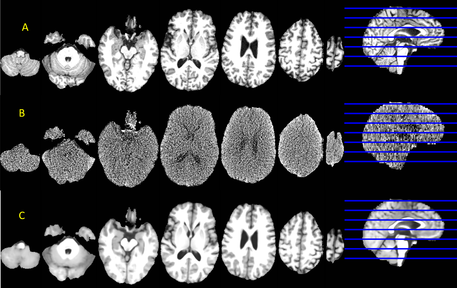
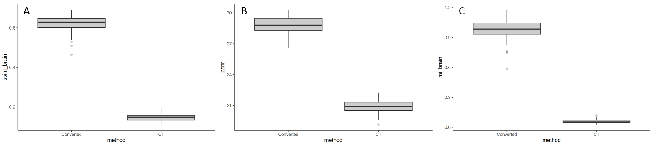
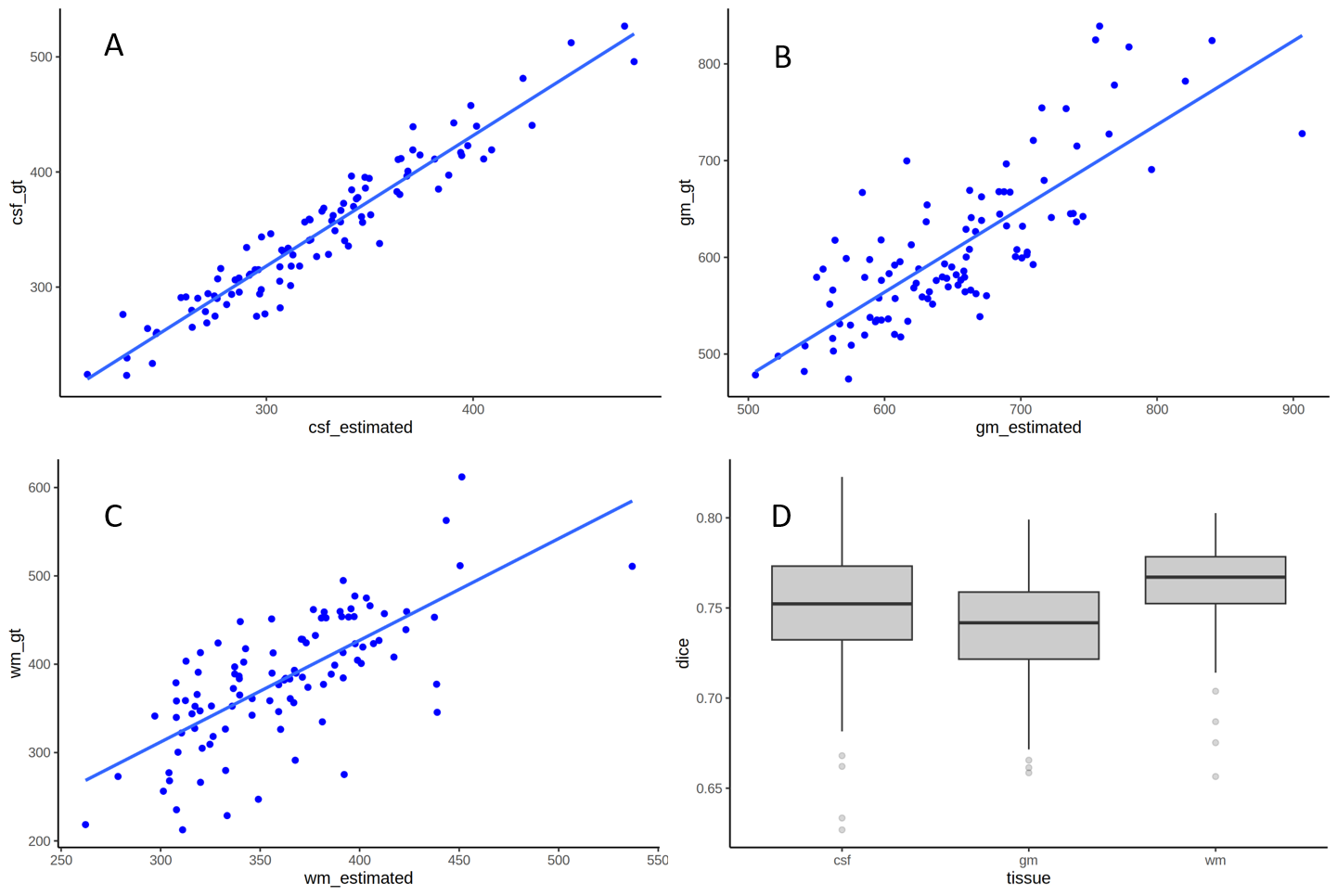
An original T1-MPRAGE, low-dose CT, and the converted T1-MPRAGE images were given in Fig. 1 for one testing subject. Even though the low-dose CT provides minimal anatomical information except ventricular CSF, this deep learning approach produced a synthetic MRI with high similarity to the original brain T1-MPRAGE image. The synthetic image has a significant improvement in image quality measured by all similarity metrics (p<10-15, Fig. 2). Moreover, the volumetric results using the converted MRI had significant correlations to brain tissue volumes in CSF (r=0.957, p<10-15), GM (r=0.789, p<10-15) and WM (r=0.706, p<10-15), and high DICE similarity coefficient with the ground-truth segmentation (Dice: 0.750+0.036 in CSF, 0.738+0.028 in GM and 0.763+0.026 in WM).Conclusion
To the best of our knowledge, this may be the first large data study converting low-dose CT to T1-MPRAGE-like images with a well-quantified validation. Despite the high noise in the low-dose CTs, our approach can provide superior brain soft-tissue contrasts, as demonstrated by the significant volume correlations and high DICE similarity coefficient in CSF, GM, and WM.Acknowledgements
This study was supported by NIH grants RF1 NS116565 and 1R01EB032713.References
[1] Yasheng Chen, Chunwei Ying, Michael M Binkley, Meher R Juttukonda, Shaney Flores, Richard Laforest, Tammie L S Benzinger, Hongyu An. Deep learning-based T1-enhanced selection of linear attenuation coefficients (DL-TESLA) for PET/MR attenuation correction in dementia neuroimaging. MRM 86(1):499-512 (2021)Figures