2230
Neural Network-based MR Elastography Wave Inversion Using Physics-based Simulations and Uncertainty Quantification1Charité - Universitätsmedizin Berlin, Berlin, Germany, 2Institute of Computer-Assisted Cardiovascular Medicine, Deutsches Herzzentrum der Charité (DHZC), Berlin, Germany, 3Berlin Institute of Health at Charité – Universitätsmedizin Berlin, BIH Biomedical Innovation Academy, BIH Charité Digital Clinician Scientist Program, Berlin, Germany, 4Fraunhofer MEVIS, Berlin, Germany, 5Institute of Medical Informatics, Berlin, Germany, 6DZHK (German Center for Cardiovascular Research), Partner Site Berlin, Berlin, Germany
Synopsis
Keywords: AI/ML Image Reconstruction, Elastography
Motivation: In Magnetic Resonance Elastography (MRE), accurate reconstruction of stiffness maps is essential for medical diagnosis. Traditional inversion techniques are limited by noise, discretization and/or low wavenumbers.
Goal(s): We aim to overcome these limitations using a neural network-based wave inversion (ElastoNet) with integrated uncertainty quantification ensuring reliable predictions with high detail resolution.
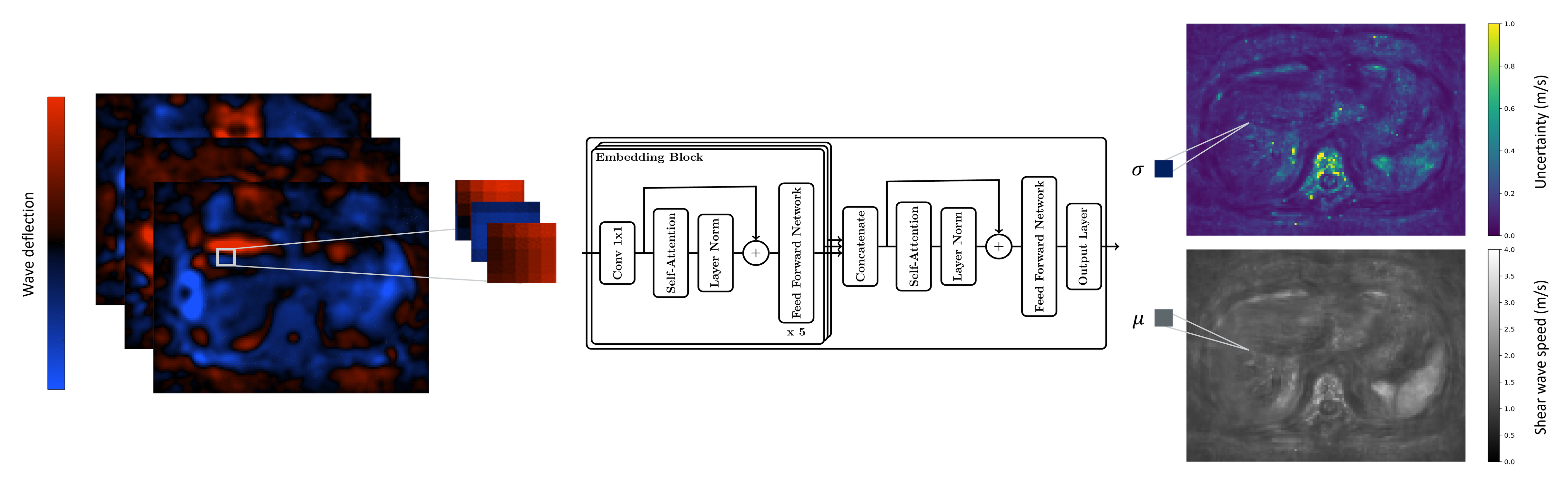
Approach: We trained ElastoNet on simulated wave patches. For inference, we combined all 3 motion encoding directions as input and used evidential deep learning as an uncertainty quantification method.
Results: ElastoNet achieves a substantial improvement in detail resolution compared to current neural network approaches and shows promising results in the low-frequency domain.
Impact: Our MR elastography neural network-based wave inversion is a promising method for enhanced accuracy and reliability in tissue property characterization. It effectively addresses challenges in reconstruction of stiffness maps, expanding the potential of MR elastography for medical diagnosis.
Introduction
Magnetic Resonance Elastography (MRE) is a well-established non-invasive technique for quantifying mechanical properties of soft tissues1. Accurate wave inversion algorithms used for the reconstruction of elastograms are crucial for quantitative medical diagnosis. Traditional approaches encounter significant challenges when dealing with noisy images or low-frequency excitations, and require careful parameter tuning. Recent neural network approaches2,3,4 have attempted to tackle these issues but are still sensitive to noise and reduced detail resolution. To overcome these limitations, we propose ElastoNet, a neural network-based inversion that combines multiple motion encoding gradient (MEG) directions as input and includes a measure of uncertainty using evidential deep learning5 for reliable wave inversion and stiffness reconstruction.Methods
We trained a neural network on 5x5 pixel simulated wave patches using the travelling wave expansion model4,6. Assuming a homogeneous, isotropic, and viscoelastic material, we expanded previous use of this model4 by including the compression wave in the simulations. The complex wave displacement $$$\mathbf{u}$$$ at a given location $$$\mathbf{r}$$$ is expressed as a superposition of shear waves, compression waves, and noise:$$u(\mathbf{r}) = \sum_{j=1}^{N_s} a_j e^{-i(k_{sj}\mathbf{r}+\phi_j)} + \sum_{j=1}^{N_c} a_j e^{-i(k_{cj}\mathbf{r}+\phi_j)} + \eta(\mathbf{r})$$
where $$$a_j$$$ and $$$\phi_j$$$ are the amplitude and phase of the wave, $$$N_s$$$ and $$$N_c$$$ are respectively the number of superimposed shear and compression wave sources located in 3D, $$$k_s$$$ and $$$k_c$$$ are respectively the wavenumbers of the shear and compression waves. The noise term $$$\eta(\mathbf{r})$$$ was chosen as zero-mean Gaussian noise.
The neural network was trained for 10000 epochs. Each input sample consisted of three randomly simulated wave patches with identical wavenumbers, generated during training. In the model, the patches were sequentially passed through an embedding block and then aggregated (see Figure 1). The output consisted of the shear wave speed prediction and corresponding epistemic uncertainty. The negative log-likelihood5 was used as loss function.
During inference, following phase unwrapping, the vibration frequency was selected using a temporal Fourier transform, and the images were smoothed. 5x5 image patches were then sampled from the wavefield for all MEG components and passed as input to the network. Multiple vibration frequencies were processed separately and finally averaged. A simple average is used in ElastoNet and an uncertainty-weighted average in w-ElastoNet.
We tested our neural network on phantom data7 and in-vivo data of the abdomen of a healthy volunteer acquired with a 1.5T-MRI scanner (Magnetom Aera, Siemens, Erlangen) using a single-shot spin-echo planar imaging sequence and multifrequency vibration from 10Hz to 80Hz.
Results
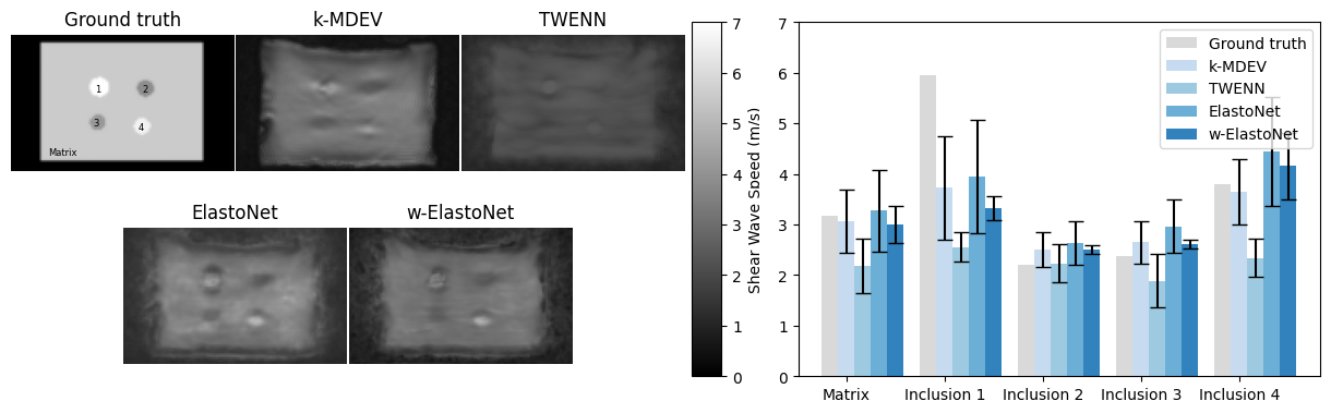
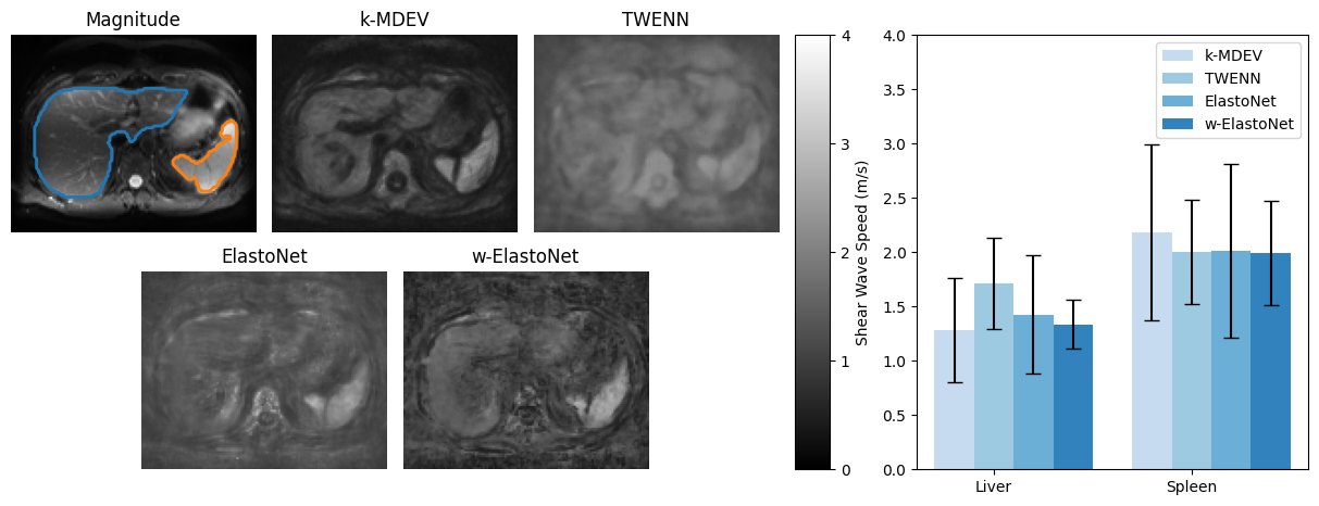
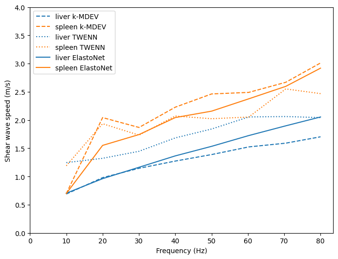
We compared ElastoNet and w-ElastoNet with two existing approaches, a classical wavenumber-based k-MDEV8, and a neural network-based TWENN4. Obtained stiffness maps for both datasets averaged across frequencies are compared in Figures 2 and 3. The proposed models provided better detail resolution compared to TWENN and close performance to k-MDEV regarding quantitative results in both datasets. w-ElastoNet additionally provides more stable stiffness maps as seen in the lower standard deviation. Additionally, frequency-resolved stiffness maps show better stability over frequencies for ElastoNet compared to the reference methods (Figure 4).Discussion
We developed a machine learning-based inversion ElastoNet that computes stiffness maps with high detail resolution and reliable stiffness estimation even at low frequencies.Simultaneous training on 3 MEG directions provided better input features to the model resulting in an overall better performance. By using a larger patch size than in the TWENN model, the resulting stiffness maps were less noisy and did not require smoothing.
Frequency-resolved analysis (Figure 4) showed that ElastoNet achieved better stability for longer wavelengths compared to other reference methods. This is evident in the spleen, where ElastoNet demonstrated a more consistent slope for changes in stiffness across frequencies.
One notable feature of our approach is the direct quantification of the uncertainty. This was achieved without incurring additional computational costs during inference as it is learned during the training.
However, our method still faces certain limitations. It was highly sensitive to noise at higher frequencies and was still outperformed by k-MDEV in this domain. Furthermore, due to the limited field of view of 5x5 patches, very long wavelengths were still challenging to distinguish as can be seen for the spleen at 10Hz in Figure 4.
Conclusion
In summary, our neural network-based wave inversion method presents promising improvements compared to existing neural network approaches and provides a direct measure of uncertainty of the predictions. While our approach closely matches state-of-the-art methods and proves more stable at lower frequencies, noise sensitivity remains a challenge at higher frequencies. Accommodating longer waves beyond the current capabilities of classical wave inversion methods offers a promising way for wave inversion and tissue properties characterization.Acknowledgements
Rolf Reiter is a participant of the BIH-Charité Digital Clinician Scientist Program funded by Charité – Universitätsmedizin Berlin, Berlin Institute of Health and the DFG.Financial support of the German Research Foundation (CRC 1340 Matrix in Vision, GRK 2260 BIOQIC) is gratefully acknowledged.
Special thanks to Yuang Feng from Shanghai Jiao Tong University, China for the provision of TWENN and helpful discussions.
References
1. Sack I. Magnetic resonance elastography from fundamental soft-tissue mechanics to diagnostic imaging. Nat Rev Phys. 2022;5(1):25-42. doi:10.1038/s42254-022-00543-2.
2. Murphy MC, Manduca A, Trzasko JD, et al. Artificial neural networks for stiffness estimation in magnetic resonance elastography. Magnetic Resonance in Med. 2018;80(1):351-60. doi:10.1002/mrm.27019.
3. Scott JM, Arani A, Manduca A, et al. Artificial neural networks for magnetic resonance elastography stiffness estimation in inhomogeneous materials. Medical Image Analysis. 2020;63:101710. doi:10.1016/j.media.2020.101710.
4. Ma S, Wang R, Qiu S, et al. MR Elastography With Optimization-Based Phase Unwrapping and Traveling Wave Expansion-Based Neural Network (TWENN). IEEE Trans Med Imaging. 2023;42(9):2631-42. doi:10.1109/TMI.2023.3261346.
5. Amini A, Schwarting W, Soleimany A, et al. Deep Evidential Regression. arXiv. 2019. doi:10.48550/arXiv.1910.02600.
6. Baghani A, Salcudean S, Honarvar M, et al. Travelling Wave Expansion: A Model Fitting Approach to the Inverse Problem of Elasticity Reconstruction. IEEE Trans Med Imaging. 2011;30(8):1555-65. doi:10.1109/TMI.2011.2131674.
7. Papazoglou S, Hirsch S, Braun J, et al. Multifrequency inversion in magnetic resonance elastography. Phys Med Biol. 2012;57(8):2329-46. doi:10.1088/0031-9155/57/8/2329.
8. Tzschätzsch H, Guo J, Dittmann F, et al. Tomoelastography by multifrequency wave number recovery from time-harmonic propagating shear waves. Medical Image Analysis. 2016;30:1-10. doi:10.1016/j.media.2016.01.001.
Figures