2210
Brain Iron Deposition in the Posterior Part of the Right Hippocampus Associated with Cognitive Functions in Cerebral Small Vessel Disease1Department of Radiology, Shandong Provincial Hospital Affiliated to Shandong First Medical University, Jinan, Shandong, China., Jinan, Shandong, China, China, 2Shandong First Medical University, Jinan, Shandong, China., Jinan, Shandong, China., China, 3Department of Psychiatry and Psychotherapy, Jena University Hospital, Jena, Germany., Jena, Germany., Germany, 4Center for Intervention and Research on adaptive and maladaptive brain Circuits underlying mental health (C-I-R-C), Jena-Magdeburg-Halle, Germany., Jena, Germany., Germany, 5Department of Radiology, Beijing Tsinghua Changgung Hospital, Beijing, China., Beijing, China., China, 6School of Medical Imaging, Binzhou Medical University, Yantai, Shandong, China., Yantai, Shandong, China., China
Synopsis
Keywords: Blood Vessels, Quantitative Susceptibility mapping, cerebral small vessel disease, hippocampus, iron deposition, cognition.
Motivation: The deposition of excess iron in subcortical nuclei may be linked to the burden of cerebral small vessel disease (CSVD) and could contribute to decreased cognitive performance.
Goal(s): To investigate the relationship between iron deposition in subcortical nuclei and CSVD severity, as well as cognitive impairment.
Approach: Brain iron deposition was assessed by quantitative susceptibility mapping (QSM).
Results: Susceptibility in the posterior part of the right hippocampus (pHIP-R) was negatively correlated with cognitive performance and positively correlated with CSVD severity.
Impact: Iron deposition in pHIP-R could be an early biomarker of CSVD-related cognitive impairment in future research, providing new evidence for investigating the mechanism of cognitive impairment in CSVD.
Introduction
Cerebral small vascular disease (CSVD), a leading cause of stroke (25%) and dementia (45%), causes cognitive impairment, which is thought to be related to unusual brain iron distribution and accumulation, especially in deep gray matter1. Subcortical nuclei serve a critical function in motion, memory, navigation, and cognition and play key roles in cognitive impairment in neurodegenerative diseases with increased iron deposition2, and likely, got impaired along with CSVD progression and cognitive impairment. The iron concentration in human brain, as one of the tissue magnetic susceptibility sources, could be reflected noninvasively by quantitative susceptibility mapping (QSM)3. Using susceptibility values extracted from QSM as a marker of iron, this study aimed to reveal the relationship between iron deposition in hippocampal subregions and CSVD progression, as well as cognitive impairment in CSVD.Methods
As a cross-sectional study, MRI data including QSM, cognitive tests, and blood samples, were collected from 72 healthy controls and 205 participants suffering from CSVD. Based on the total CSVD score, all participants were separated into three groups: total CSVD score = 0 (HCs), total CSVD score = 1 (CSVD-m), and total CSVD score ≥2 (CSVD-s). The subcortical regions were segmented automatically into 32 subregions (16 subregions were defined on each side of the cerebrum) by the Melbourne Subcortex Atlas4.One-way analysis of variance (ANOVA) was used to find the difference in the susceptibility values within the 32 subregions among the three groups. Pearson correlation analysis was performed, followed by multiple linear stepwise regression to investigate the relationship between susceptibility values and clinical variables. In addition, Spearman’s correlation analysis was performed, followed by multiple linear stepwise regression analysis to explore the factors influencing cognitive scores.
Results
The clinical characteristics of the participants were summarized (Figure 1). There were significant differences in the mean susceptibility of the posterior part of the right hippocampus (pHIP-R) (P = 0.032, FDR correction) and the posterior part of the right caudate nucleus (pCAU-R) (P = 0.048, FDR correction) among the three groups (Figure 2, Figure 3).In multiple linear regression, the susceptibility of the pHIP-R correlated with sex (t = -2.879, P < 0.05) and total CSVD score (t = 2.579, P < 0.05). Susceptibility in pCAU-R was related to age (t = 3.721, P < 0.05) and diabetes (t = 3.705, P < 0.05) (Figure 4). Furthermore, the susceptibility values in pHIP-R were positively correlated with the scores obtained from the Stroop color-word Test (SCWT) (t = 2.575, P < 0.05), but their correlations with scores from the Auditory Verbal Learning Test (AVLT) were not detected. And the susceptibility values in pCAU-R were negatively correlated with the scores obtained from the Montreal Cognitive Assessment (MoCA) (t = -2.477, P < 0.05) and the Symbol Digit Modalities Test (SDMT) (t = -1.992, P < 0.05), positively correlated with the Train Making Test (TMT) scores (t = 2.115, P < 0.05) (Figure 5).
Discussion
Using QSM as a marker of iron deposition and the Melbourne Subcortex Atlas to define subregions, our results showed that only the changes in susceptibility values in pHIP-R correlated with both CSVD severity and cognitive performance, presenting a promising diagnostic performance for CSVD severity. The mechanism of iron deposition in pHIP-R remains unclear, but its relationship to sex may be reasonable since hippocampal structure and function are different between males and females5. Given that the posterior hippocampus performs primarily cognitive functions, compared to the anterior hippocampus, which relates to stress, emotion, affect, and verbal memory6,7, it can be explained that there is a correlation between pHIP-R and SCWT scores but no correlation between pHIP-R and AVLT scores. Our results also indicate that the anterior and posterior hippocampus might play different roles in CSVD progression and cognitive impairment. Additionally, the susceptibility values in pCAU-R showed significant differences across the groups, correlated with age, and diabetes, but their correlations to CSVD severity were not detected. Therefore, the changes in susceptibility values in pCAU-R and related cognitive impairment may result from age.Conclusion
Our results revealed iron deposition in pHIP-R driven by CSVD and its relationship to cognitive decline. Susceptibility changes in pHIP-R measured by QSM could be a promising biomarker for cognitive decline and CSVD severity. Future studies could investigate how changes in subregional iron deposition affect cognitive function and deepen our understanding of the mechanism of cognitive impairment in CSVD.Acknowledgements
We appreciate the active cooperation of all participants and the contributions of all researchers to the data collection. Especially thanks to the esteemed professors for their guidance and revision of the abstract.
References
1. Sun, C. et al. Deep Gray Matter Iron Deposition and Its Relationship to Clinical Features in Cerebral Autosomal Dominant Arteriopathy With Subcortical Infarcts and Leukoencephalopathy Patients: A 7.0-T Magnetic Resonance Imaging Study. Stroke 51, 1750–1757 (2020).
2. Thomas, G. E. C. et al. Brain iron deposition is linked with cognitive severity in Parkinson’s disease. J. Neurol. Neurosurg. Psychiatry 91, 418–425 (2020).
3. Wang, Y. et al. Clinical quantitative susceptibility mapping (QSM): Biometal imaging and its emerging roles in patient care. J. Magn. Reson. Imaging JMRI 46, 951–971 (2017).
4. Tian, Y., Margulies, D. S., Breakspear, M. & Zalesky, A. Topographic organization of the human subcortex unveiled with functional connectivity gradients. Nat. Neurosci. 23, 1421–1432 (2020).
5. Persson, J. et al. Sex differences in volume and structural covariance of the anterior and posterior hippocampus. NeuroImage 99, 215–225 (2014).
6. Du, C., Chen, Y., Chen, K. & Zhang, Z. Disrupted anterior and posterior hippocampal structural networks correlate impaired verbal memory and spatial memory in different subtypes of mild cognitive impairment. Eur. J. Neurol. 28, 3955–3964 (2021).
7. Fanselow, M. S. & Dong, H.-W. Are the dorsal and ventral hippocampus functionally distinct structures? Neuron 65, 7–19 (2010).
Figures
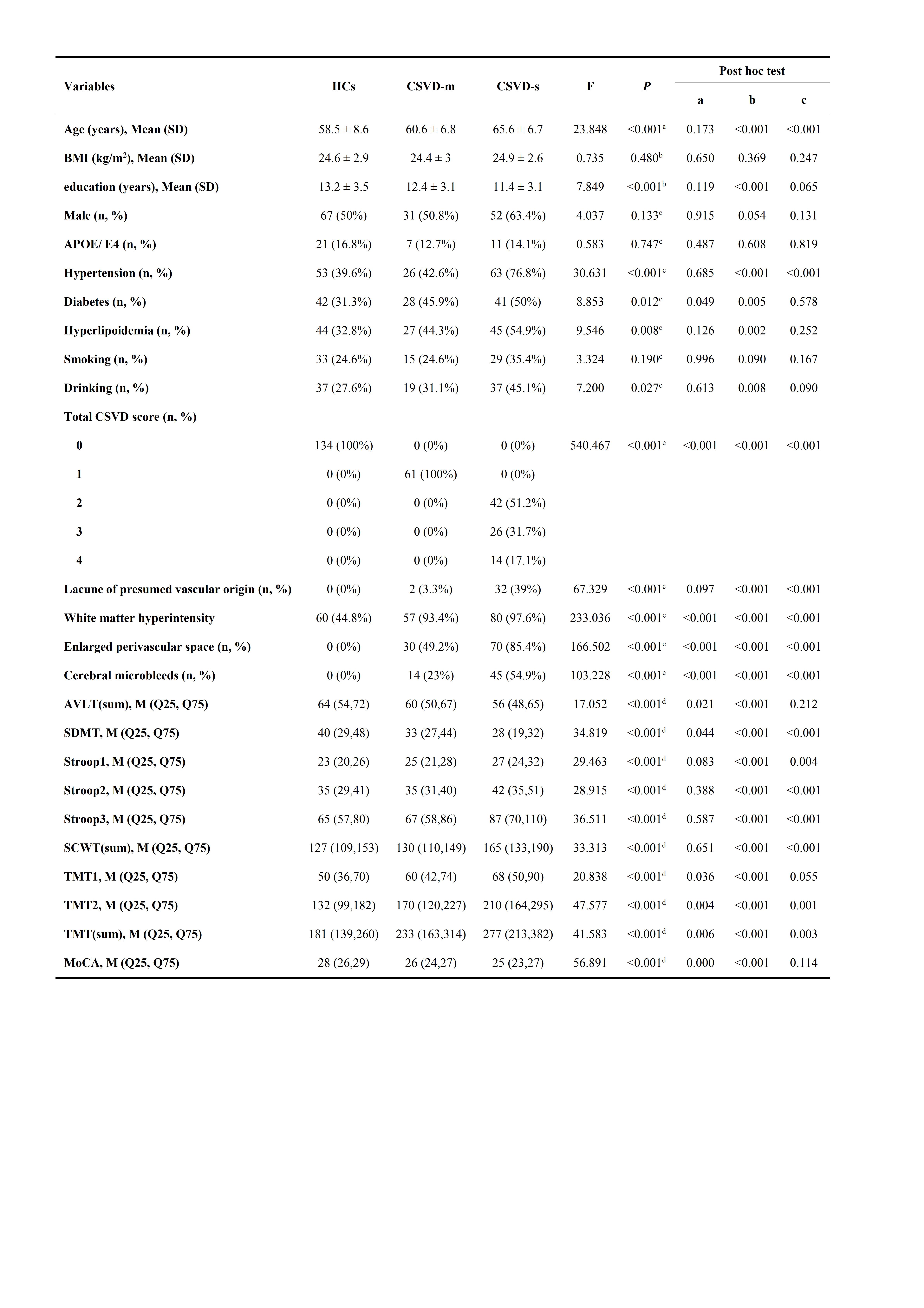
Figure 1. Clinical Characteristics of the Participant Groups
awelch test; bOne-way ANOVA analysis; cChi-quare test; dKruskal-Wallis H test; Lacune: Lacune of presumed vascular origin; WMH: white matter hyperintensity; PVS: perivascular space; CMBs: cerebral microbleeds; AVLT: auditory verbal learning test; SDMT: symbol digit modalities test; SCWT: stroop color-word test; TMT: trail making test; MoCA: montreal cognitive assessment.
Post hoc test: a = "HCs" vs "CSVD-m" ; b = "HCs" vs "CSVD-s"; c = "CSVD-m" vs "CSVD-s".
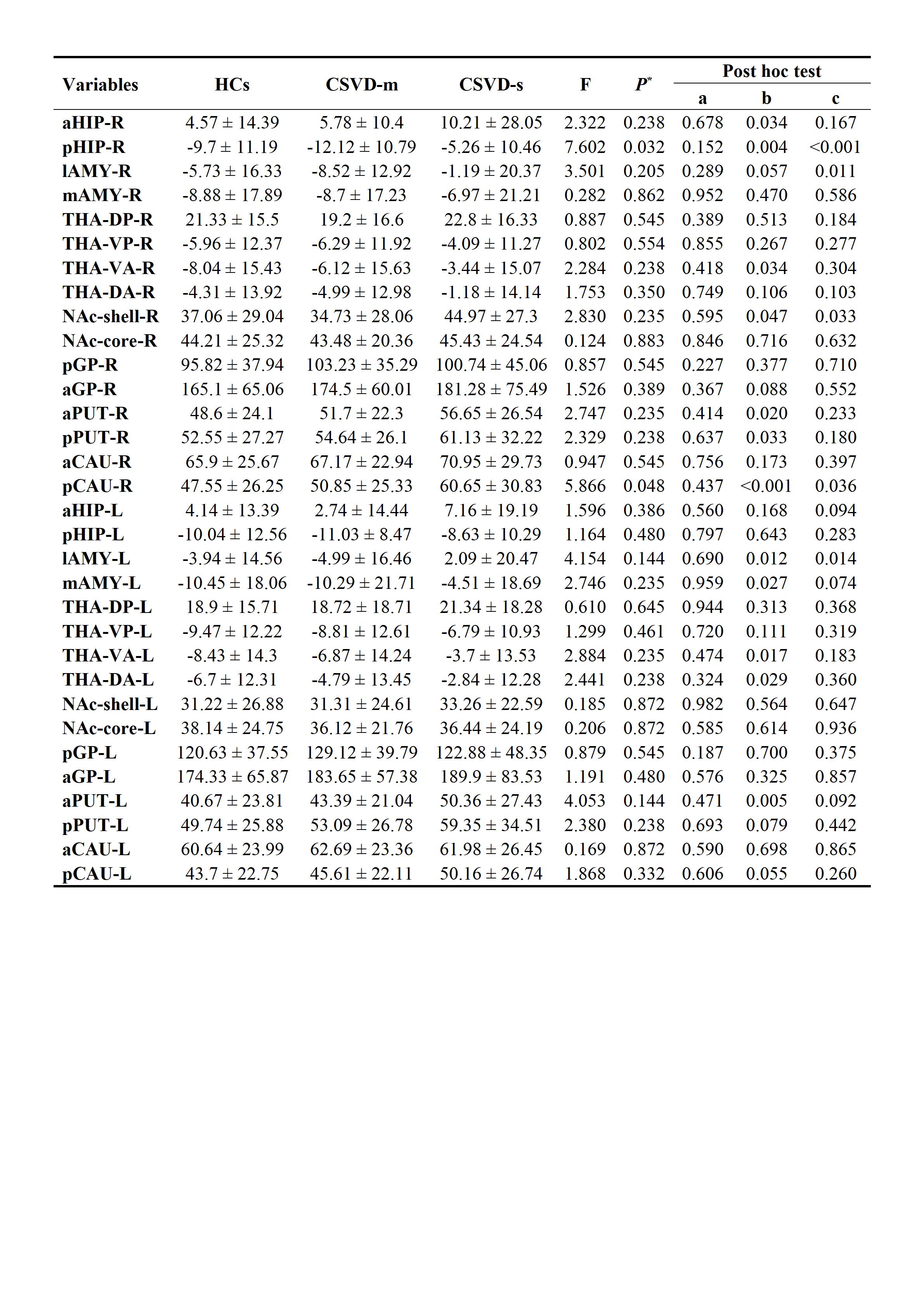
Figure 2. Susceptibility Values Differences Among the Three Groups
P*, P value of one-sample ANOVA corrected by False Discovery Rate correction, and the corrected significance value was 0.05. a, anterior; p, posterior; l, lateral; m, medial; d, dorsal; v, ventral; R, right brain; L, left brain; HIP: hippocampus; AMY, amygdala; THA, thalamus; NAc, nucleus accumbent; GP, globus pallidus; PUT, putamen; CAU, caudate nucleus.
Post hoc test: a = "HCs" vs "CSVD-m" ; b = "HCs" vs "CSVD-s"; c = "CSVD-m" vs "CSVD-s".
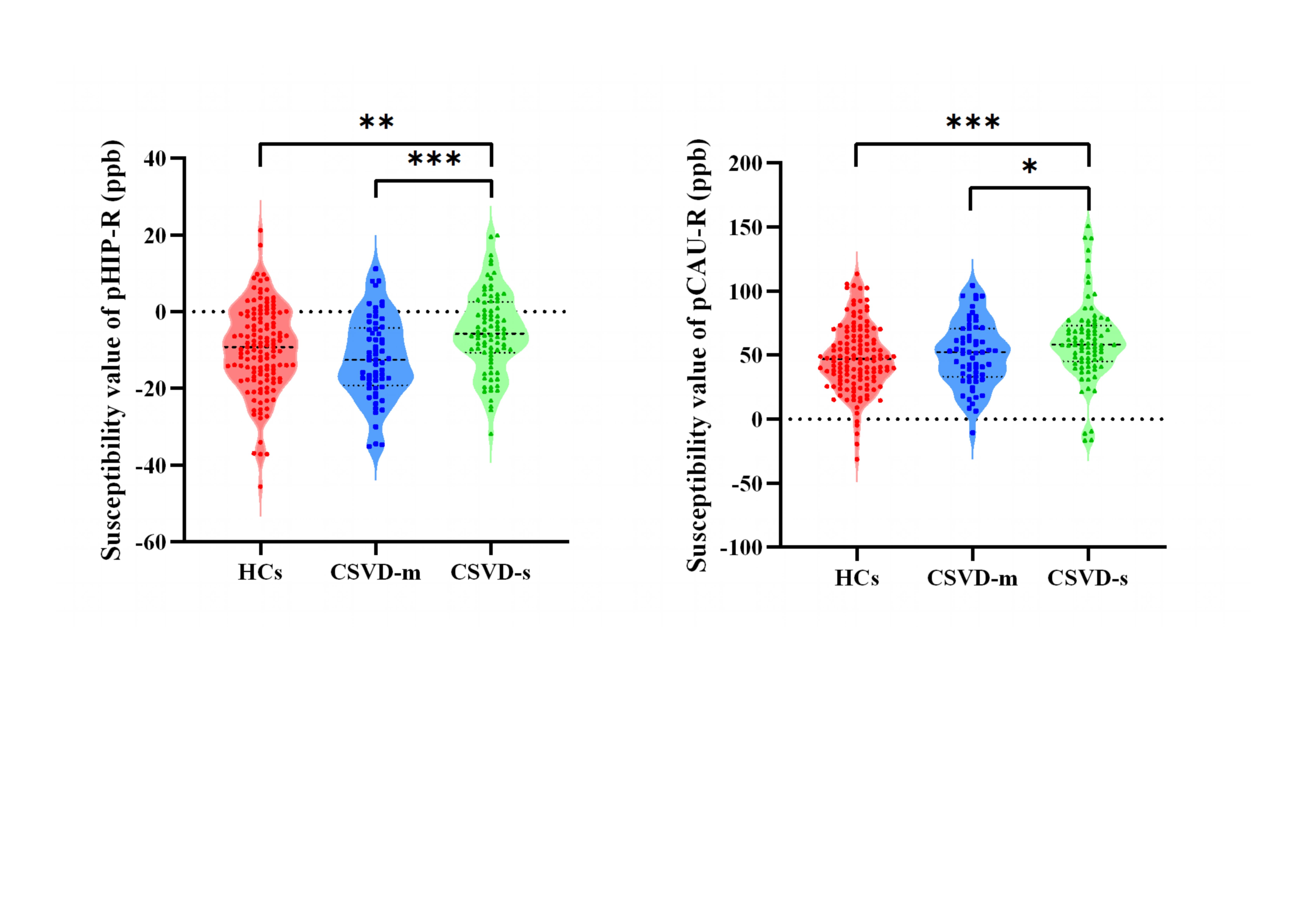
Figure 3. Susceptibility values differences in hippocampus and caudate nucleus among HCs, CSVD-m, CSVD-s groups. *, P<0.05; **, P<0.01; ***, P<0.001.
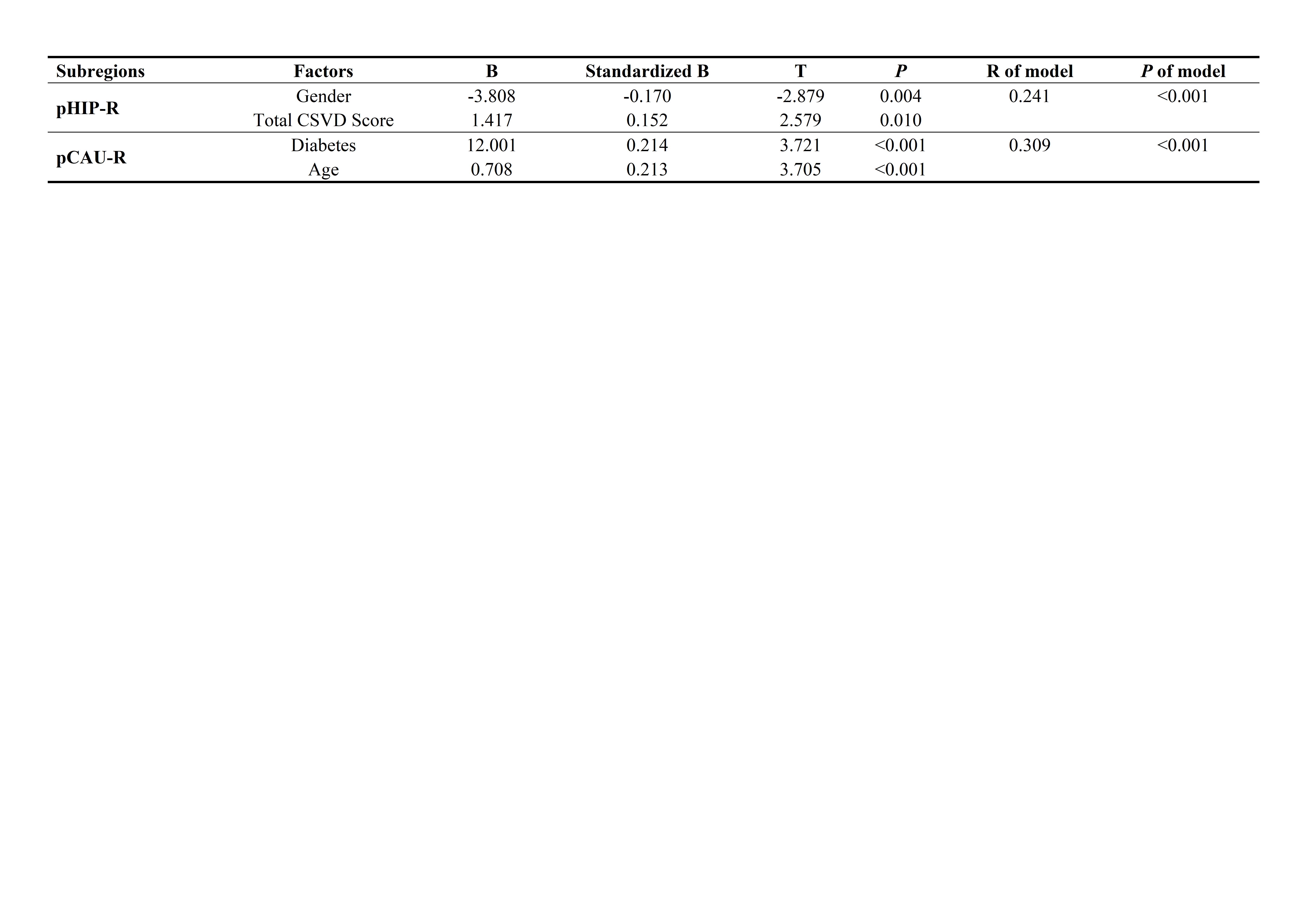
Figure 4. Determinants of Susceptibility Values: Results of Multiple Linear Stepwise Regression Analysis
p, posterior; R, right brain; HIP, hippocampus; CAU, caudate nucleus.
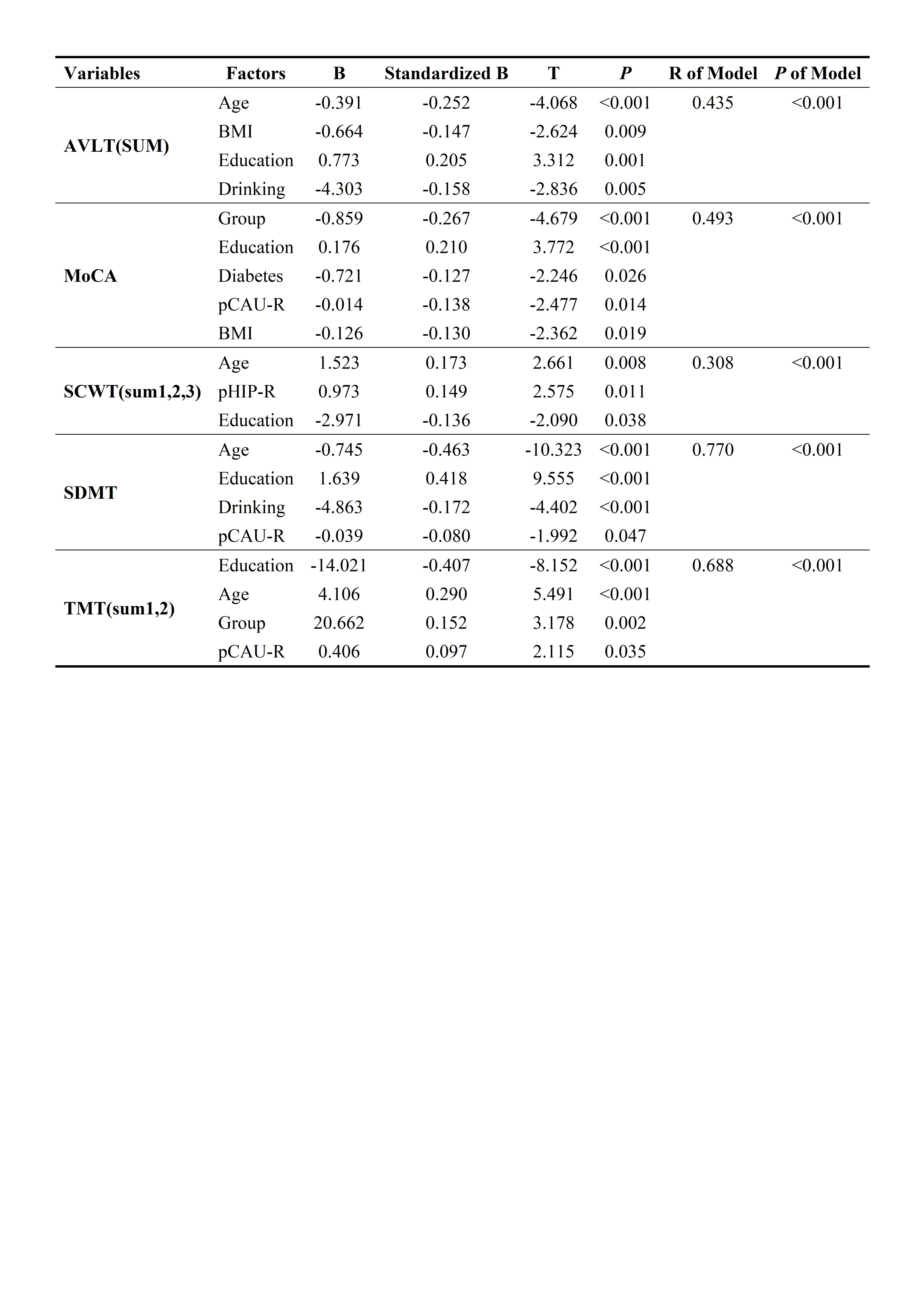
AVLT: Auditory Verbal Learning Test; SDMT: Symbol Digit Modalities Test; SCWT: Stroop color-word Test; TMT: Trail Making Test; MoCA: Montreal Cognitive Assessment; p, posterior; R, right brain; HIP, hippocampus; CAU, caudate nucleus; Group: HCs, CSVD-m, CSVD-s.