2207
LSA branches in symptomatic and asymptomatic unilateral MCA stenosis patients at 7T1Department of Radiology, Chinese PLA General Hospital, Beijing, China, Beijing, China, 2MR Collaboration, Siemens Healthineers Ltd., Beijing, China, Beijing, China
Synopsis
Keywords: Blood Vessels, Blood vessels
Motivation: The annual stroke risk of symptomatic middle cerebral artery (MCA) stenosis patients is more than four times that of asymptomatic patients, and doctors need to identify these two types for personalized treatment
Goal(s): We aim to investigate whether there is a difference in the number of stems and branches of the Lenticulostriate Artery (LSA) between symptomatic and asymptomatic unilateral MCAstenosis patients at 7T
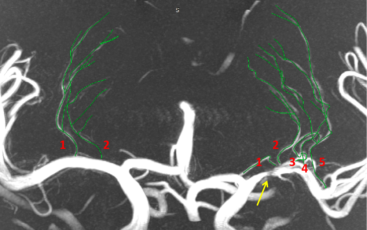
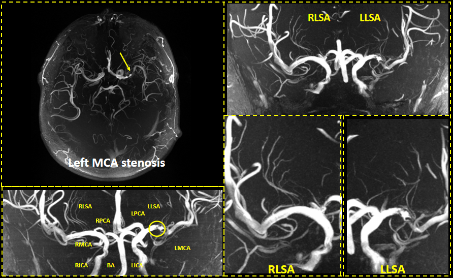
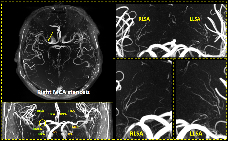
Approach: Count the number of LSA stems and branches in the patient's cerebral hemisphere using 7T TOF MRA
Results: :We found that the number of LSA branches varies between symptomatic and asymptomatic patients
Impact: LSA may serve as an imaging indicator for early diagnosis and personalized treatment of different types of MCA stenosis patients.
Introduction
Symptomatic patients with middle cerebral artery (MCA) stenosis have a higher risk of stroke than asymptomatic patients [1]. However, antithrombotic therapy is often ineffective for symptomatic patients [2], so personalized treatment is of critical clinical importance.Lenticulostriate arteries (LSA) are the main perforating artery that originate from MCA. Previous studies have shown that MCA stenosis affects the morphology of LSA [3-4],but there is limited research on the difference of LSA between symptomatic and asymptomatic patients. In this study, we used 7T MRI, a high-resolution imaging modality, to investigate the LSA morphology in patients with unilateral MCA stenosis.Methods
Ten Unilateral MCA stenosis patients, 4 asymptomatic and 6 symptomatic, were enrolled in this study. The examinations were performed on a 7T whole-body scanner (MAGNETOM Terra, Siemens Healthcare, Erlangen, Germany) equipped with an 8/32 Tx/Rx head coil. Routine examination, including T2w, T2w dark-fluid, T1w and 3D TOF MRA were performed. The acquisition parameters for TOF-MRA are: (repetition time 23.0 ms, echo time 3.42 ms, voxel size 0.3×0.3×0.3 mm3, flip angle 24°, the acquisition time is 4 minutes 52 seconds). The LSA stem and branch number in each hemisphere were counted from the generated MIP (maximum intensity projection) image of the MRA sequence.Stems were defined as the portion of the LSAs that originated directly from the middle cerebral artery,branches were defined as daughter vessels originating from common stems of parent LSAs plus stems without any branches (single vessels)[6].The number of LSA stems and branches between the affected and normal hemispheres of asymptomatic and symptomatic patients were analyzed by an unpaired Student's t test, respectively. All 50% or greater stenoses were classified as symptomatic or asymptomatic in association with the clinical presentation and results of parenchymal brain imaging in a month[7].Results
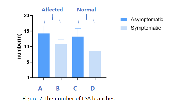
There were significant differences in the number of branches between the symptomatic and asymptomatic patients of affected hemisphere (14.25±2.36 VS 10.83±1.47,P=0.021), and there was also significant difference in the number of branches between symptomatic and asymptomatic patients of normal hemisphere(13.25±2.63 VS 8.67±1.86,P=0.012). There is a difference in the number of LSA branches between affected hemisphere and normal hemisphere in all patients (12.20±2.49 VS 10.50±3.1,P=0.028),The number of branches in the symptomatic patient is relatively small, while there is no significant difference in the number of LSA stems between symptomatic and asymptomatic patients.Discussion and conclusion
Previous studies have reported that patients with acute LSA acute infarction have fewer LSA than healthy individuals [5]. Similar to previous studies, we found that symptomatic patients have fewer branches.Whether in the affected hemisphere or the normal hemisphere, the number of LSA branches in symptomatic MCA stenosis patients is smaller than that in asymptomatic patients.Our results suggest that the number of LSA branches can be a potential imaging marker for distinguishing symptomatic and asymptomatic MCA stenosis patients. This study was exploratory and had a small sample size. In the future, more experiments are needed to confirm our findings.Summary of Main Findings
3D TOF MRA at 7T can provide detailed imaging of LSA, and the number of LSA branches can be a potential imaging marker for early diagnosis and personalized treatment of symptomatic MCA stenosis patientsAcknowledgements
This work was supported by the National Natural Science Foundation of China (No.81825012, No. 82151309 to X.L. and No. 82271952 to J.H.L)References
1. Kern, R et al. “Stroke recurrences in patients with symptomatic vs asymptomatic middle cerebral artery disease.” Neurology vol. 65,6 (2005): 859-64.
2. Thijs, V N, and G W Albers. “Symptomatic intracranial atherosclerosis: outcome of patients who fail antithrombotic therapy.” Neurology vol. 55,4 (2000): 490-7.
3. Bai, Xiaoyan et al. “Evaluating Middle Cerebral Artery Plaque Characteristics and Lenticulostriate Artery Morphology Associated With Subcortical Infarctions at 7T MRI.” Journal of magnetic resonance imaging : JMRI, 10.1002/jmri.28839
4. Wu, Fang et al. “Whole-brain magnetic resonance imaging of plaque burden and lenticulostriate arteries in patients with different types of stroke.” Therapeutic advances in neurological disorders vol. 12 1756286419833295. 26 Feb. 2019
5. Kang, Chang-Ki et al. “Lenticulostriate arteries in chronic stroke patients visualised by 7 T magnetic resonance angiography.” International journal of stroke : official journal of the International Stroke Society vol. 5,5 (2010): 374-80
6. KANG CK, PARK CA, LEE H, et al. Hypertension correlates with lenticulostriate arteries visualized by 7T magnetic resonance angiography [J]. Hypertension (Dallas, Tex : 1979), 2009, 54(5): 1050-6.
7. Hurford, Robert et al. “Prognosis of Asymptomatic Intracranial Stenosis in Patients With Transient Ischemic Attack and Minor Stroke.” JAMA neurology vol. 77,8 (2020): 947-954.
Figures