2206
Amygdala Subregion Atrophy: Early Indicator of Cognitive Impairment in Cerebral Small Vessel Disease1School of Medical Imaging, Binzhou Medical University, Yantai, Shandong, China., YAN TAI, China, 2Department of Psychiatry and Psychotherapy, Jena University Hospital, Jena, Germany, Jena, Germany, 3Department of Radiology, Beijing Tsinghua Changgung Hospital,Beijing, China., Bei jing, China, 4Key Laboratory of Endocrine Glucose & Lipids Metabolism and Brain Aging, Ministry of Education; Department of Radiology, Shandong Provincial Hospital Affiliated to Shandong First Medical University, Jinan, Shandong, China, Ji nan, China
Synopsis
Keywords: Blood Vessels, Neurodegeneration, Cerebral Small Vessel Disease
Motivation: The motivation behind this study was to delve deeper into the amygdala subregion changes in Cerebral Small Vessel Disease (CSVD) and how these changes correlate with cognitive impairment.
Goal(s): The primary goal was to determine whether specific amygdala subregions could serve as early indicator for cognitive impairment in CSVD, thereby aiding in early diagnosis and intervention.
Approach: Combined 3T MRI neuroimaging with cognitive assessments and focused statistical evaluation of amygdala subregions
Results: Our study revealed significant volume reductions in specific amygdala subregions among CSVD group, with pronounced atrophy observed in the left cortical nucleus.
Impact: This research introduces a novel indicator that utilizes neuroimaging techniques for the early prediction of CSVD progression and associated cognitive impairment, which could significantly enhance the precision of diagnostics and inform effective management strategies for CSVD.
Introduction
Cerebral small vessel disease (CSVD) is a prevalent vascular disorder that has been consistently associated with cognitive decline1. Previous research had linked amygdala volume to CSVD-related cognitive impairment2. However, most existing research treats the amygdala as a whole structure, leading to research focusing primarily on changes in total amygdala volume. This approach overlooks the potential differential impact of volumetric changes within its subregions. Therefore, our study investigates the relationship between CSVD progression and specific amygdala subregion volumes, as determined by FreeSurfer, to better understand their role in cognitive impairment. Furthermore, we examine how changes in these volumes affect cognition, highlighting potential avenues for early intervention in cognitive impairment related to CSVD.Methods
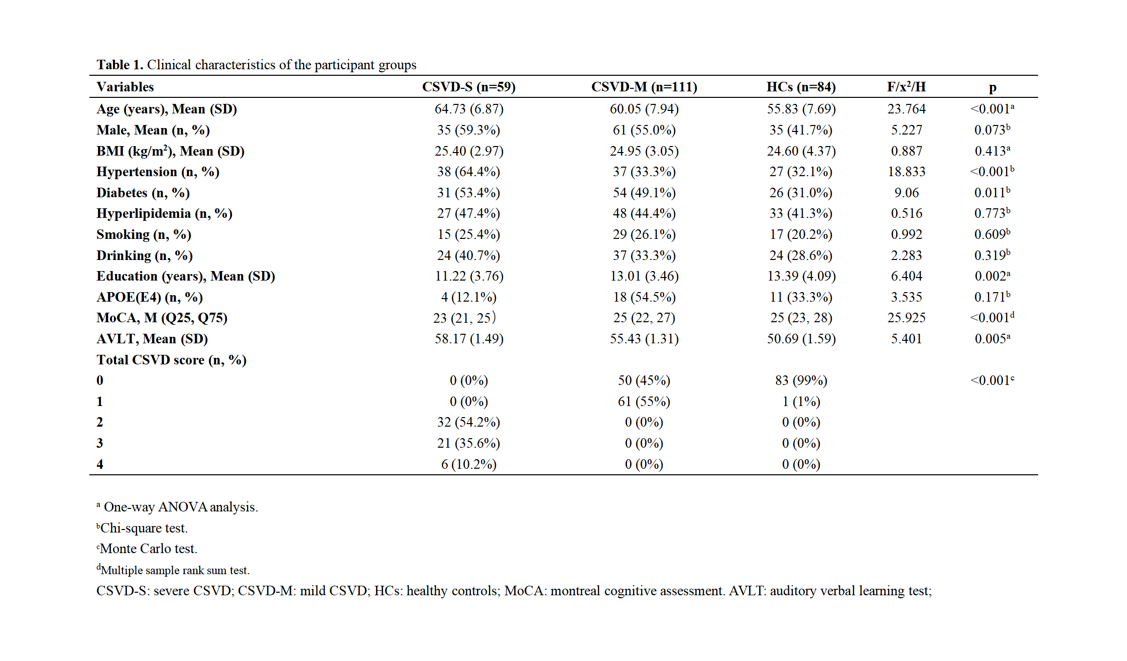
170 participants diagnosed with CSVD and 84 healthy controls aged 40-76 years were divided into three groups based on total CSVD score: HCs (CSVD score=0), CSVD-m (CSVD score=1), CSVD-s (CSVD score>1)3. All participants were scanned by 3T MRI and relevant cognitive tests. Using FreeSurfer, we automatically segmented the amygdala into nine subregions and assessed cognitive status utilizing the montreal cognitive assessment (MoCA) and the auditory verbal learning test (AVLT) scores. A multivariate linear regression was conducted with the MoCA score as the dependent variable and demographic and clinical factors as covariates to explore the correlation between CSVD severity and cognitive function. We also performed univariate ANOVA and Spearman correlation analyses on the amygdala subregions to identify those associated with CSVD severity. Linear regression analysis further examined whether amygdala subregion atrophy could serve as an early indicator of CSVD, considering subregion volumes and covariates such as age, gender, and education.Results
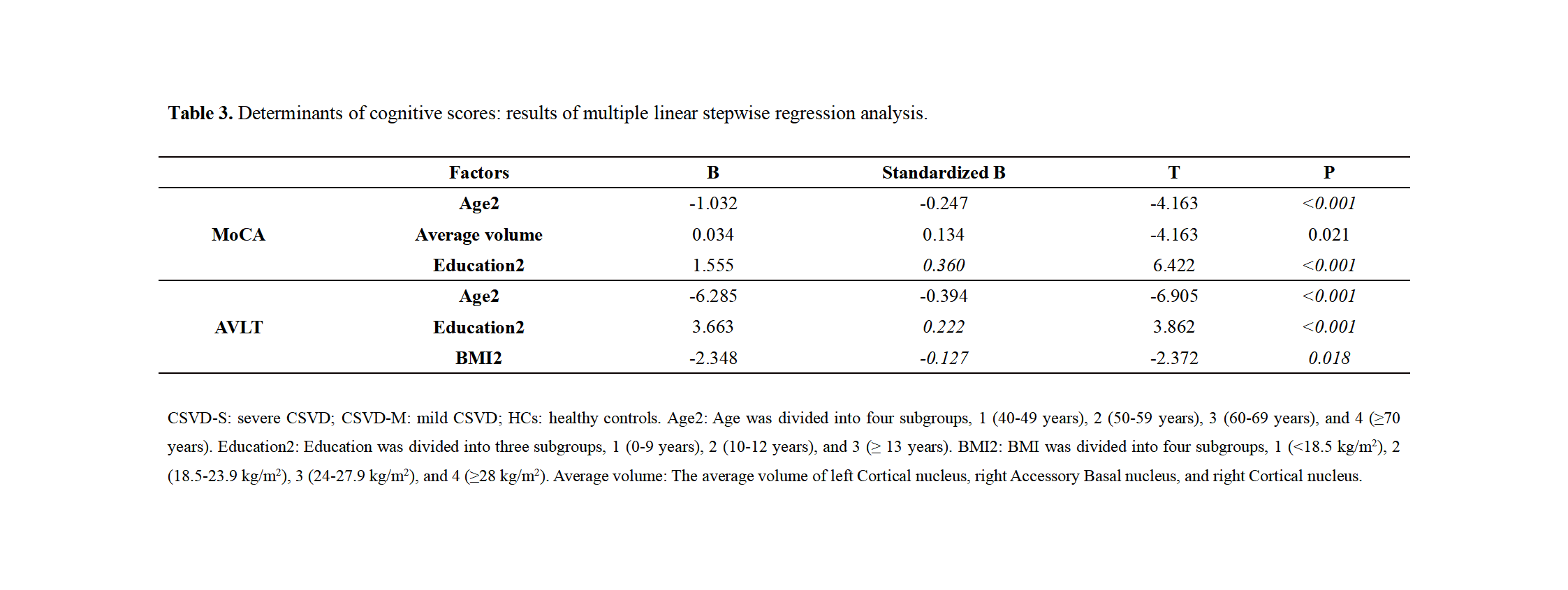
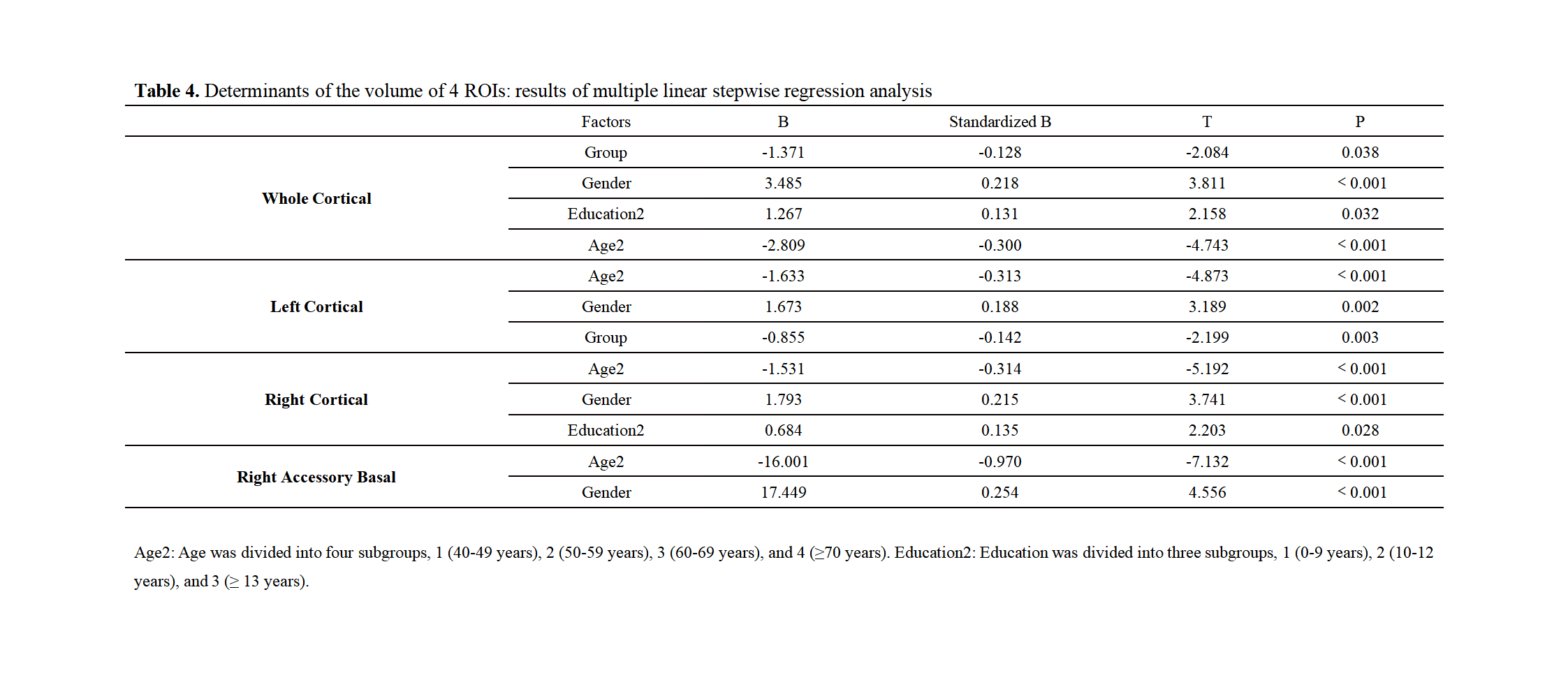
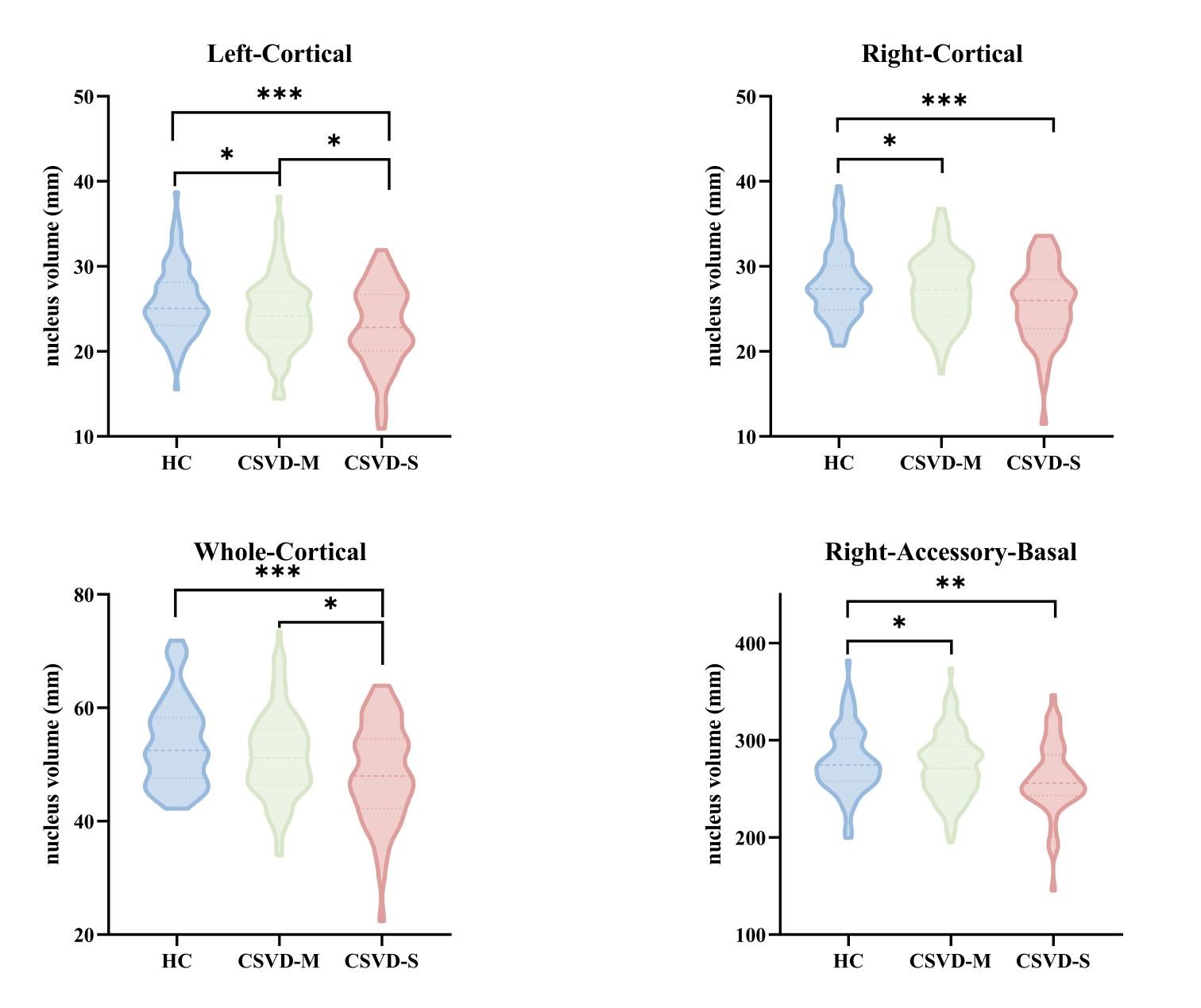
Significant volume loss was observed in the left cortical, right accessory basal, and right cortical regions among the three groups (P < 0.05, FDR correction) (Figure 5). Specifically, the volume of the left cortical nuclei was significantly different among groups with varying CSVD severity (P = 0.032) (Figure 2). As the burden of CSVD increases, the volume of the left cortical nucleus gradually decreases. Multivariable linear regression analysis revealed a positive correlation between MoCA scores and the average volume of the three amygdala subregions (P = 0.021) (Figure 3). However, no correlation was found with AVLT scores (Figure 3). Further regression analysis investigating the factors influencing subregion volumes found a significant association between the severity of CSVD and the volume of the cortical subregion (P = 0.038) (Figure 4). Specifically, the impact of CSVD severity on the volume of the left cortical nucleus was significant (P = 0.003) (Figure 4), whereas it was not significant for the right cortical nucleus.Discussion
This study was designed to elucidate the relationship between amygdala subregion volume changes and the progression of CSVD, particularly its influence on cognitive impairment. Our findings provide compelling evidence that the left cortical nucleus of the amygdala exhibits significant volumetric changes across different stages of CSVD progression. This pattern of atrophy suggests that the left cortical nucleus may be particularly sensitive to the pathophysiological processes of CSVD, reinforcing its potential role as a biomarker for the disease's progression. Additionally, the observed lateralization in atrophy aligns with the functional asymmetry of the cerebral hemispheres, implicating the left amygdala in cognitive functions such as language and memory, which are often affected by CSVD. The differential atrophy between the left and right cortical nuclei could reflect distinct neuroanatomical networks and their susceptibility to CSVD. While our study provides evidence of the relationship between amygdala subregion volumes and CSVD stages, longitudinal research is essential to establish the temporal sequence of these changes and their direct impact on cognitive impairment. Considering the implications for clinical practice, our research suggests that detailed amygdala subregion analysis should be incorporated into CSVD diagnostic protocols. This approach could facilitate earlier and more precise interventions, potentially mitigating the cognitive effects of CSVD.Conclusion
Our research highlights the critical role of atrophy in amygdala subregions, particularly the left cortical nucleus, as an indicator of CSVD progression and the resulting cognitive impairment. These findings support the early use of neuroimaging to identify potential cognitive impairment in CSVD, emphasizing the importance of prompt detection and targeted treatment at the disease's outset. Further research is necessary to explore the complex interactions among amygdala subregions, which will enhance our understanding of CSVD's pathophysiology and its impact on cognitive functions.Acknowledgements
We express our profound gratitude to all the participants who dedicated their time and effort to partake in this study. Their contribution has been invaluable and is the foundation upon which our research findings rest. We also extend our heartfelt thanks to our colleagues, whose unwavering support and collaboration have been instrumental in the execution of this project. Special appreciation is directed to the esteemed professors who provided guidance and insight that greatly enriched our work.References
1. Zanon Zotin, M. C., Sveikata, L., Viswanathan, A. & Yilmaz, P. Cerebral small vessel disease and vascular cognitive impairment: from diagnosis to management. Curr. Opin. Neurol. 34, 246–257 (2021).
2. van Uden, I. W. M. et al. Depressive Symptoms and Amygdala Volume in Elderly with Cerebral Small Vessel Disease: The RUN DMC Study. Journal of Aging Research 2011, 647869 (2011).
3. Pasi, M. et al. Association of Cerebral Small Vessel Disease and Cognitive Decline After Intracerebral Hemorrhage. Neurology 96, e182–e192 (2021).
Figures
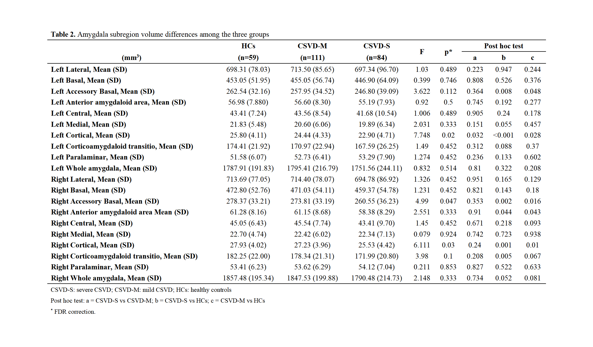
Amygdala subregion volume differences among the three groups