2203
Hemodynamic Alterations of Carotid Siphons after Revascularization is Associated with Outcomes in Moyamoya Angiopathy: A 4D Flow MRI Study1Department of Radiology, West China Hospital, Sichuan University, Chengdu, China, 2Research Unit of Psychoradiology, Chinese Academy of Medical Sciences, Chengdu, China, 3Department of Biomedical Engineering, Tsinghua University, Beijing, China, 4Department of Neurosurgery, West China Hospital, Sichuan University, Chengdu, China, 5Department of Neurosurgery, The First Affiliated Hospital of Xi’an Jiaotong University, Xi’an, China
Synopsis
Keywords: Blood Vessels, Velocity & Flow, Cerebrovascular disease, moyamoya angiopathy
Motivation: Current evidence is insufficient to help better understand the interactions between hemodynamics of carotid siphons in moyamoya angiopathy (MMA) and outcomes of revascularization.
Goal(s): To evaluate hemodynamic alterations of carotid siphons after revascularization for MMA using 4D Flow MRI and to explore its association with outcomes.
Approach: MMA patients receiving revascularization were prospectively enrolled. We used VesselExplorer2 software to analyze 4D Flow MRI data and measure hemodynamic parameters of carotid siphons. Relationships between hemodynamic parameters of carotid siphons and surgical outcomes were explored.
Results: Flowmean and Flowmax of bilateral ICAs in MMA decreased at follow-up, which was associated with outcomes of revascularization surgery.
Impact: We identified new hemodynamic parameters that might be used to predict the prognosis of MMA. 4D Flow MRI is a promising tool for quantitatively detecting hemodynamics in intracranial vessels of MMA patients, providing valuable hemodynamic parameters for evaluating surgical outcomes.
Introduction
Moyamoya angiopathy (MMA), a chronic cerebrovascular disease, is characterized by progressive stenosis or occlusion of the distal part of large intracranial arteries1. Revascularization is one of the most important treatments for MMA2, where timely postoperative evaluation of hemodynamic changes is crucial. Postoperative changes in hemodynamics are found to be linked to the prognosis of this disease3,4. However, no consensus has been reached on which hemodynamic parameters may alter after revascularization and what relationships are between these alterations and surgical outcomes in MMA. More recently, 4D Flow MRI enables noninvasive analysis of the time-resolved 3D dynamics of blood flow5. Our hypothesized that 4D Flow MRI could be employed to detect hemodynamic alterations in carotid siphons where may exhibit hemodynamic abnormalities in MMA. In this study, we aimed to quantitatively detect the 4D Flow MRI-based hemodynamic characteristics of carotid siphons in MMA before and after revascularization, to reveal its relationships with treatment outcomes.Methods
In this prospective, observational, and longitudinal study, patients with MMA-related diagnoses were consecutively collected from West China Hospital, Sichuan University between July 2018 and January 2020. All patients underwent 4D Flow MRI at baseline and follow-up. All MRI examinations were performed on a 3.0T MRI scanner (Skyra, Siemens Medical Systems, Germany) with a 20-channel head coil. 4D Flow MRI was acquired using a free-breathing, peripheral pulse-gated, multi-shot turbo filed echo sequence with TR/TE=45/2.7ms; slice thickness=1mm; FOV=200mm; resolution matrix=256×216. Raw data of 4D Flow MRI were imported into VesselExplorer2 software (TSimaging Healthcare, China) to measure hemodynamic parameters by two experienced neuroradiologists, with 6 and 8 years of diagnosis experience. Mean and maximum values of flow (Flowmean and Flowmax), velocity (Vmean and Vmax), and WSS (WSSmean and WSSmax) were calculated on the plane of the posterior bend of carotid siphons. Paired t-test was performed to compare baseline and follow-up hemodynamic parameters in bilateral carotid siphons. The associations between hemodynamics of bilateral carotid siphons and clinical and imaging outcomes at follow-up were assessed with a paired-samples t-test. The intraclass correlation coefficient (ICC) was performed to evaluate the inter-observer consistency of hemodynamic parameters.Results
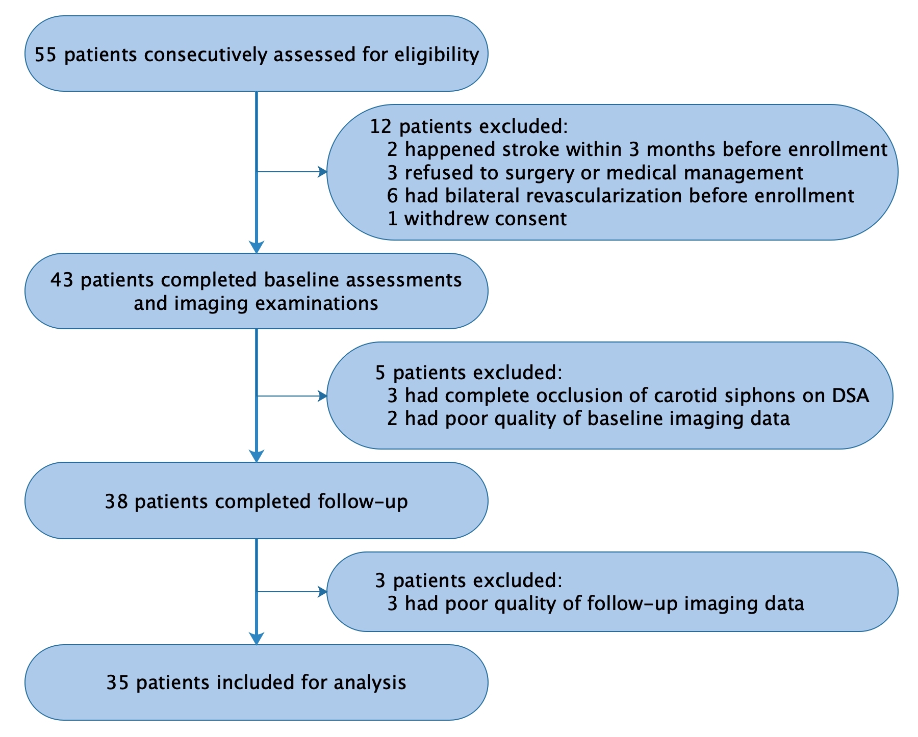
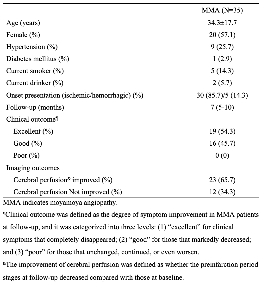
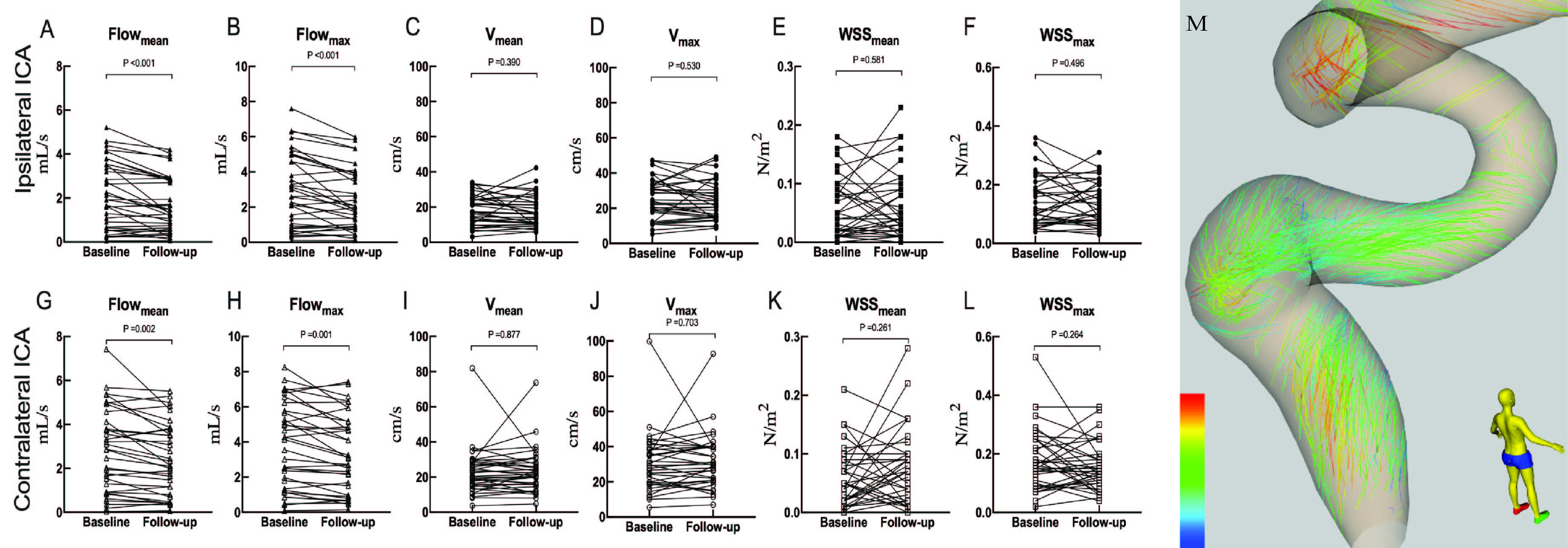
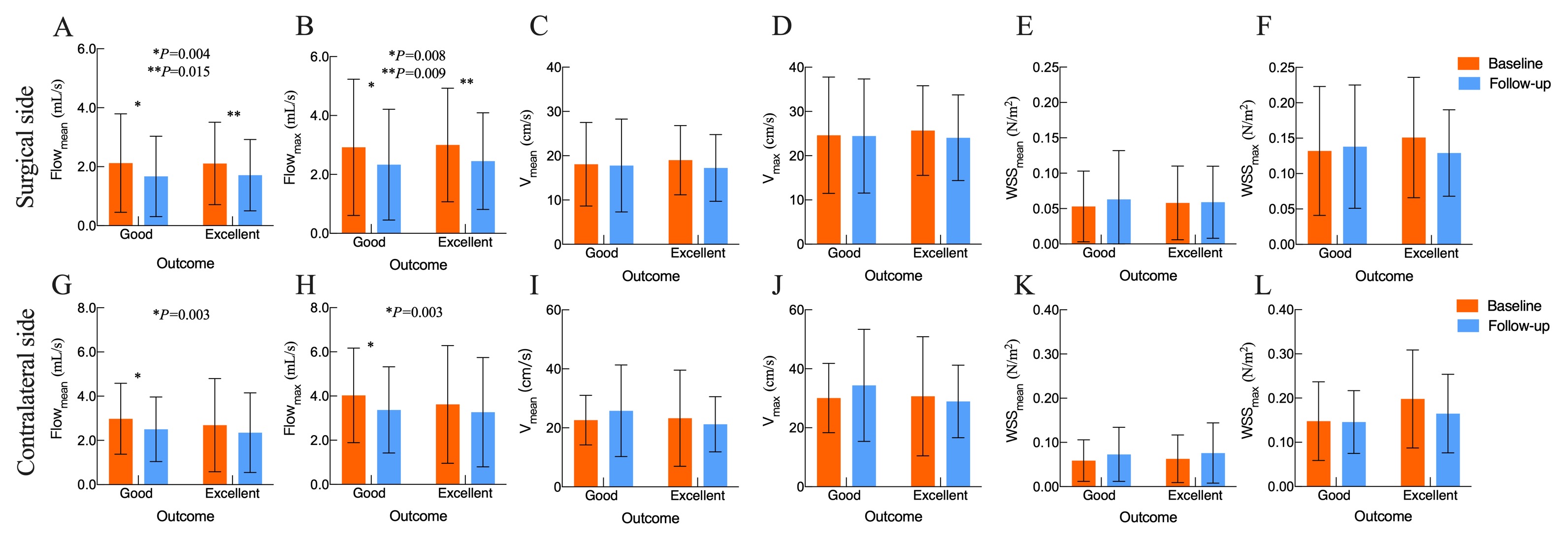
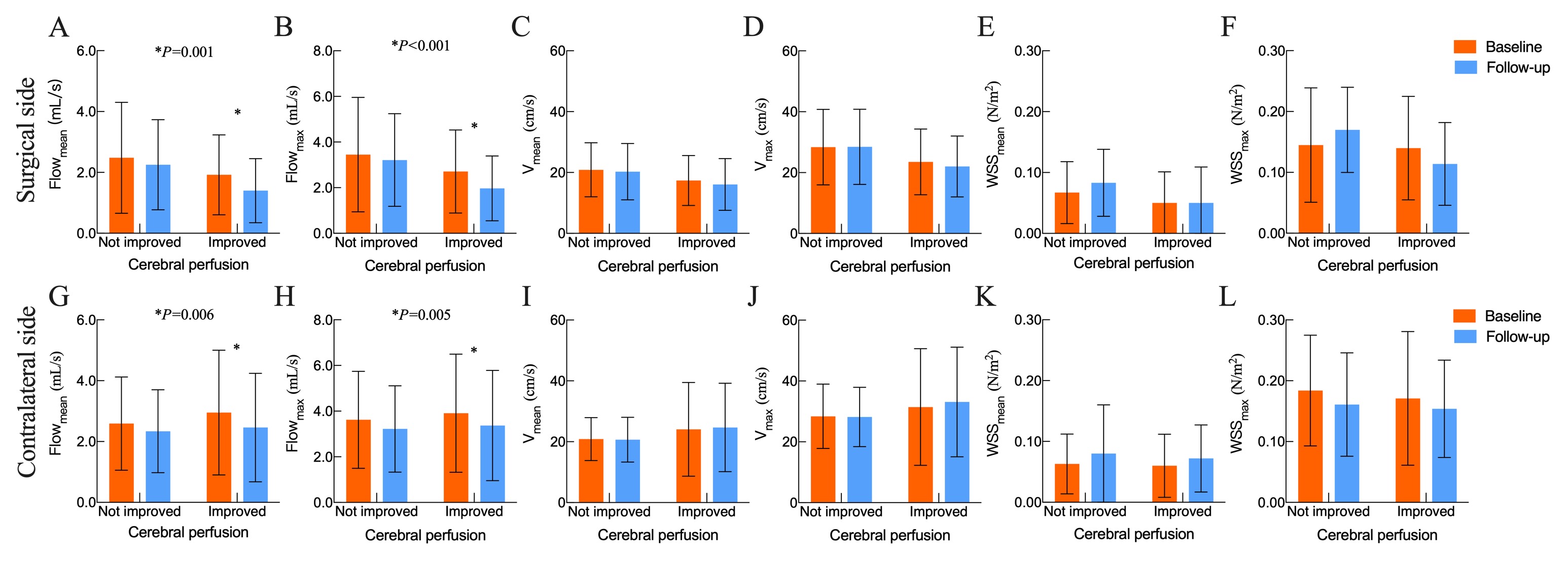
The ICC of the APTw value assessment of the two neuroradiologists was pretty good (ICC value>0.85). Thirty-five patients (range, 6-69 years) were enrolled in this study, and the duration of follow-up was 7 (5-10) months (Figure 1). The demographic and clinical characteristics of patients are shown in Table 1. Compared to those at baseline, Flowmean/ Flowmax of bilateral carotid siphons decreased at follow-up (P<0.01). No significant changes were found in Vmean/Vmax and WSSmean/WSSmax on either surgical or contralateral side (Figure 2). In patients with good clinical outcome, Flowmean/Flowmax of bilateral carotid siphons reduced significantly at follow-up (P<0.01), though those with excellent outcome only had a lower Flowmean/Flowmax on the surgical side (P<0.05) (Figure 3). Significant reductions of Flowmean/Flowmax of bilateral carotid siphons in patients with improved cerebral perfusion were found at follow-up (P<0.01) (Figure 4).Discussion
In this study, we found significant diminutions of Flowmean and Flowmax of bilateral ICAs at follow-up compared to their baseline levels. Flowmean and Flowmax were related to clinical symptom improvement and cerebral perfusion improvement, indicating that these hemodynamic parameters were associated with treatment outcomes. Although these results corroborated with an earlier hemodynamic study on vessel remodeling of MMA conducted by Zhu F et al.6, hemodynamics measured by 4D Flow MRI with a simpler data analysis were more accurate than those simulated by computational fluid dynamics technique. These results clarified that unilateral revascularization not only affected cerebral hemodynamics of surgical-side hemispheres but also influenced contralateral hemispheres, owing to the connection of bilateral ICAs through the circle of Willis, bilateral superficial temporal arteries through the external carotid system, and collateral formation6,7. To our knowledge, this was the first study to estimate associations of hemodynamic changes with both clinical and imaging outcomes after revascularization in MMA via 4D Flow MRI technique. We found that apparent reductions of Flowmean/Flowmax of bilateral ICAs were associated with surgical outcomes in MMA, including the improvement of clinical symptoms and the improvement of cerebral perfusion status at follow-up. However, previous studies with small sample sizes found that some different hemodynamic parameters, such as pressure drop, pressure drop difference, pressure drop index, and flow rate of posterior communicating arteries, may be associated with surgical outcomes3,8,9.Conclusion
Flowmean and Flowmax of bilateral ICAs in MMA decreased after revascularization, which was associated with the prognosis of combined revascularization surgery. 4D Flow MRI is a promising tool for directly quantitatively detecting hemodynamic status in intracranial vessels, providing valuable hemodynamic parameters for evaluating surgical outcomes of MMA.Acknowledgements
The authors thanked the patients for participating in this study. This study was supported by grants from China Postdoctoral Science Foundation (2022M722270); the Youth Science Fund of the Natural Science Foundation of Sichuan Province, China (2022NSFSC1435); Sichuan University Postdoctoral Interdisciplinary Innovation Fund (JCXK2209); and Fund of the Beijing Medical Award Foundation (YXJL-2022-0665-0189).References
1. Burke GM, et al. Moyamoya disease: A summary. Neurosurg Focus. 2009;26:E11.
2. Fujimura M, et al. 2021 Japanese Guidelines for the Management of Moyamoya Disease: Guidelines from the Research Committee on Moyamoya Disease and Japan Stroke Society. Neurol Med Chir (Tokyo). 2022;62:165-70.
3. Zhu F, et al. Assessing surgical treatment outcome following superficial temporal artery to middle cerebral artery bypass based on computational haemodynamic analysis. J Biomech. 2015;48(15):4053-8.
4. Jamil M, et al. Changes to the geometry and fluid mechanics of the carotid siphon in the pediatric Moyamoya disease.Comput Methods Biomech Biomed Engin. 2016;19(16):1760-71.
5. Soulat G, et al. 4D Flow with MRI. Annu Rev Biomed Eng. 2020;22:103-26.
6. Zhu F, et al. Quantitative assessment of changes in hemodynamics of the internal carotid artery after bypass surgery for moyamoya disease. J Neurosurg. 2018;129(3): 677-83.
7. Gao F, et al. Non-Invasive Evaluation of Cerebral Hemodynamic Changes After Surgery in Adult Patients With Moyamoya Using 2D Phase-Contrast and Intravoxel Incoherent Motion MRI. Front Surg. 2022;9:773767.
8. Karunanithi K, et al. Identification of a hemodynamic parameter for assessing treatment outcome of EDAS in Moyamoya disease. J Biomech. 2015;48(2):304-9.
9. Sun W, et al. Quantifying Hemodynamic Changes in Moyamoya Disease Based on Two-Dimensional Cine Phase-Contrast Magnetic Resonance Imaging and Computational Fluid Dynamics. World Neurosurg. 2018;120:e1301-9.
Figures