2202
Alterations of amide proton transfer weighted (APTw) signal after revascularization in patients with moyamoya disease1Department of Radiology, West China Hospital, Sichuan University, Chengdu, China, 2Research Unit of Psychoradiology, Chinese Academy of Medical Sciences, Chengdu, China, 3Department of Neurosurgery, West China Hospital, Sichuan University, Chengdu, China, 4Philips Healthcare, Xi’an, China
Synopsis
Keywords: Blood Vessels, CEST & MT, Cerebrovascular disease
Motivation: Assessing alterations of the brain microenvironment in patients with moyamoya disease after revascularization (MMD) would develop optimal postoperative management and improve the prognosis.
Goal(s): This study aimed to explore alterations of amide proton transfer weighted (APTw) signal in patients with MMD after receiving revascularization surgery.
Approach: MMD patients who underwent unilateral revascularization surgery were prospectively collected. Baseline computed tomography perfusion (CTP) and APTw imaging were performed. Patients were followed up with CTP and APTw imaging about 1 year after revascularization.
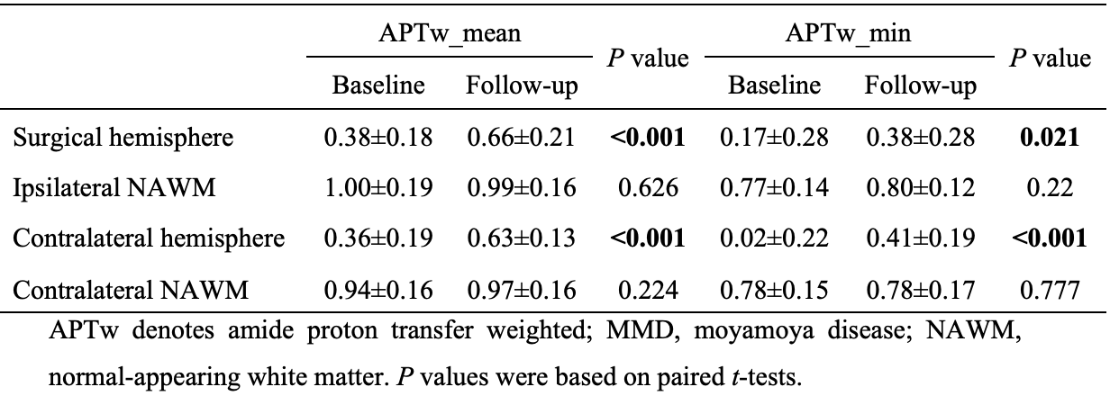
Results: APTw values in both surgical and contralateral hemispheres at follow-up were found to be significantly higher than those at baseline.
Impact: APTw imaging is a potential assessment tool to quantitatively evaluate alterations of acid-base metabolism in the brain microenvironment of patients with MMD after revascularization, which provides additional information to help neurosurgeons develop optimal postoperative management and follow-up protocols for MMD.
Introduction
Moyamoya disease (MMD) is a chronic cerebrovascular disease characterized by progressive stenosis or occlusion of the distal part of large intracranial arteries1. Although revascularization is currently one of the most important treatments for moyamoya disease2, there are still some patients whose symptoms or signs are not improved or alleviated after surgery. Amide proton transfer weighted (APTw) imaging is a novel technique to detect endogenous dissociated peptides and proteins in tissue, which are highly sensitive to changes of pH in the internal environment3. Although previous studies have reported the possibility of pH-weighted APTw imaging in detecting acidosis penumbra in acute ischemic stroke4-6, how APTw signal changes in MMD patients has not been discussed yet. Additionally, the effect of revascularization on acid-base metabolism in brain microenvironment of MMD remains unknown. In this study, we utilized APTw imaging to evaluate alterations of APTw signal in patients with MMD receiving revascularization surgery.Methods
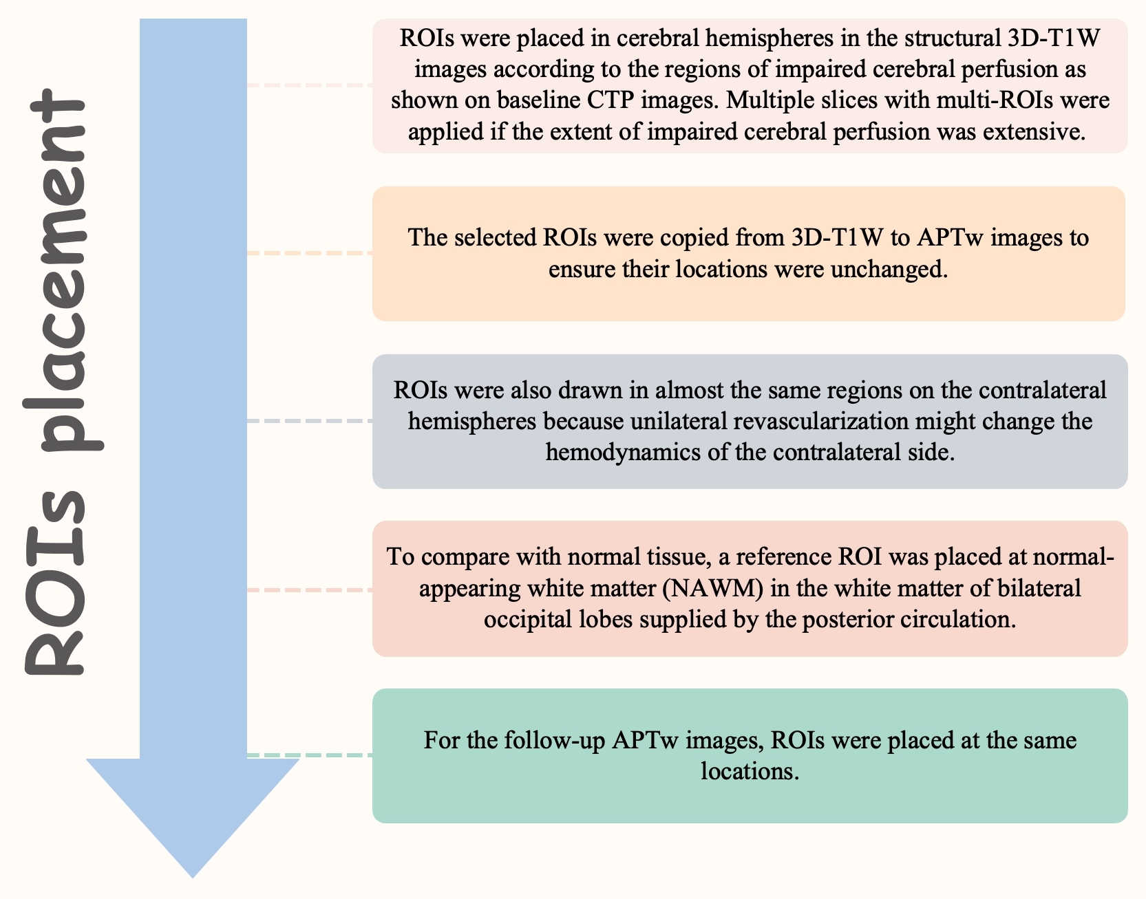
Patients with MMD who underwent unilateral revascularization surgery were prospectively enrolled at West China Hospital, Sichuan University from July 2021 to June 2023. All patients underwent routine computed tomography perfusion (CTP), conventional magnetic resonance imaging (MRI), and APTw imaging at baseline. Patients were followed up with CTP and MRI examinations about 1 year after revascularization. All patients underwent MRI examination on a 3.0T scanner (Ingenia Elition, Philips Healthcare, the Netherlands) with a 32-channel dedicated head coil. The structural images were acquired by using a high-resolution three-dimensional turbo field echo T1-weighed (3D-T1-TFE) sequence. The APTw images were acquired by using a 3D TSE-DIXON sequence with TR/TE=6,120/7.8ms; FOV=230*180mm2; slice thickness=4.0mm; voxel=1.8*1.8*4.0mm3; flip angle=90 degrees; radiofrequency power=2μT; duration=2,000ms. B0 corrected APTw images were transferred to the post-processing workstation (Philips InteliSpace Portal) to do the automatic calculation of APTw values. Before the region of interests (ROIs) drawing, APTw images were automatically co-registered to 3D-T1W images by performing a rigid transformation of the datasets. The flowchart of ROIs placement is shown in figure 1. Two experienced neuroradiologists, with 6 and 8 years of diagnosis experience, manually drew ROIs (50~60mm2 each) independently to obtain APTw values. The intraclass correlation coefficient (ICC) was performed to evaluate the inter-observer consistency of APTw values. The difference between APTw values at baseline and those at follow-up was analyzed by the paired t-test.Results
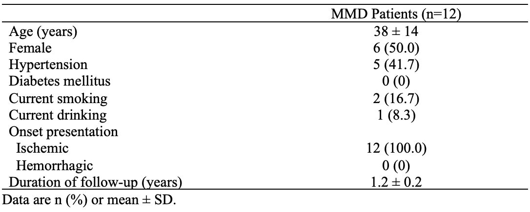
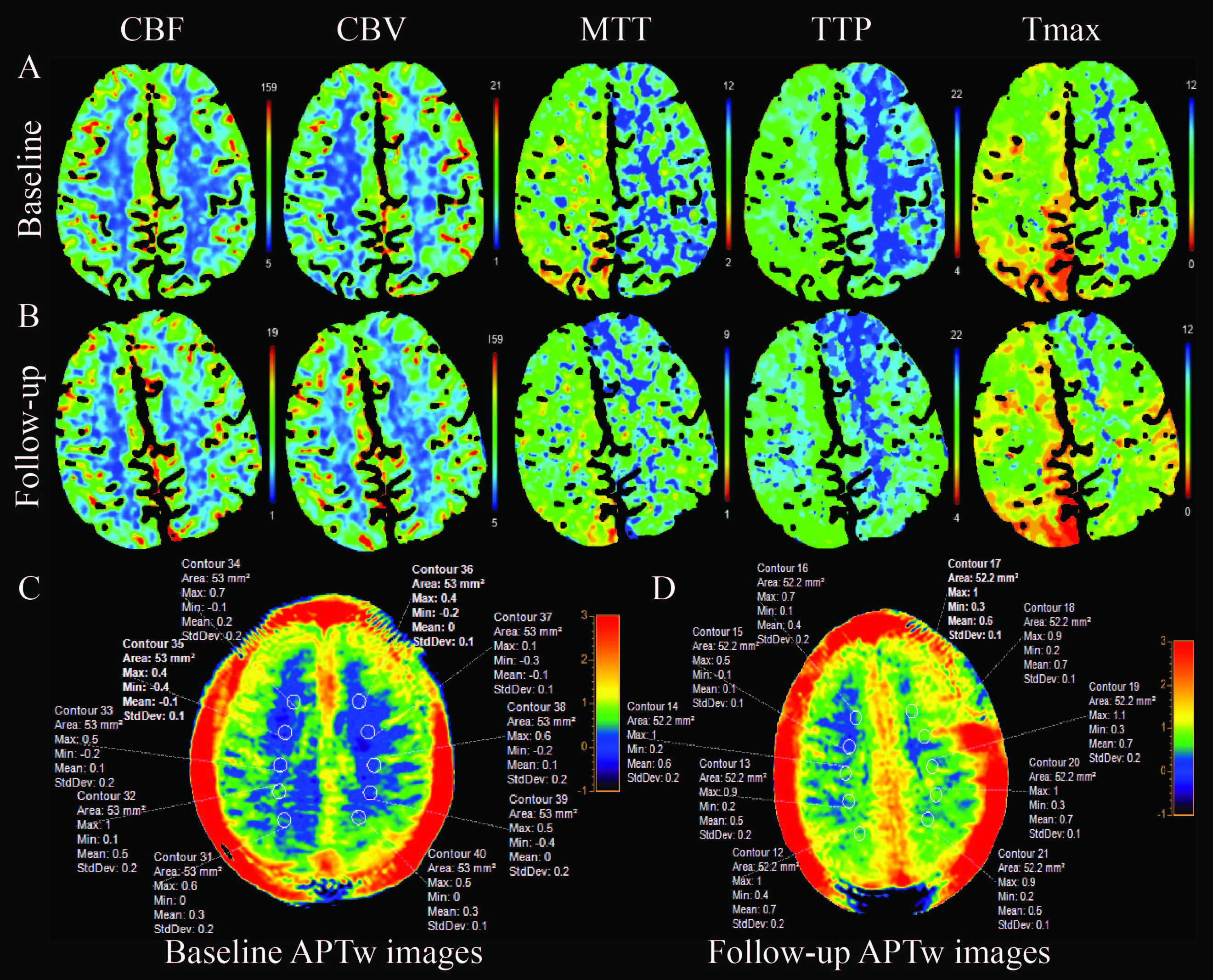
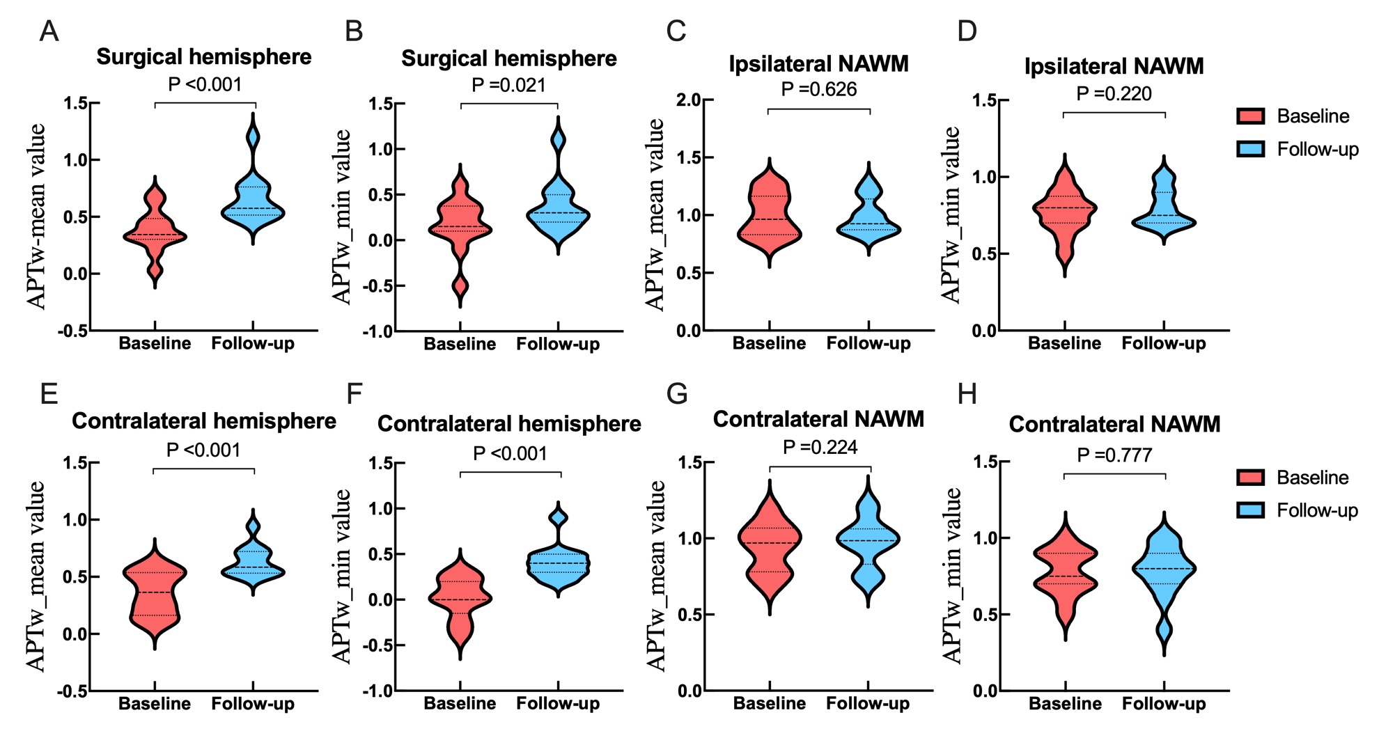
The ICC of the APTw value assessment of the two neuroradiologists was pretty good (ICC value>0.85). Twelve patients with MMD (range, 21-64 years) were enrolled in this study, and the duration of follow-up was 1.2±0.2 years. The demographic and clinical characteristics of patients with MMD are shown in Table 1. Figure 2 showed baseline and follow-up CTP and MRI images of a 24-year-old patient receiving left revascularization surgery and the related ROIs placement of APTw signal in the cerebral hemispheres. Table 2 and figure 3 showed that both APTw_mean and APTw_min in the white matter of surgical hemispheres at follow-up were significantly higher than their baseline levels (P<0.001 and P=0.021, respectively). Additionally, APTw_mean and APTw_min in the contralateral hemispheres also significantly increased at follow-up compared to those at baseline (P<0.001 and P<0.001, respectively). However, APTw values of NAWM in both surgical and contralateral occipital lobes supplied by the posterior circulation were maintained at follow-up (P>0.05) (Table 3). Moreover, we also observed that some patients (eg. the patient in Figures 2) had normal or slightly impaired cerebral perfusion in the contralateral hemisphere on CTP images. However, the degree and extent of APTw signal reduction were comparable to those in the surgical hemisphere.Discussion
In this study, we found significant diminutions of APTw values of bilateral hemispheres in MMD patients receiving revascularization at follow-up compared to their baseline levels. The hemodynamics and cerebral perfusion in MMD patients were redistributed after revascularization, and the metabolisms including acid-base metabolism in brain tissue subsequently improved7, 8. This finding suggested that unilateral revascularization surgery would alleviate the acidosis of not only surgical hemispheres but also contralateral hemispheres. Our study also demonstrated that APTw imaging is highly sensitive to ischemia and acidosis, making it a superior tool for assessing chronic ischemic cerebrovascular diseases compared to CTP. This sensitivity was evident in both surgical and contralateral hemispheres, despite some patients having normal or slightly impaired cerebral perfusion on CTP images.Conclusion
APTw-based pH evaluation can effectively reflect brain tissue microenvironment changes of post-revascularization in MMD patients. It is a promising tool for quantitatively tracking acid-base metabolism dynamics in MMD, offering valuable neuroimaging biomarkers for perioperative and follow-up protocols.Acknowledgements
The authors thank the patients for participating in this study. This study was supported by grants from China Postdoctoral Science Foundation (2022M722270); the Youth Science Fund of the Natural Science Foundation of Sichuan Province, China (2022NSFSC1435); Sichuan University Postdoctoral Interdisciplinary Innovation Fund (JCXK2209); and Fund of the Beijing Medical Award Foundation (YXJL-2022-0665-0189).References
1. Burke GM, et al. Moyamoya disease: A summary. Neurosurg Focus. 2009;26:E11.
2. Fujimura M, et al. 2021 Japanese Guidelines for the Management of Moyamoya Disease: Guidelines from the Research Committee on Moyamoya Disease and Japan Stroke Society. Neurol Med Chir (Tokyo). 2022;62:165-70.
3. Togao O, et al. Amide proton transfer imaging of adult diffuse gliomas: correlation with histopathological grades. Neuro Oncol. 2014;16:441-8.
4. Yu L, et al. Amide Proton Transfer MRI Signal as a Surrogate Biomarker of Ischemic Stroke Recovery in Patients with Supportive Treatment. Front Neurol. 2019;10:104.
5. Sun PZ, et al. Demonstration of magnetization transfer and relaxation normalized pH‐specific pulse‐amide proton transfer imaging in an animal model of acute stroke. Magn Reson Med. 2020;84:1526-33.
6. Wang E, et al. Mapping tissue pH in an experimental model of acute stroke - Determination of graded regional tissue pH changes with non-invasive quantitative amide proton transfer MRI. Neuroimage. 2019;191:610-7.
7. Zhang K, et al. Angiographic Characteristics of Cerebral Perfusion and Hemodynamics of the Bridging Artery After Surgical Treatment of Unilateral Moyamoya Disease. Front Neurosci. 2022;16: 922482.
8. Zhu F, et al. Quantitative assessment of changes in hemodynamics of the internal carotid artery after bypass surgery for moyamoya disease. J Neurosurg. 2018;129(3): 677-83.
Figures