2201
Cerebral arterial venous blood flow dynamics under the influence of free and deep breathing1Amiens Picardy University Hospital, CHIMERE UR.7516, Amiens, France, 2Jules Verne University of Picardy, CHIMERE UR 7516, Amiens, France, 3Amiens Picardy University Hospital, Neurosurgery Department, Amiens, France, 4Amiens Picardy University Hospital, Radiology Department, Amiens, France
Synopsis
Keywords: Blood Vessels, Neurofluids, respiratory effects, CBF, cerebral blood flow, phase contrast, real time phase contrast
Motivation: The hydrodynamic characteristics of arterial venous cerebral blood flow (AVCBF) in different breathing patterns remain insufficiently understood.
Goal(s): Quantifying the effects of free and deep breathing on multiple parameters of CBF using real-time phase contrast MRI.
Approach: We applied RT-PC on 14 healthy volunteers to continuously quantify AVCBF dynamics in the main cerebral vessels during free and deep breathing. A time-domain analysis investigated the influence of breathing change on the AVCBF parameters: mean flow, stroke volume, cardiac period.
Results: Deep-breathing decreased global AVCBF dynamics. Mean flows, stroke volumes and cardiac periods decreased but their percentage change between inspiratory and expiratory periods increased.
Impact: This study quantified the effects of free- and deep-breathing on cerebral blood flow dynamics. It should contribute to a better understanding of cerebral hemodynamics and its relation with breathing, providing a valuable reference for clinical applications and physiological studies.
Introduction
Cerebral blood flow (CBF) is the main driver of brain metabolic activity and CSF oscillations1,2. Using real-time phase-contrast (RT-PC) MRI, it has been shown that breathing can significantly affect CBF3-6. However, systematic studies of the effects of free- versus deep-breathing on various parameters of CBF remain limited. This study aimed to analyze the specific effects of different breathing patterns on CBF parameters, including cerebral blood inflow and outflow.Methods
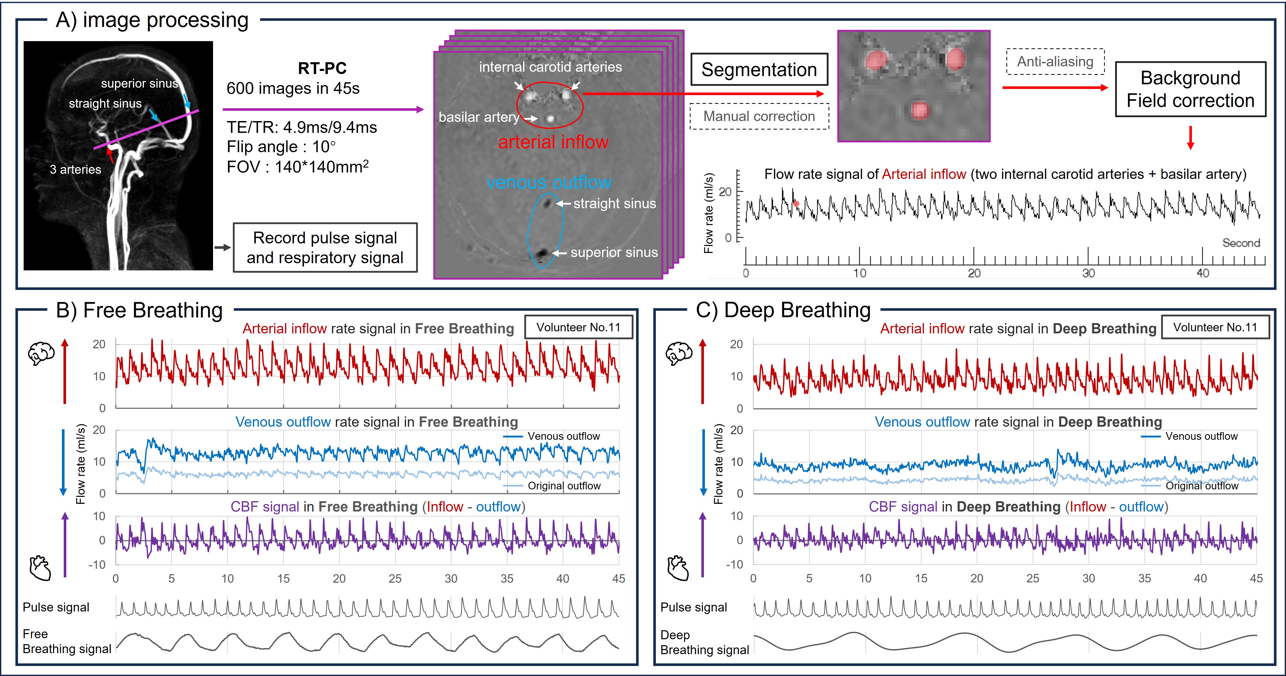
− Image acquisitionDue to the higher compliance and significant variability in internal jugular veins morphology, we selected the intracranial level to quantify the arterial and venous CBF (AVCBF).
Fourteen healthy volunteers (age: 20~34) were examined using a clinical 3T scanner and a 32-channel head coil. Pulse and breathing signals were recorded simultaneously using a finger plethysmograph and a chest sensor during two acquisitions: under free-breathing and deep-breathing conditions.
The RT-PC used in this study was a multi-shot, gradient-recalled echo-planar imaging sequence with parallel acquisition technology. Parameters were as follows: SENSE=2.5, EPI-factor=7, spatial resolution=2*2mm2, and temporal resolution=75ms/image (Fig.1-A).
− Image processing
All image and signal processing was performed using in-house software – Flow 2.07,8. Inflow and outflow were extracted through post-processing steps, including image segmentation, background field correction, and de-aliasing.
The sum of flows from the left/right internal carotid arteries and the basilar artery was considered the intracranial inflow. Similarly, the sum of flows from the superior sagittal sinus and the straight sinus was considered as the intracranial outflow.
The venous outflow curve was adjusted by multiplying the measured venous flow curve by a factor λ to account for unconsidered peripheral venous drainage. Then λ = mean arterial flow divided by mean measured venous flow. The AVCBF curve was calculated by subtracting the venous outflow curve from the arterial inflow curve (Fig.1-B&C). The oscillating positive and negative segments of the AVCBF curve represent the increase and decrease in intracranial blood volume during the cardiac cycle.
− Effect of breathing conditions on cerebral blood flow
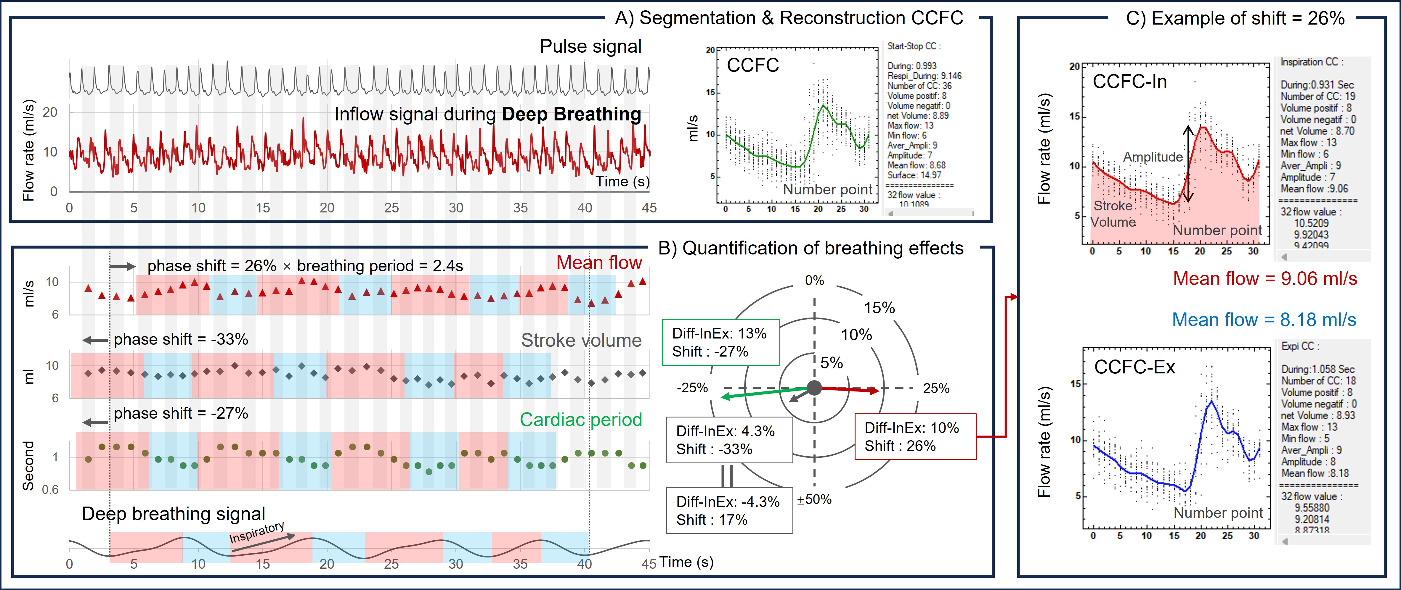
Time-domain analysis was used to individually segment cardiac cycle on RT-PC to quantify the effects of breathing on measured flows9 (Fig.2):
A) The software segmented each flow signal into multiple independent cardiac cycle flow curves (CCFCs).
B) The inspiration and expiration phases were determined by the respiratory signal. The corresponding CCFCs for inspiration and expiration were labeled as CCFC_In and CCFC_Ex, respectively.
C) Average of the CCF_ In and CCFC_Ex was then reconstructed to calculate their mean flow, amplitude, cardiac period and stroke volume (SV).
D) Diff_InEx% was calculated to quantify the percentage of change between inspiration and expiration period for all the flow parameters.
E) By shifting the respiratory signal with RT-PC signal, the maximum change of Diff-InEx% was determined. For example, in Figure 2-C, the highest Diff-InEx%, for the mean flow, occurred when the respiratory phase window was shifted 26%. This indicates a 10% increase in mean flow from mid-inspiration to mid-expiration.
Results
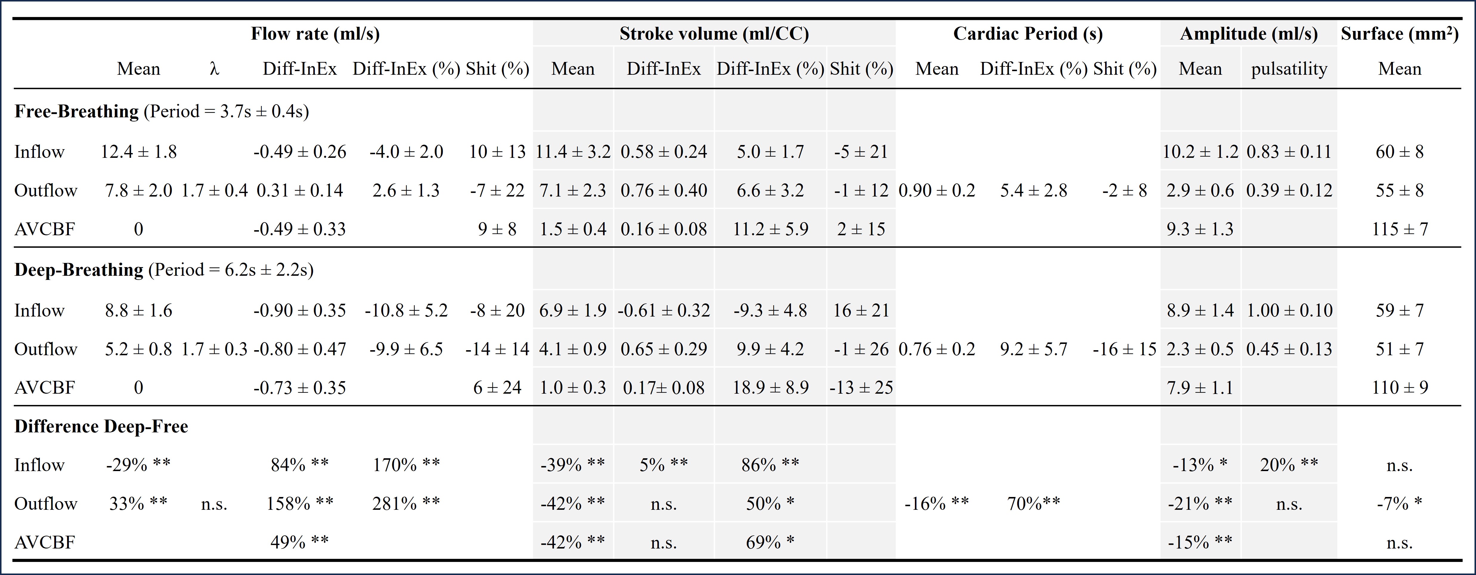
The changes of AVCBF, inflow, and outflow signals between inspiration and expiration periods during free- and deep-breathing acquisitions are presented in Fig.3.During inspiration (free&deep), SV and cardiac periods of AVCBF increased.
Deep-breathing significantly reduces AVCBF SV and cardiac period by 42% and 16%, respectively, compared to free-breathing. Furthermore, deep-breathing increases Diff-InEx of mean flow by 49% and Diff-InEx% of SV by 69% in CBF, with no significant impact on Diff-InEx of SV.
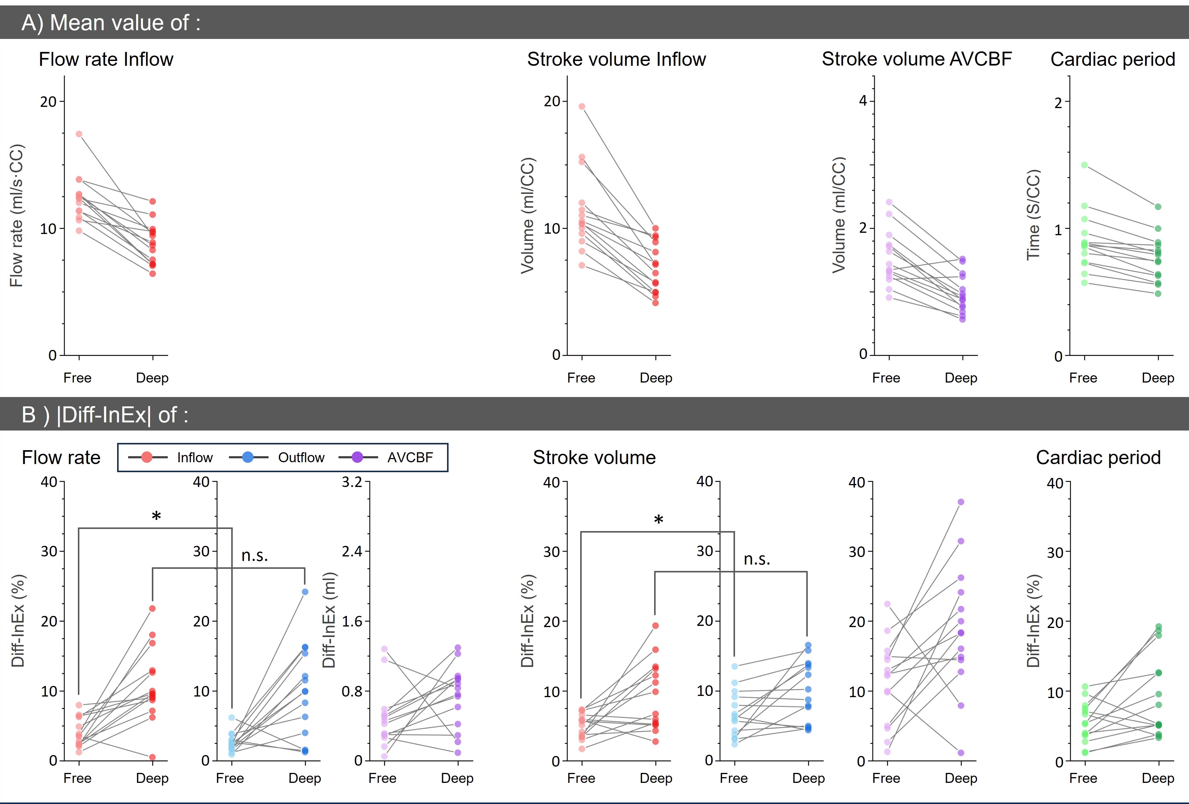
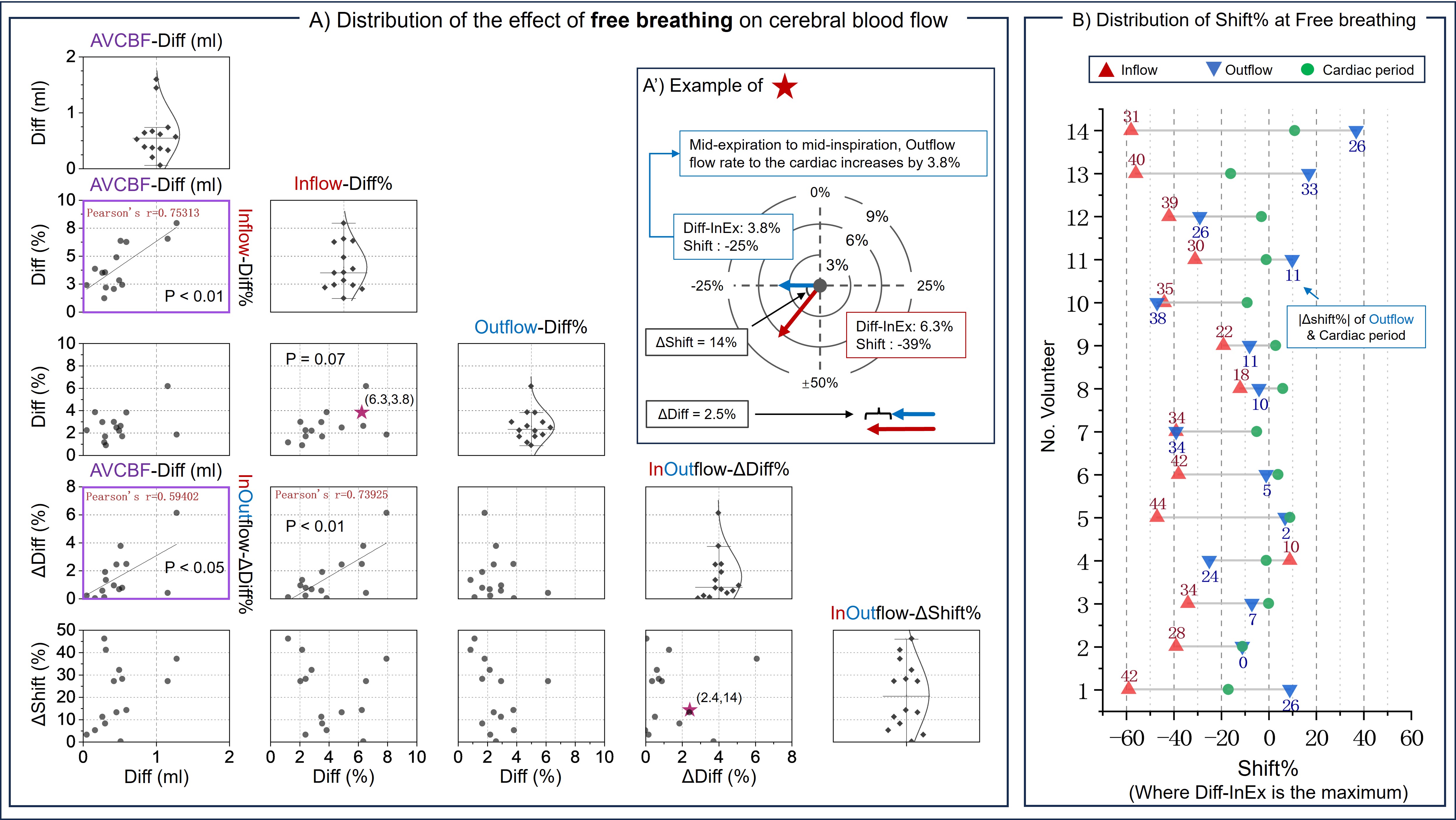
Fig.4 illustrates the distribution of the effect of free- and deep-breathing on each parameter.
Fig.5-A demonstrates the correlation, specifically regarding mean flow, between the Diff-InEx in CBF and the Diff-InEx% in inflow during free-breathing. Fig.5-B shows that during free-breathing, compared to inflow, the shift% of mean flow in outflow is closer to the shift% of cardiac period.
Discussion
The mean CBF (669±97 ml/min) measured by RT-PC in young healthy population is in agreement with previous studies using conventional PC-MRI with higher spatial resolution10.During inspiration, the cardiac period increases while mean inflow decreases, somewhat reducing the SV variation (Fig.5-B).
The Diff-InEx% of all the parameters increased significantly during deep-breathing. Conversely, the cardiac period and mean values of inflow and outflow decreased, which, to some extent, counteracted the effect of the increase in Diff-InEx. For example, the SV in CBF increased significantly in Diff-InEx% during deep-breathing, but not in Diff-InEx.
Conclusion
Deep-breathing decreased global AVCBF dynamics. Mean flows, SV and cardiac periods decreased but their percentage change between inspiratory and expiratory periods increased. This study can enhance our comprehension of fluid dynamics associated with CBF and CSF oscillations.Acknowledgements
This research was supported by EquipEX FIGURES (Facing Faces Institute Guilding Research), Hanuman ANR-18-CE45-0014 and Region Haut de France. Thanks to the staff members at the Facing Faces Institute (Amiens, France) for technical assistance. Thanks to David Chechin from Phillips industry for his scientific support.References
1. Balédent O, Idy-peretti I. Cerebrospinal fluid dynamics and relation with blood flow: a magnetic resonance study with semiautomated cerebrospinal fluid segmentation. Investigative radiology. 2001 Jul 1;36(7):368-77.
2. Alperin N, Lee SH, Loth F, Raksin P, Lichtor T. MR-Intracranial Pressure (ICP): A method for noninvasive measurement of intracranial pressure and elastance. Baboon and Human Study. Radiology. 2000;217(3):877-85. https://doi.org/10.1148/radiology.217.3.r00dc42877.
3. Liu P, Fall S, Balédent O. Use of real-time phase-contrast MRI to quantify the effect of spontaneous breathing on the cerebral arteries. NeuroImage. 2022 Jun 7:119361. https://doi.org/10.1016/j.neuroimage.2022.119361.
4. Liu P, Monnier H, Owashi K, Constant JM, Capel C, Balédent O. “The Effects of Free Breathing on Cerebral Venous Flow: a real-time phase contrast MRI study in healthy adults”. Journal of neuroscience, Accepted, in production.
5. Laganà MM, Pirastru A, Ferrari F, Di Tella S, Cazzoli M, Pelizzari L, Jin N, Zacà D, Alperin N, Baselli G, Baglio F. Cardiac and Respiratory Influences on Intracranial and Neck Venous Flow, Estimated Using Real-Time Phase-Contrast MRI. Biosensors. 2022 Aug 8;12(8):612. https://doi.org/10.3390/bios12080612.
6. Kollmeier, J. M., Gürbüz-Reiss, L., Sahoo, P., Badura, S., Ellebracht, B., Keck, M., ... & Dreha-Kulaczewski, S. (2022). Deep breathing couples CSF and venous flow dynamics. Scientific reports, 12(1), 2568. https://doi.org/10.1038/s41598-022-06361-x.
7. Liu P, Lokossou A, Fall S, Makki M and Bamendent O, 2019. Post Processing Software for Echo Planar Imaging Phase Contrast Sequence. ISMRM 27th, (4823). https://archive.ismrm.org/2019/4823.html
8. Liu P, Fall S, Balédent O. Flow 2.0-a flexible, scalable, cross-platform post-processing software for realtime phase contrast sequences. In ISMRM 2022-International Society for Magnetic Resonance in Medicine 2022 May 7. https://archive.ismrm.org/2022/2772.html
9. Liu P, Monnier H, Owashi K, Metanbou S, Capel C, Balédent O. Effects of Free-Breathing on Cerebral Veins: study by Real-Time Phase-Contrast MRI. In ISMRM 2023-International Society for Magnetic Resonance in Medicine 2023 (No. Oral# 0938).
10. Owashi, K. P., Capel, C., & Balédent, O. (2023). Cerebral arterial flow dynamics during systole and diastole phases in young and older healthy adults. Fluids and Barriers of the CNS, 20(1), 65.
Figures