2200
Cerebrovascular Flow and White Matter Microstructural Integrity in the Presence of Amyloid and Tau Biomarkers1University of Wisconsin Madison, Madison, WI, United States
Synopsis
Keywords: Blood Vessels, Alzheimer's Disease, White Matter; Velocity & Flow
Motivation: This study is driven by the need to understand the intricate relationship between cerebrovascular disease (CVD) and Alzheimer's disease (AD) pathology.
Goal(s): In this study, we aim to investigate the correlations between CVD markers, AD biomarkers and white matter (WM) microstructure.
Approach: To achieve these goals, we used 4D flow MRI and neurite orientation dispersion and density imaging (NODDI) and utilized statistical models to examine the relationships between vascular flow and WM neurite density index.
Results: Our results identified significant correlations between AD biomarkers, WM integrity and cerebrovascular flow in specific vessels.
Impact: Our findings motivate further investigations into the intricate relationship between cerebrovascular disease and Alzheimer's disease (AD) pathology. Better understanding of this relationship may improve early AD detection and therapeutic strategies.
Introduction
Recent studies suggest potential interactions between cerebrovascular disease (CVD) and Alzheimer's disease (AD) pathology, by investigating correlations between AD biomarkers, hypoperfusion1 , white matter hyperintensity (WMH) burden2, and WM abnormalities3 . However, hypoperfusion and WMH are not specific to CVD pathology4, limiting the potential to identify CVD and AD interaction pathways. More specific CVD markers, such as intracranial vessel flow rates and velocities, can be quantified using 4D flow MRI.Our study examines correlations between CVD markers and WM microstructure across the AD clinical spectrum, utilizing 4D flow MRI and neurite orientation dispersion and density imaging (NODDI)5. This research aims to bridge existing knowledge gaps by examining the connections between CVD, AD, and WM alterations.
Methods
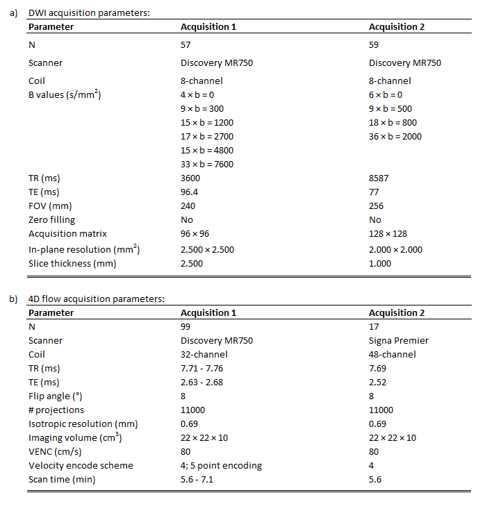
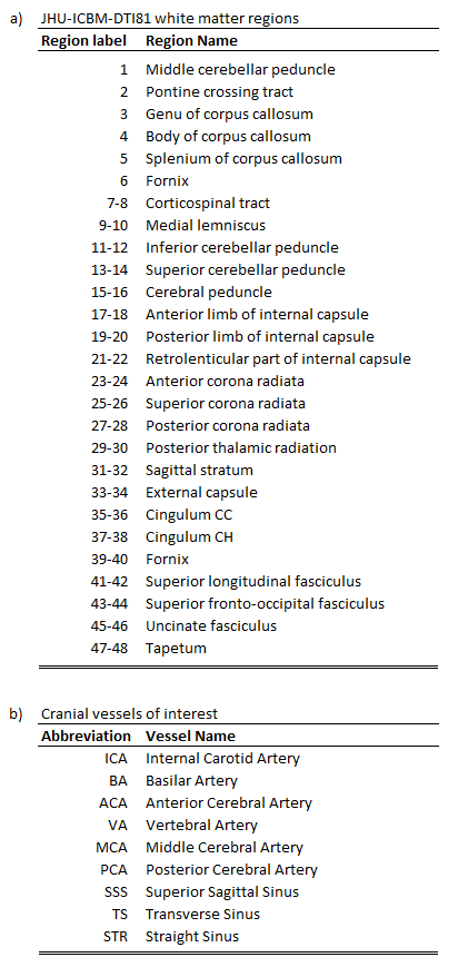
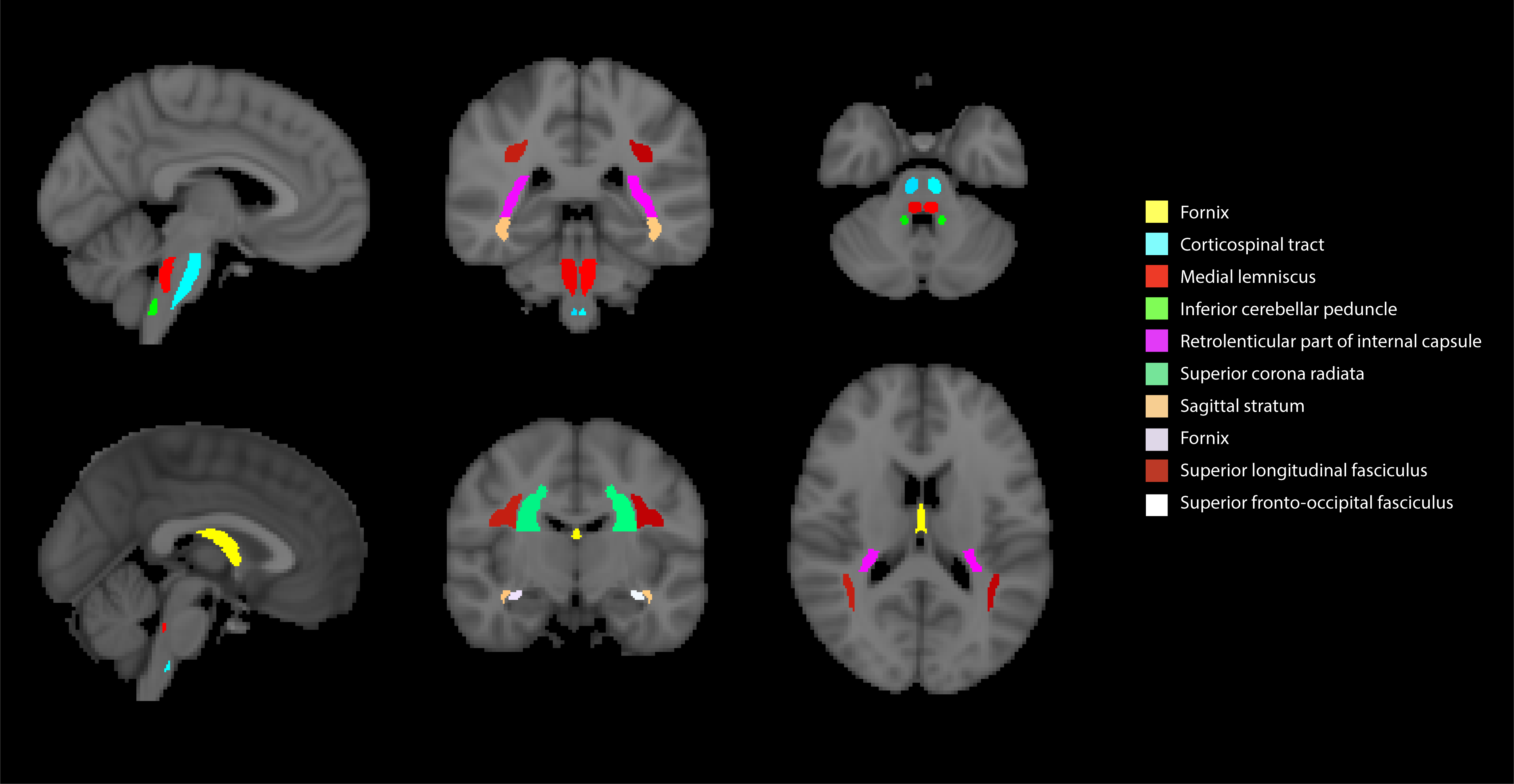
The cohort consisted of 116 subjects (65F/51M, age=70.9±5.4 years). Three groups were defined based on PET biomarker status: amyloid-positive/tau-positive (A+/T+;N=30), amyloid-positive/tau-negative (A+/T-;N=30), and amyloid-negative/tau-negative (A-/N-;N=56).All MRI data were acquired on 3T scanners (Fig. 1). Diffusion-weighted imaging (DWI) data was acquired using two spin-echo echo-planar protocols and pre-processed using FSL6 and MRtrix37, including denoising8, Gibb’s ringing correction9, brain extraction10, motion and eddy current correction11, and diffusion tensor estimation12. Neurite density index (NDI) parametric maps were generated using the NODDI MATLAB Toolbox13 and nonlinearly registered to 1mm-FMRIB58_FA standard space14. Mean NDI value was calculated for 48 regions of interest (ROIs), obtained from the JHU-ICBM-DTI-81 WM atlas15 (Fig. 2a). Finally, neuroCombat16 was used to harmonize the ROI data from different acquisition protocols using sex as covariate.
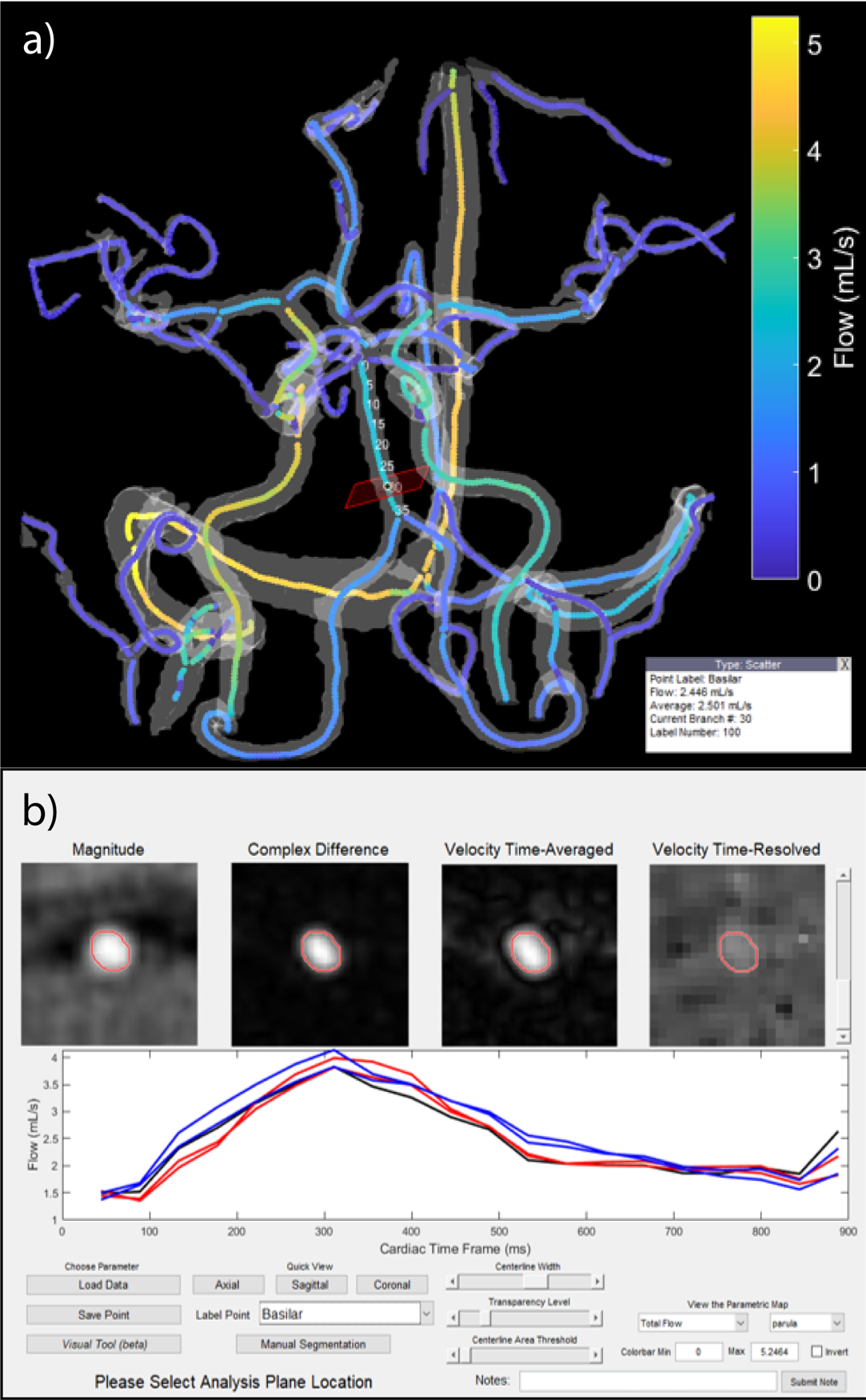
4D flow data was acquired using a 3D radially-undersampled sequence17,18 (Fig. 1b) and reconstructed into 20 cardiac frames using retrospective gating and temporal radial view sharing19 . A 4D flow processing tool20 (Fig. 3) was used to measure mean volumetric flow rates (mL/min) in 16 cerebral vessels (Fig. 2b). To reduce multiple comparisons, hemispheric structures were averaged into unified bilateral measurements resulting in 27 WM ROIs and 9 vessels of interest.
Ten multivariate general linear models (GLMs) were used to determine if age, sex, Aβ, or tau are predictors of vessel flow. Ten additional GLMs, simultaneously testing all WM ROIs, were used to determine the correlation between NDI and vessel flow while controlling for sex and age.
Results
All DWI datasets were successfully processed. Due to poor image quality, flow was not measured in some subjects: posterior cerebral arteries (PCA;n=1), middle cerebral arteries (MCA;n=2), vertebral arteries (VA;n=7), and transverse sinuses (TS;n=1).Linear models show significant inverse correlation between age and mean flow in PCA only (b=-0.43;p=0.01). Sex was not a predictor of flow in any of the measured vessels. Ab pathology was directly correlated with flow in PCAs (b=6.14;p=0.005), basilar artery (BA;b=18.75;p=0.022), straight sinuses (STR;b=8.19;p=0.034), and VA (b=10.09;p=0.041). Tau presence showed a strong inverse correlation with flow in STR (b=-11.86;p=0.008) and VA (b=-18.59;p=0.002). Weak inverse correlation was observed between tau presence and flow in PCA (b=-4.85;p=0.052) and BA (b=-18.18;p=0.054).
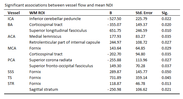
Linear models that tested for correlations between vessel flow and NDI values in different WM ROIs reached significance with at least one WM ROI in all vessels except in the VA. Significant ROI results for this analysis are summarized in Fig. 4.
Discussion
Our study aimed to improve our understanding of the interaction between CVD and AD pathologies.A significant correlation between AD biomarkers and flow in smaller arteries and STR suggests potential interactions that may alter vascular dynamics. The inverse correlation between tau and flow in STR and VA aligns with expectations21, as tau-related neuronal loss may reduce vascular output. Moreover, our vessel-specific analysis indicates a significant relationship between flow and WM integrity in transentorhinal region associated with early tau accumulation22,23. However, the observed direct relationship between flow and Aβ lacks a clear rationale without further analysis. Our study did not provide a broad age range to detect age-related flow effects shown by others24. Lack of gender-related flow differences aligns with some studies24 but may be due to a small sample size. More advanced statistical models and inclusion of other risk factors (APOEε4, AD parental history, cardiovascular risk effects) may improve understanding of observed correlations and their implications.
Conclusion
This is the first study that characterizes the relationship between 4D flow–based vessel-specific flow and NODDI-based WM neurite density in a cohort with known AD biomarker status. Positive correlation was observed between Aβ presence and flow in inferior and posterior arteries as well as the straight sinuses. Tau presence exhibited an inverse correlation with flow in the straight sinuses and vertebral arteries. Our data further suggests interaction mechanisms between cerebrovascular flow and NDI alterations in localized WM microstructure.Acknowledgements
Research reported in this abstract was supported by the National Institutes of Health (NIH) under award numbers F31AG071183, KL2TR002374, UL1TR002373, R01AG075788, RF1AG027161, P30AG062715, R21AG077337, TL1TR002375, and the Alzheimer’s Association under award number AARFD-20-678095. The content is solely the responsibility of the authors and does not necessarily represent the official views of these institutions. We would like to thank GE Healthcare for their continued technical assistance and product support. We also gratefully acknowledge the researchers and staff at the Wisconsin Institutes for Medical Research, Wisconsin Alzheimer’s Disease Research Center, and Waisman Brain Imaging Core for assistance in recruitment, data collection, and data analysis. Lastly, the authors extend their most sincere thanks to all participants involved in this study.References
1. Clark, L. R. et al. Macrovascular and microvascular cerebral blood flow in adults at risk for Alzheimer’s disease. Alzheimers Dement (Amst) 7, 48–55 (2017).
2. Birdsill, A. C. et al. Regional white matter hyperintensities: aging, Alzheimer’s disease risk, and cognitive function. Neurobiol Aging 35, 769–76 (2014).
3. Vemuri, P. et al. White matter abnormalities are key components of cerebrovascular disease impacting cognitive decline. Brain Commun 3, fcab076 (2021).
4. Alber, J. et al. White matter hyperintensities in vascular contributions to cognitive impairment and dementia (VCID): Knowledge gaps and opportunities. Alzheimers Dement (N Y) 5, 107–117 (2019).
5. Zhang, H., Schneider, T., Wheeler-Kingshott, C. A. & Alexander, D. C. NODDI: practical in vivo neurite orientation dispersion and density imaging of the human brain. Neuroimage 61, 1000–16 (2012).
6. Jenkinson, M., Beckmann, C. F., Behrens, T. E. J., Woolrich, M. W. & Smith, S. M. FSL. Neuroimage 62, 782–790 (2012).
7. Tournier, J.-D. et al. MRtrix3: A fast, flexible and open software framework for medical image processing and visualisation. Neuroimage 202, 116137 (2019).
8. Veraart, J. et al. Denoising of diffusion MRI using random matrix theory. Neuroimage 142, 394–406 (2016).
9. Kellner, E., Dhital, B., Kiselev, V. G. & Reisert, M. Gibbs-ringing artifact removal based on local subvoxel-shifts. Magn Reson Med 76, 1574–1581 (2016).
10. Smith, S. M. Fast robust automated brain extraction. Hum Brain Mapp 17, 143–155 (2002).
11. Andersson, J. L. R. & Sotiropoulos, S. N. An integrated approach to correction for off-resonance effects and subject movement in diffusion MR imaging. Neuroimage 125, 1063–1078 (2016).
12. Basser, P. J., Mattiello, J. & Lebihan, D. Estimation of the Effective Self-Diffusion Tensor from the NMR Spin Echo. J Magn Reson B 103, 247–254 (1994).
13. http://mig.cs.ucl.ac.uk.
14. Andersson, J. L. R., Jenkinson, M. & Smith, S. Non-linear registration aka spatial normalisation. (2007).
15. Oishi, K. et al. Atlas-based whole brain white matter analysis using large deformation diffeomorphic metric mapping: application to normal elderly and Alzheimer’s disease participants. Neuroimage 46, 486–99 (2009).
16. Fortin, J.-P. et al. Harmonization of multi-site diffusion tensor imaging data. Neuroimage 161, 149–170 (2017).
17. Johnson, K. M. et al. Improved 3D phase contrast MRI with off-resonance corrected dual echo VIPR. Magn Reson Med 60, 1329–1336 (2008).
18. Gu, T. et al. PC VIPR: a high-speed 3D phase-contrast method for flow quantification and high-resolution angiography. AJNR Am J Neuroradiol 26, 743–9 (2005).
19. Liu, J. et al. Generation and visualization of four-dimensional MR angiography data using an undersampled 3-D projection trajectory. IEEE Trans Med Imaging 25, 148–57 (2006).
20. Roberts, G. S. et al. Automated hemodynamic assessment for cranial 4D flow MRI. Magn Reson Imaging 97, 46–55 (2023).
21. Rivera-Rivera, L. A. et al. Cerebrovascular stiffness and flow dynamics in the presence of amyloid and tau biomarkers. Alzheimers Dement (Amst) 13, e12253 (2021).
22. Schöll, M. et al. PET Imaging of Tau Deposition in the Aging Human Brain. Neuron 89, 971–982 (2016).
23. Thal, D. R., Attems, J. & Ewers, M. Spreading of Amyloid, Tau, and Microvascular Pathology in Alzheimer’s Disease: Findings from Neuropathological and Neuroimaging Studies. Journal of Alzheimer’s Disease 42, S421–S429 (2014).
24. Roberts, G. S. et al. Normative Cerebral Hemodynamics in Middle-aged and Older Adults Using 4D Flow MRI: Initial Analysis of Vascular Aging. Radiology 307, e222685 (2023).
Figures