2196
Detecting Microvascular Abnormalities in Neurodegenerative Diseases using MICRO at 3T1Neurology, Wayne State University, DETROIT, MI, United States, 2Clinical Neurological Sciences, Western University, London, ON, Canada, 3Radiology, New York University Grossman School of Medicine, New York, NY, United States, 4Neurology, Wayne State University, Detroit, MI, United States, 5Radiology, Wayne State University, DETROIT, MI, United States
Synopsis
Keywords: Blood Vessels, Blood vessels, Microvasculature mapping, vascular abnormalities
Motivation: There is limited understanding of microvascular alterations in neurovascular diseases such as multiple sclerosis (MS) and Parkinson’s disease (PD) using in vivo imaging techniques.
Goal(s): To detect microvascular abnormalities using the novel imaging technique MICRO (Microvascular In-vivo Contrast Revealed Origins).
Approach: MICRO imaging involves high resolution susceptibility weighted imaging (SWI) acquired in the presence of ultrasmall superparamagnetic iron oxides (USPIO).
Results: As compared to non-contrast conventional vascular imaging, MICRO drastically improves the detection of microvascular abnormalities that can help in identifying the most vulnerable structures in vascular etiology of MS and PD.
Impact: This multicentre study demonstrates that MICRO imaging has the potential to comprehensively study vascular pathology something that is often ignored in evaluating neurodegenerative diseases.
Introduction
Cadaver brain studies are essential in capturing vessels at a micrometer level, but can only evaluate a single timepoint (that is, an end timepoint) for a given patient with lack of blood flow, large brain coverage and vessel from post-mortem fixation of the brain. Therefore, understanding spatial and time domains of the microvascular abnormalities and their contributions to neurological development and progression is crucial. Conventional vascular MRI techniques are limited to their low resolution and sensitivity in detecting small vessels. There is an inherent trade off between the imaging resolution and signal-to-noise ratio (SNR) in mapping the vasculature using these methods. Higher imaging resolution will certainly help in visualizing smaller vessels, but at the expense of reduced SNR (and contrast-to-noise (CNR)) and longer scan times. Furthermore, the sensitivity of the imaging methods limits them to major vessels due to the low resolution for dynamic susceptibility contrast data, slow blood flow rate in small vessels for time-of-flight based data1,2 and a relatively low magnetic susceptibility effect in veins (0.45 ppm for 70% oxygenation level) for pre-contrast SWI data. To overcome the abovementioned limitations of current technology, we developed a new technique: ‘Microvascular In-vivo Contrast Revealed Origins (MICRO)’ using an ultrasmall superparamagnetic iron oxide (USPIO) and utilized it for our multicenter study on patients with relapsing-remitting multiple sclerosis (RRMS) and Parkinson’s disease (PD) to reveal microvascular abnormalities3–5.Materials and Methods
An USPIO agent, Ferumoxytol (Feraheme, AMAG Pharmaceuticals, Waltham, MA) was used in this study. Six RRMS subjects (scanned at Wayne State University, USA) and 31 PD patients (scanned at Western University, Canada) were scanned at 3T with high resolution (0.22×0.44×1 mm3) SWI (TE = 15 ms, flip angle = 12°) acquired before and after the infusion of Ferumoxytol. Composite data was generated by registering the FLAIR data to the high resolution SWI data in order to highlight the vascular information in MS lesions. Any vascular abnormalities were visually identified in the MS lesions, and white matter hyperintensities (WMH) in PD patients, and surrounding tissues on pre- and post-contrast SWI.Results
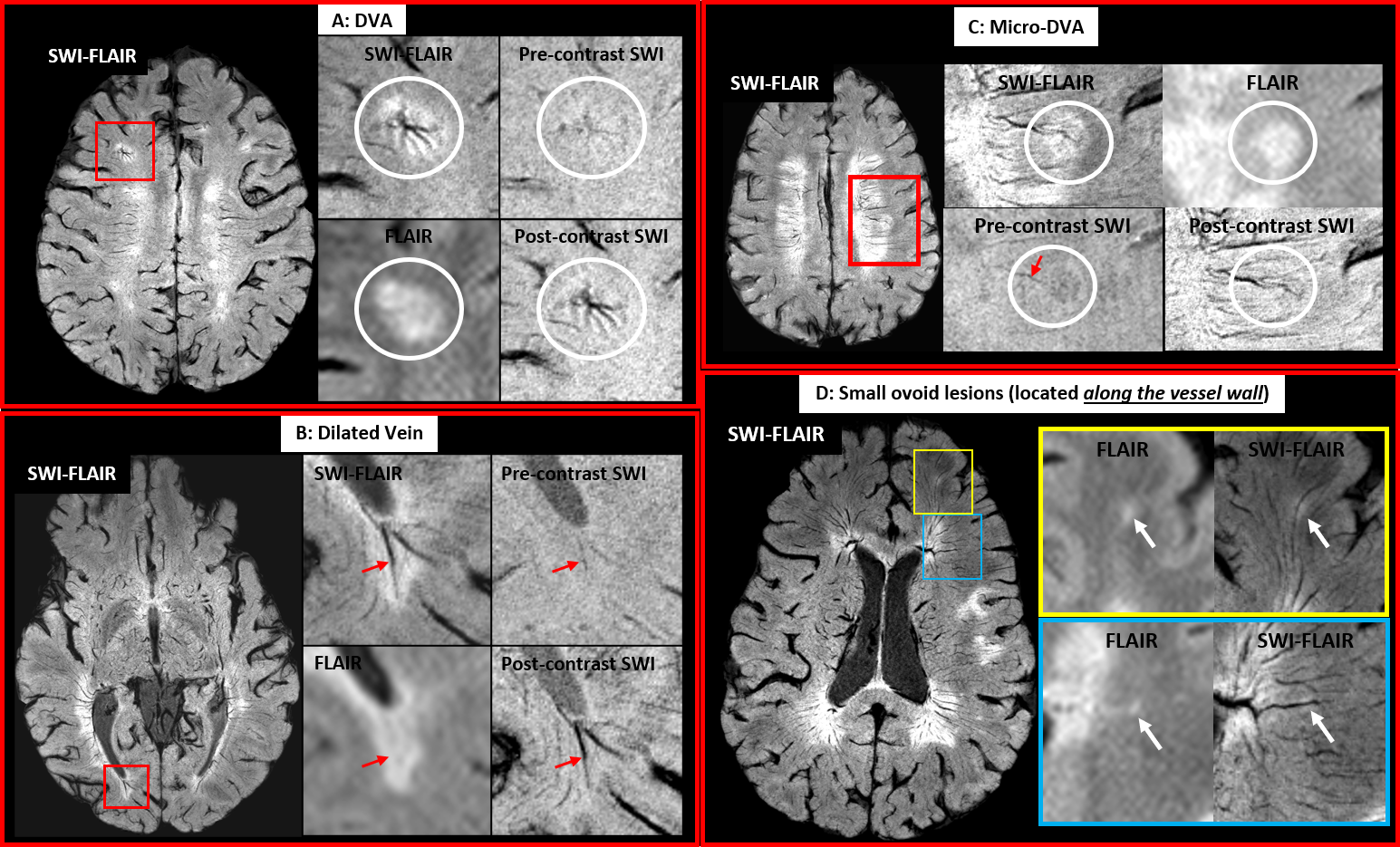
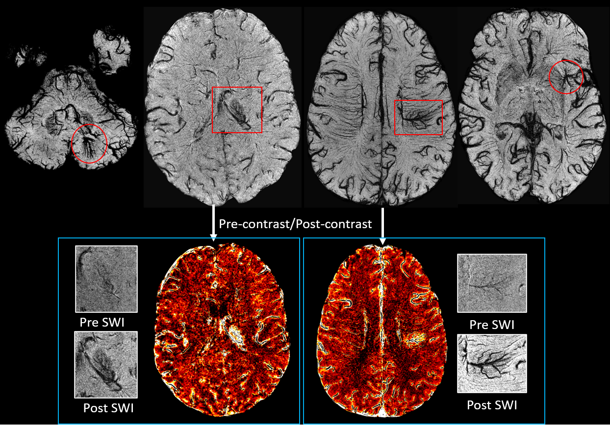
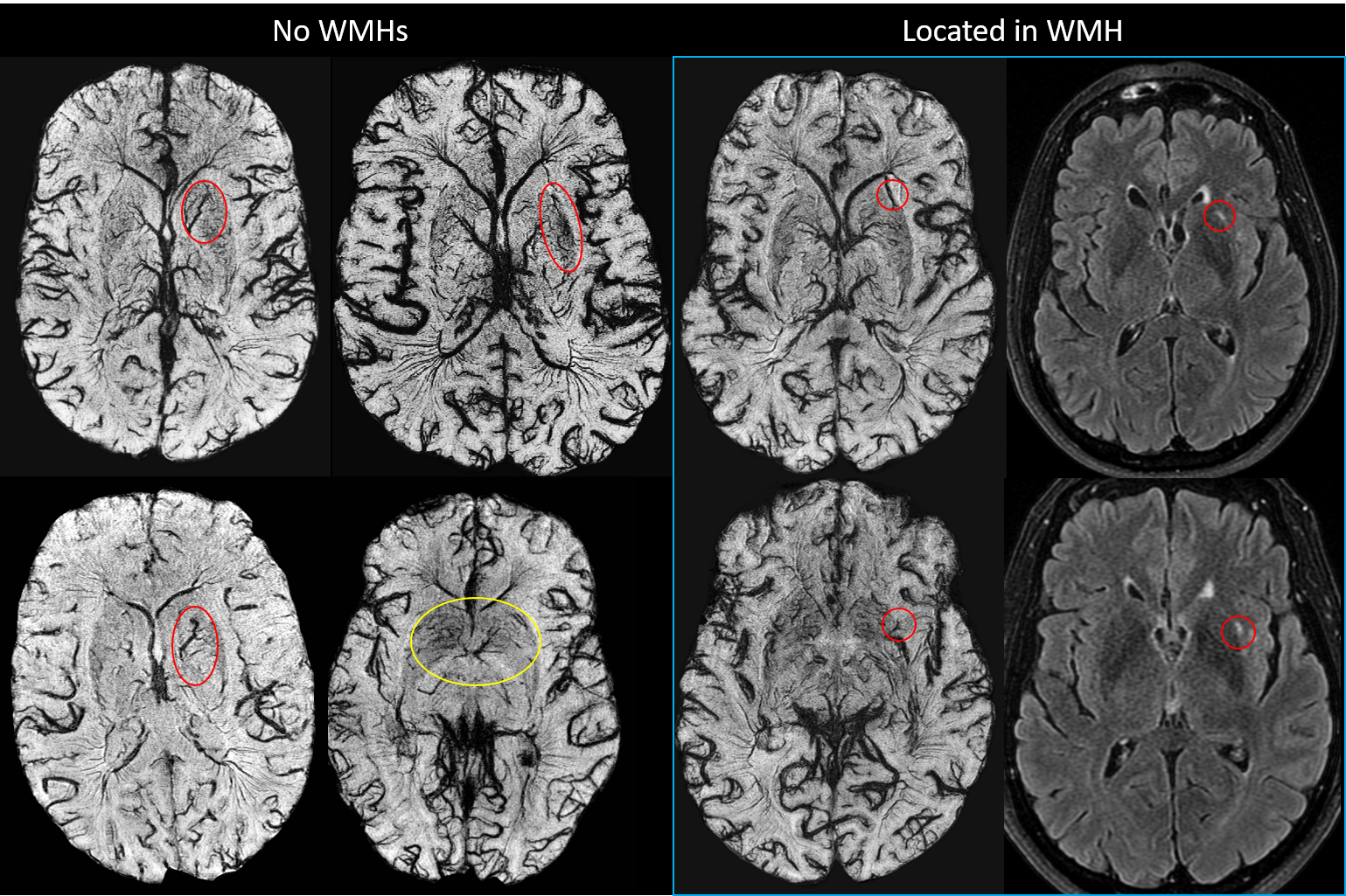
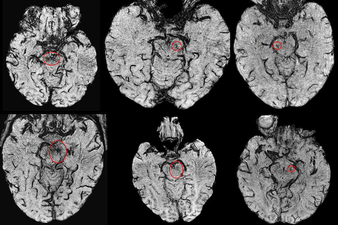
By inducing superparamagnetic susceptibility into the blood, both small arteries and veins at the level of 50μm to 100μm can be seen on MICRO MRI (Figure 1). In both patient cohorts, several developmental venous anomalies (DVA) were revealed that were undetectable on conventional T1 and SWI data. For MS patients, several abnormal microvasculature abnormalities within the lesions can now be seen in vivo (Figure 2) on SWI-FLAIR3,6 data. Lesions with abnormal vascular behavior were broken up into the following categories for MS patients: small lesions appearing only at the vessel boundary; dilated vessels within the lesions; and developmental venous angiomas. These vessel abnormalities observed within lesions increased from 55 on pre-contrast data to 153 on post-contrast data for the RRMS patients. For PD patients, the major vascular malformations were revealed in four patients and two exhibited the retention of contrast in the surrounding tissues at the capillary level (n=4, Figure 3). Moreover, PD patients also exhibited abnormally dense and engorged microvasculature particularly in the midbrain (n=6, Figure 4) and basal ganglia (n=5, Figure 5). The remaining vascular abnormalities were found in different regions of white matter (n=8).Discussion and Conclusion
MICRO imaging has shown potential in detecting microvascular abnormalities that previously could only be seen in cadaver brain studies and has the potential to study the vascular etiology of MS and PD in living brains3,7. While vascular abnormalities in histopathological studies are long known in neurovascular diseases, its in vivo characterization has never been well elucidated besides blood brain barrier breakdown using gadolinium-based MRI. For RRMS subjects, MICRO not only detected lesion-centric abnormal networks (such as DVAs and engorged vessels), but also small WMHs (<3mm) that are located along the vessel wall on the post-contrast SWI-FLAIR data7. This approach has the potential to monitor the venous vasculature present in MS lesions, catalogue their characteristics and compare the vascular structures spatially to the presence of lesions. Interestingly, the DVAs in MS patients were WMH-centric and small whereas the major malformations in PD patients were not enclosed in WMH. Finally, the distinctive microvasculature in the midbrain (particularly in the substantia nigra) and basal ganglia (particularly in the putamen) in PD patients may suggest vulnerability of the basal ganglia and midbrain to chronic ischemia facilitated by the presence of poor flow caused by abnormal vasculature that could be contributory to PD pathogenesis. The enhanced vascular features provided by MICRO imaging should provide new insight into the pathophysiology of MS and PD.Acknowledgements
This work was supported in part by the sub-organizations of National Institutes of Health (NIH): National Institute on Aging (grant numbers: R56-AG060822), National Institute of Neurological Disorders and Stroke (grant numbers: R01-NS108491). Dr. LeWitt is the Sastry Foundation Endowed Chair in Neurology at Wayne State University School of Medicine and is supported by Parkinson Disease and Movement Disorder funding from Henry Ford Hospital. This work was also supported, in part, by the Silverman Endowment Fund at Wayne State University and by the Office of the Vice President for Research at Wayne State University for their support of the MR Research Facility.References
1. Haacke EM, Masaryk TJ, Wielopolski PA, et al. Optimizing blood vessel contrast in fast three-dimensional MRI. Magn Reson Med. 1990;14(2):202-221. doi:10.1002/mrm.1910140207
2. Bosmans H, Marchal G, Lukito G, et al. Time-of-flight MR angiography of the brain: comparison of acquisition techniques in healthy volunteers. AJR Am J Roentgenol. 1995;164(1):161-167. doi:10.2214/ajr.164.1.7998531
3. Buch S, Wang Y, Park MG, et al. Subvoxel vascular imaging of the midbrain using USPIO-Enhanced MRI. NeuroImage. 2020;220:117106. doi:10.1016/j.neuroimage.2020.117106
4. Liu S, Brisset JC, Hu J, Haacke EM, Ge Y. Susceptibility weighted imaging and quantitative susceptibility mapping of the cerebral vasculature using ferumoxytol. J Magn Reson Imaging. 2018;47(3):621-633. doi:10.1002/jmri.25809
5. Shen Y, Hu J, Eteer K, et al. Detecting sub-voxel microvasculature with USPIO-enhanced susceptibility-weighted MRI at 7 T. Magnetic Resonance Imaging. 2020;67:90-100. doi:10.1016/j.mri.2019.12.010
6. Buch S, Chen Y, Jella P, Ge Y, Haacke EM. Vascular Mapping of the Human Hippocampus Using Ferumoxytol-Enhanced MRI. NeuroImage. Published online February 2, 2022:118957. doi:10.1016/j.neuroimage.2022.118957
7. Buch S, Subramanian K, Jella PK, et al. Revealing vascular abnormalities and measuring small vessel density in multiple sclerosis lesions using USPIO. Neuroimage Clin. 2021;29:102525. doi:10.1016/j.nicl.2020.102525
Figures
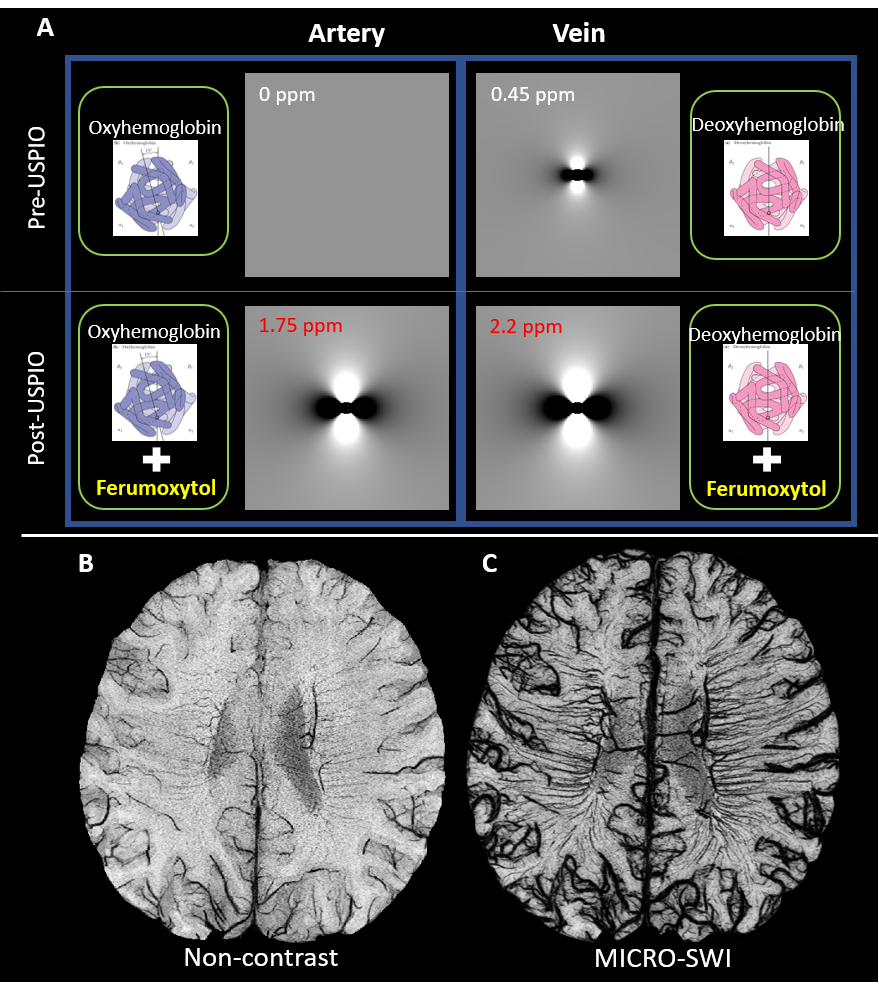
Figure 1. (A) Illustration of the changes in field surrounding the cross-section of a cylinder (representing the arteries and veins) before and after the injection of Ferumoxytol. Using high-resolution SWI, the non-contrast SWI (B) already shows the medullary veins in the white matter, but the vascular contrast increases significantly for post-contrast MICRO-SWI (C) taking advantage of the T2* related signal loss caused by low-dose (4 mg/kg) Ferumoxytol.