2194
Temporal changes in tissue plasticity and microvasculature as a gauge of recovery in stroke1Department for physics and image analysis, Oslo University Hospital, Oslo, Norway, 2LVTS, INSERM U1148, Paris, France, 3Department of Neuroradiology, Heidelberg University Hospital, Heidelberg, Germany
Synopsis
Keywords: Vascular, Stroke
Motivation: Stroke is major global cause of death. However markers of recovery are still lacking.
Goal(s): Vasculature affects shear waves propagation acting as a scattering source, ultimately affecting waves dispersion properties measurable with MRE.
Approach: In this study we use multi frequency MRE to find the fingerprint of vascular remodelling after stroke in the dispersion properties of waves, and explored the temporal profile of vascular and tissue remodelling and its relashionship with recovery.
Results: We show for the first time in-vivo that MRE senses tissue integrity as well as vascular organization. We show that such parameters have a good prognostic value.
Impact: We show that MRE could disentangle tissue constitutive properties and vasculature, and thereby could provide new insights in the understanding of tissue plasticity after stroke and potentially constitute a relevant marker in the context of stroke therapy.
Introduction
Stroke is a major global cause of death [1]. The abrupt reduction in blood flow triggers a series of dynamic events known as ischemic cascade leading to morphological and functional changes in the tissue and in the vascular network. [2, 3]. Magnetic resonance imaging (MRI) is the gold standard to accurately diagnose and assess the size and age of the lesion [4, 5]. However, while MRI-derived biomarkers provide critical information for diagnosis, markers of recovery are still lacking. There is growing evidence that vascular remodelling has important prognostic implications in stroke, however the relationship between the temporal profile of such changes and the recovery is still elusive [6]. Magnetic Resonance Elastography (MRE) makes use of shear wave to probe tissue rheology. Vasculature, one of the stiffest component of tissue, affects shear waves propagation acting as a scattering source, ultimately affecting waves dispersion properties [7]. In this study we use multi frequency MRE to find the fingerprint of vascular remodelling after stroke in the dispersion properties of waves. Additionally, we explored the temporal profile of vascular and tissue remodelling and its relationship with the stroke recovery.Methods
Five mice underwent stroke induction through intra-arterial suture occlusion of the middle cerebral artery (MCAo) [8]. Imaging was conducted on a 7T preclinical MRI scanner (Bruker, Ettlingen, Germany). Anaesthesia, induced with isoflurane delivered via a nose cone, was maintained throughout the imaging session with constant monitoring of respiration rate. Shear wave vibrations were generated using a a linear motor connected through a cantilever assembly to a custom-built bed where the head of the mice were fixated. MRE was performed at different vibration frequencies (600-700-900-1000 Hz) using a multi-slice, single spin echo MRE sequence (TR/TE 1600/26 ms; FOV 19.2 mm; matrix 64 × 64; 1 average; 6 wave phases; 18 slices; isotropic resolution 0.250 mm) [9]. Post-acquisition, the MRE data were reconstructed following Sinkus et al. [10]. This allowed to calculate the complex shear modulus G*=G’ +iG’’, where G’ (Gd) is the shear stiffness and G’’ (Gl) is the shear viscosity, and the phase angle Y=2/π*atan(Gl/Gd) in [0,1]. Additionally, dispersion properties were assessed by studying power law behaviour of the shear wavelength λ≈ωκ where ω is the frequency and κ is the dispersion exponent. Brain biomechanics was assessed prior to the stroke induction and longitudinally 1 day and 7 days after the ischemic event.Results and discussion
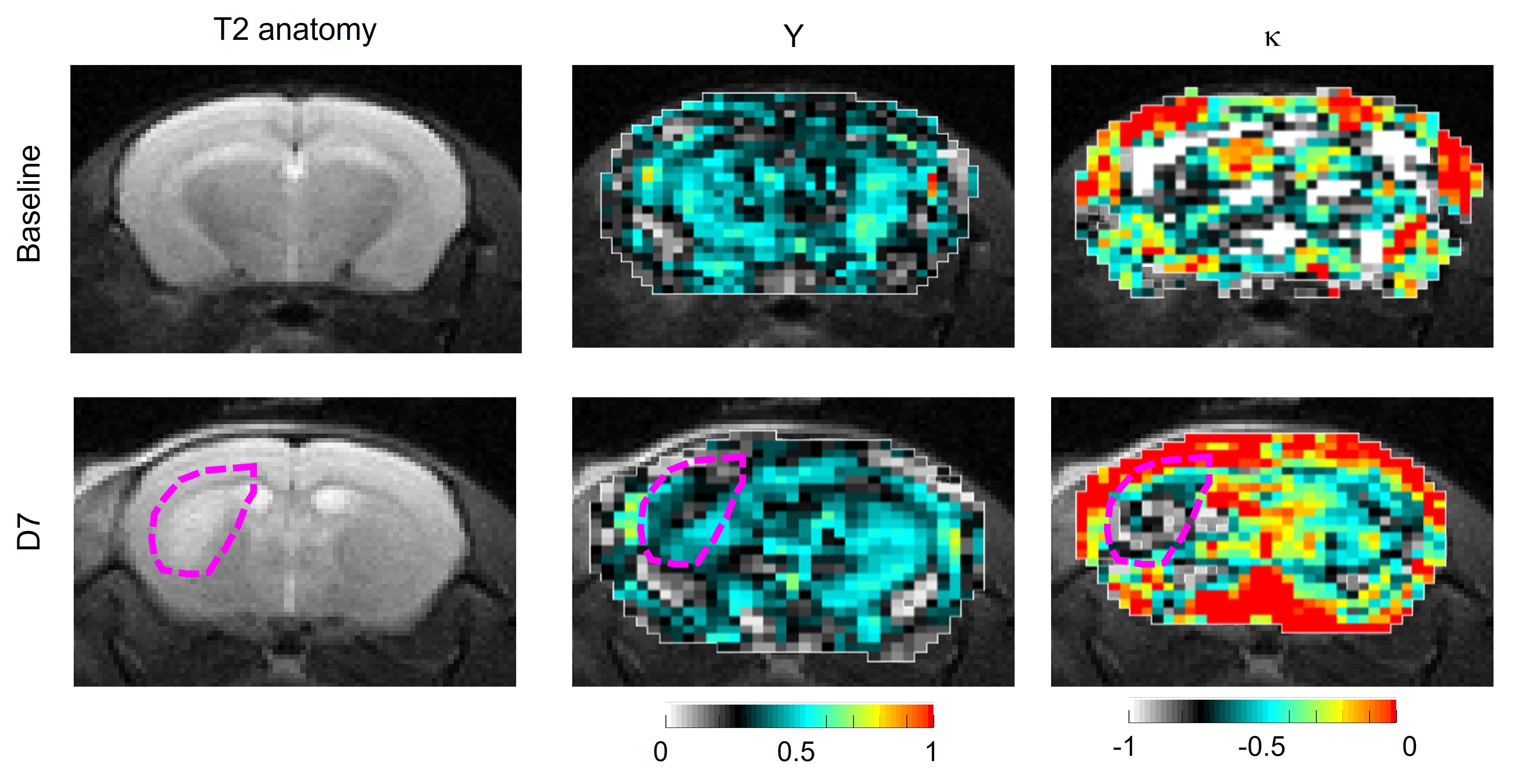
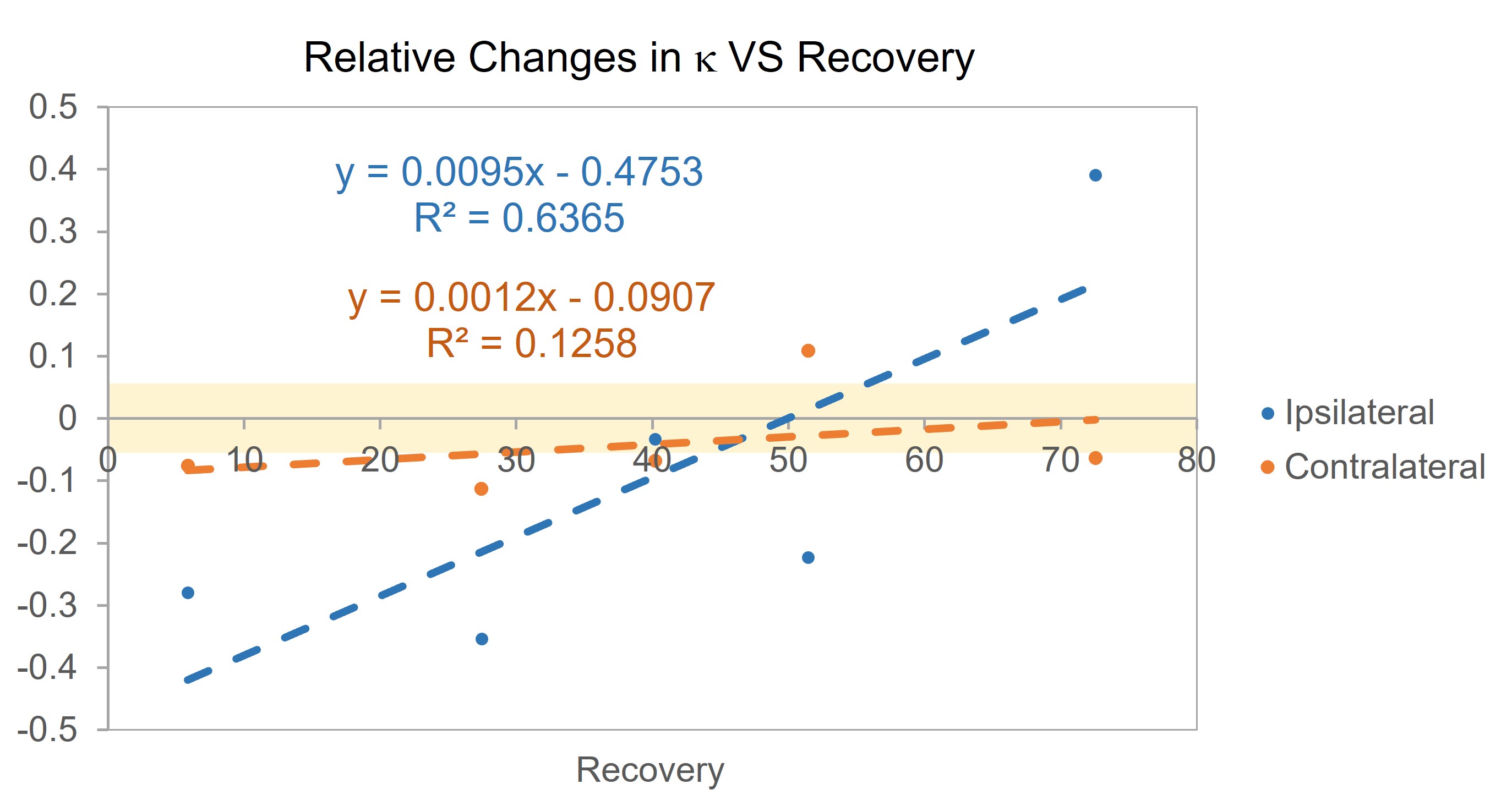
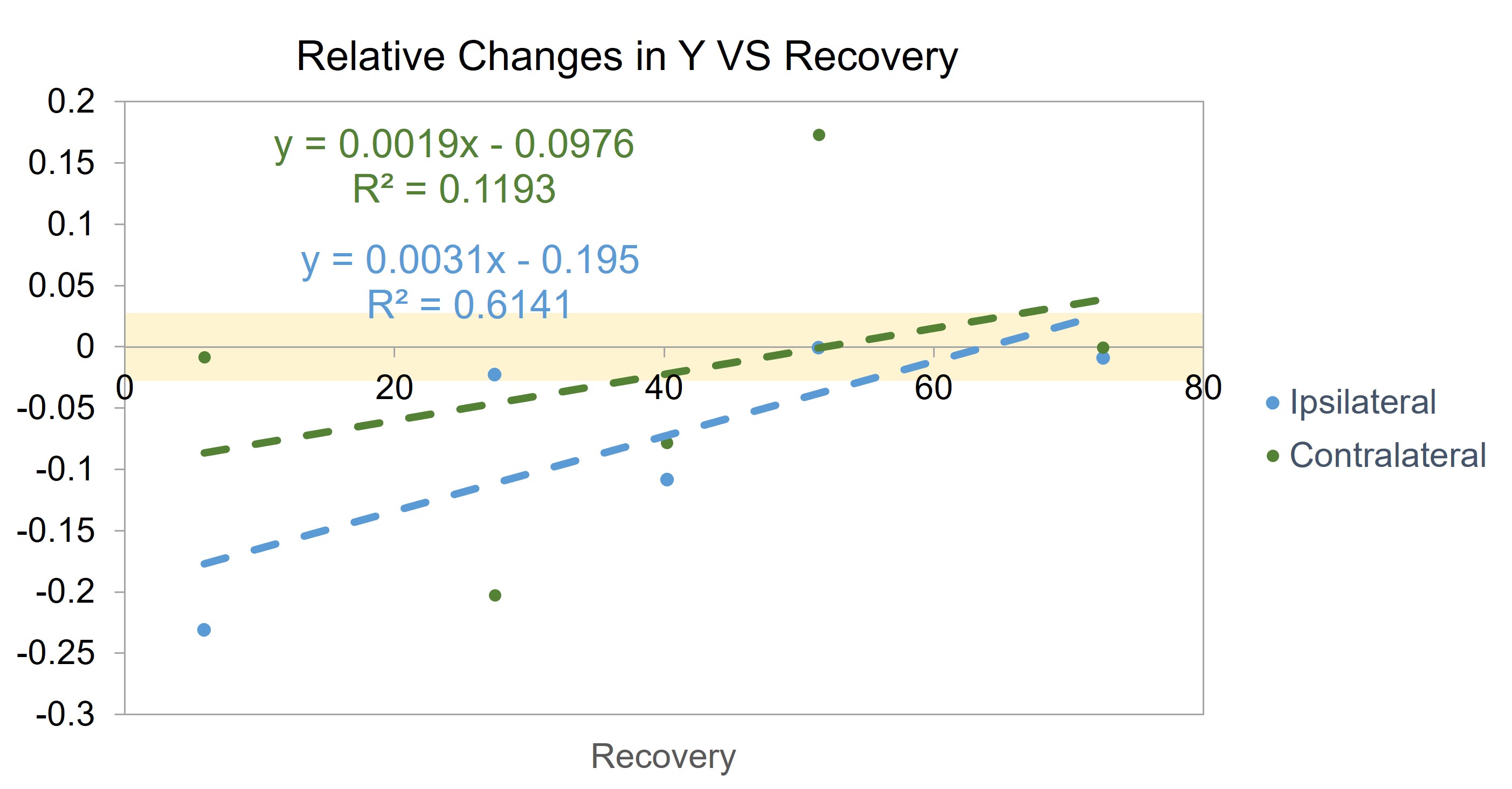
In Figure 1 Y and κ are shown before and 7 days after stroke with the corresponding T2 anatomical image. Brain biomechanics at baseline shows high symmetry between left and right both in the tissue constitutive properties – Y – and in the vascular organization – κ. Contrarily, marked asymmetry is visible at day 7 between ipsilateral and contralateral side in Y and κ. The former reflects the disruption of neuronal integrity and extracellular matrix, and the necrosis [2], the latter results from the vascular remodeling in the infarcted area. Furthermore, we evaluated the correlation between recovery and the relative change of κ (Figure 2) and Y (Figure 3) in the ipsilateral and contralateral region. Recovery was calculated as the relative change from day 1 to day 7 in the lesion sizes (in pixels, from MRE magnitude images) [12, 13]. Averages and standard deviations of Y and κ were calculated for the baselines in ipsilateral and contralateral regions and used to compute the significative relative change in those parameters. κ and Y relative change correlate with recovery in the ipsilateral region ONLY [11]. While a temporal increase in κ results in a higher recovery, its decrease implies a lower recovery. Oppositely, small changes in Y correlate with higher recovery, while a decrease in Y results in a poorer outcome. These trends demonstrate that tissue damage – Y – and pronounced vascular plasticity – κ - are both vital for recovery.Conclusions
In this work we investigated the time evolution of brain biomechanics after an ischemic event, using MRE in mice. Our preliminary data show for the first time in-vivo that the dispersive properties of shear waves gauge vascular organization in tissue. We found a correlation between lesion recovery and time dependent relative changes in tissue biomechanics and vasculature. This newfound possibility of disentangling vasculature changes from tissue remodeling could provide new insights in the understanding of tissue plasticity after stroke and potentially constitute a relevant marker in the context of stroke angiogenic therapies.Acknowledgements
No acknowledgement found.References
1. Lopez, A.D., et al., Global and regional burden of disease and risk factors, 2001: systematic analysis of population health data. The lancet, 2006. 367(9524): p. 1747-1757.
2. Freimann, F.B., et al., MR elastography in a murine stroke model reveals correlation of macroscopic viscoelastic properties of the brain with neuronal density. NMR in Biomedicine, 2013. 26(11): p. 1534-1539.
3. Fluri, F., M.K. Schuhmann, and C. Kleinschnitz, Animal models of ischemic stroke and their application in clinical research. Drug design, development and therapy, 2015. 9: p. 3445.
4. Wouters, A., et al., Association between time from stroke onset and fluid-attenuated inversion recovery lesion intensity is modified by status of collateral circulation. Stroke, 2016. 47(4): p. 1018-1022.
5. Kao, Y.-C.J., et al., Role of genetic variation in collateral circulation in the evolution of acute stroke: a multimodal magnetic resonance imaging study. Stroke, 2017. 48(3): p. 754-761.
6. Liu, J., et al., Vascular remodeling after ischemic stroke: mechanisms and therapeutic potentials. Progress in neurobiology, 2014. 115: p. 138-156.
7. Jugé, L., et al., Microvasculature alters the dispersion properties of shear waves–a multi‐frequency MR elastography study. NMR in Biomedicine, 2015. 28(12): p. 1763-1771.
8. Chiang, T., R.O. Messing, and W.-H. Chou, Mouse model of middle cerebral artery occlusion. Journal of visualized experiments: JoVE, 2011(48).
9. Schregel, K., et al., Characterization of glioblastoma in an orthotopic mouse model with magnetic resonance elastography. NMR in Biomedicine, 2018. 31(10): p. e3840.
10. Sinkus, R., et al., Rheological determinants for simultaneous staging of hepatic fibrosis and inflammation in patients with chronic liver disease. NMR Biomed, 2018. 31(10): p. e3956.
11. Saunders, D.E., A.G. Clifton, and M.M. Brown, Measurement of infarct size using MRI predicts prognosis in middle cerebral artery infarction. Stroke, 1995. 26(12): p. 2272-2276.
12. Beloosesky, Y., et al., The importance of brain infarct size and location in predicting outcome after stroke. Age and ageing, 1995. 24(6): p. 515-518.
13. Cirillo, C., et al., Post-stroke remodeling processes in animal models and humans. Journal of Cerebral Blood Flow & Metabolism, 2020. 40(1): p. 3-22.
Figures