2192
Chemical exchange saturation transfer for detecting altered APT in arterial stenosis at 7T1Department of Radiology, Chinese PLA General Hospital, Beijing, China, 2Research Collaboration Team, Siemens Healthineers, Beijing, China, 3Research Collaboration Team, Siemens Healthineers, Erlangen, Germany
Synopsis
Keywords: Blood Vessels, Brain, 7T-CEST
Motivation: The incidence of middle cerebral arterial (MCA) stenosis increases yearly, molecular biomarkers for this condition are needed. Amide proton transfer imaging is a potential imaging marker.
Goal(s): To investigate the APT changes in MCA stenosis patients.
Approach: We quantitatively evaluated both the affected and healthy middle cerebral arterial blood-supplying brain regions in eight patients with unilateral middle cerebral arterial stenosis using CEST-APT at 7T.
Results: The CEST values of the affected MCA-supplied brain regions were higher than those of the healthy MCA-supplied brain regions, suggesting metabolic alterations in patients with MCA stenosis.
Impact: CEST values for vascularized brain regions can be quantitatively measured using 7T-CEST and can provide new markers for mechanistic and intervention studies in patients with MCA stenosis. This can contribute to a better understanding of the pathogenesis of MCA stenosis.
Introduction
Amide proton transfer (APT) imaging is a type of chemical exchange saturation transfer (CEST) that can detect intracranial metabolic changes at the molecular level. APT has been applied in studies of brain injury, brain tumors, and neurodegenerative diseases.1,2. The incidence of middle cerebral arterial (MCA) stenosis increases yearly, molecular biomarkers for this condition are needed. APT benefits from increased field strength, which improves signal-to-noise ratio, and widens frequency distance between compounds3,4. Currently, few studies have applied CEST to investigate the changes of APT in MCA stenosis patients. This study aims to explore the value of APT in patients with unilateral MCA stenosis, by analyzing both the affected side and the unaffected sides of the brain.Methods
Eight patients (aged 59.50±10.35 years, five men and three women), diagnosed with unilateral MCA stenosis or occlusion, were enrolled in this study. All the patients underwent routine MR examinations including T2w, T1w, time-of-flight MR angiography (TOF-MRA), and CEST. The examination was performed at a 7.0T whole-body scanner (MAGNETOM Terra, Siemens Healthcare, Erlangen, Germany) equipped with an 8Tx/32Rx head coil. 3D fast spoiled gradient echo was used in CEST imaging, with the following parameters: field of view 220 mm, acquisition matrix 128x128, slice thickness 5 mm, number of slices 12, flip angle 5-degree, echo time of 3.09 ms and repetition time of 5 ms. Fifty-six offset frequencies were sampled, using the recommended frequency table listed on the website [https://www.cest-sources.org]. Two B1 amplitude, 0.6uT and 0.9uT were acquired with an extra B1 map acquired to correct for B1 inhomogeneity5. The APT map was analyzed from the original CEST images and corrected for B1 inhomogeneity. The MCA supply region was outlined in ITK-SNAP and the mean APT value of the corresponding region was calculated. Statistical analyses were performed using SPSS version 26.0 (IBM Corporation, Armonk, NY, USA). Paired t-tests were utilized to compare changes in APT values between vessels on the affected and normal sides. Interobserver agreement of the MRI readings was assessed using the intraclass correlation coefficient6,7.Results
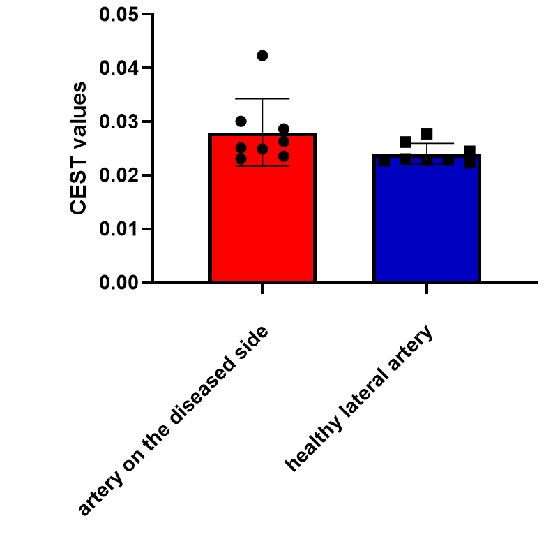
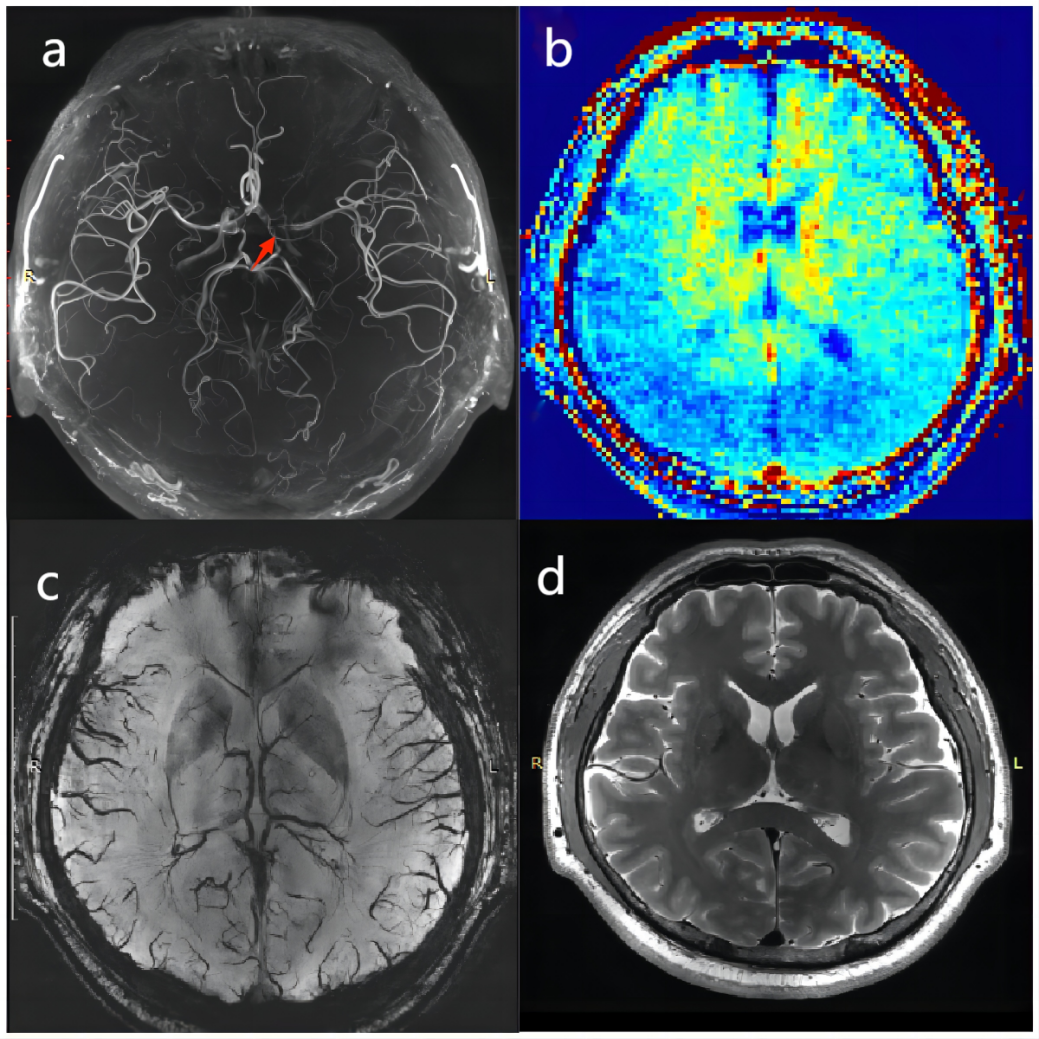
The APT values of the affected MCA blood-supplying brain regions in the eight patients with unilateral MCA stenosis or occlusion were 0.028±0.006 %, and those of the healthy MCA blood-supplying brain regions were 0.024±0.002 %. Figure 1 shows a comparison of the APT values. Figure 2 shows CEST and conventional MRI images of a patient with MCA stenosis occlusion. Although t-tests revealed no statistically significant differences in APT values (P=0.108), the results show that the APT values were higher for the affected MCA supplied brain area than for the healthy MCA-supplied brain area.Discussion and Conclusion
We quantitatively evaluated the APT of the affected and healthy hemisphere in patients with unilateral MCA stenosis. Our results indicate that the APT value of the affected side is markedly higher than that of the healthy side, indicating that metabolic changes occur intracranially in patients with MCA stenosis, thus providing a new imaging marker for patients with MCA stenosis. Whether these abnormalities precede the development of intracranial complications requires further exploration.Acknowledgements
This work was supported by the National Natural Science Foundation of China (No.81825012, No. 82151309 to X.L. and No. 82271952 to J.H.L)References
1.Zhou J, Lal B, Wilson DA, et al. Amide proton transfer (APT) contrast for imaging of brain tumors. Magn Reson Med. 2003;50(6):1120-1126.2. Van Zijl PC, Zhou J, Mori N, et al. Mechanism of magnetization transfer during on-resonance water saturation. A new approach to detect mobile proteins, peptides, and lipids. Magn Reson Med. 2003;49(3):440-449.
3. Wang Y, Zhao X, Liu L, et al; CICAS Study Group. Prevalence and outcomes of symptomatic intracranial large artery stenoses and occlusions in China: the Chinese Intracranial Atherosclerosis (CICAS) Study. Stroke. 2014 Mar;45(3):663-669.
4. Saber H, Amiri A, Thrift AG, et al. Epidemiology of Intracranial and Extracranial Large Artery Stenosis in a Population-Based Study of Stroke in the Middle East. Neuroepidemiology. 2017;48(3-4):188-192.
5. Windschuh, J., Zaiss, nd Bachert, et al., Correction of B1-inhomogeneities for relaxation-compensated CEST imaging at 7T. NMR Biomed. 2015, 28: 529–537.
6. Springer E, Dymerska B, Cardoso PL, et al. Comparison of routine brain imaging at 3 T and 7 T. Invest Radiol. 2016;51(8):469-482.
7. Stahl R, Krug R, Kelley DA, et al. Assessment of cartilage-dedicated sequences at ultra-high-field MRI: comparison of imaging performance and diagnostic confidence between 3.0 and 7.0 T with respect to osteoarthritis-induced changes at the knee joint. Skeletal Radiol. 2009;38(8):771-783.
Figures