2191
4D flow analysis of whole-brain cerebral blood flow in patients with Moyamoya disease before and after direct revascularization—A pilot study1Radiology, Sichuan Provincial People's Hospital, Chengdu, China, 2Neurosurgery, Sichuan Provincial People's Hospital, Chengdu, China, 3MR Research Collaboration, Siemens Healthineers, Chengdu, China
Synopsis
Keywords: Blood Vessels, Velocity & Flow
Motivation: The hemodynamic changes after superficial temporal artery-middle cerebral artery (STA-MCA) bypass in patients with Moyamoya disease (MMD) remain unclear. 4D flow is expected to provide more information about the hemodynamic parameters of patients with MMD before and after surgery.
Goal(s): To clarify the hemodynamics by comparing flow parameters obtained by 4D flow magnetic resonance imaging (MRI).
Approach: Patients with MMD were prospectively recruited for 4D flow magnetic resonance scanning before and after bypass surgery.
Results: After bypass surgery, the contralateral internal carotid artery blood flow decreased and cerebral blood flow was redistributed.
Impact: The results indicate that 4D flow MRI is a feasible, noninvasive alternative for evaluating bypass grafts in the early postoperative period. These flow parameter values may help improve the way patients are evaluated after bypass surgery.
Introduction
Moyamoya disease (MMD) is a steno-occlusive disease of the cerebral arteries, causing chronic progressive stenosis in the terminal portions of bilateral internal carotid arteries (ICAs). This disease mechanism, in turn, stimulates the compensatory development of collateral vessels at the base of the brain 1,2. Cerebrovascular events in patients with MMD often present as transient ischemic attacks, cerebral infarction, or intracerebral hemorrhage 3. Generally, surgical revascularization prevents ischemic attacks by improving the cerebral blood flow in patients with MMD 4. However,after surgical revascularization, the patient must be monitored to assess the patency of the bypass and to evaluate the changes in vascularization state.Non-contrast 4D flow MRI has recently been used to evaluate cerebrovascular hemodynamics 5,6. It can provide a variety of hemodynamic parameters, such as blood flow volume (BFV), peak velocity, wall shear force, etc. However, there is no conclusion on the hemodynamics and redistribution of whole-brain blood flow in patients with MMD after direct revascularization surgery.Thus, the aims of this study were to characterize intracranial vascular hemodynamics in patients with MMD by 4D flow MRI.Methods
Ten patients (aged 24-60 years) diagnosed with MMD by DSA were prospectively recruited. All patients underwent conventional head magnetic resonance scan and 4D flow magnetic resonance scan before and after bypass surgery. 4D flow MRI was performed using a 3T scanner (Vida, Siemens, Germany). The brief summary of 4D flow MRI parameters was as follows: VENC =120 cm/s, voxel size = 1x1x1 mm3 isotropic, 15 cardiac phases, sensitivity encoding (SENSE) factor 2 and acquisition time approximately 12 mins. The MASS research software (MASS; Version 2019-EXP, Leiden University Medical Center, Leiden, The Netherlands) was used to obtain the BFV and peak velocity of the internal carotid artery (ICA), anterior cerebral artery (ACA), middle cerebral artery (MCA), posterior cerebral artery (PCA), basilar artery (BA), and bypass artery (STA-MCA) (Figure 1). The side of the brain that underwent bypass surgery was taken as the ipsilateral. The difference of BFV in each artery, the change of total cerebral BFV before and after surgery, and the difference of BFV between extracranial segment and intracranial segment of bypass artery was compared. For all analysis, P < 0.05 was considered to be statistically significant.Results
As show in Table 1, the BFV of contralateral ICA decreased significantly after the bypass surgery (101.49±67.41 ml/min after surgery vs. 129.94±68.74 ml/min before surgery, P=0.011). There was no significant change in the BFV of other measured arteries. The total cerebral blood flow before and after surgery was 440.85±204.04 and 509.73±172.66 ml/min, respectively (P=0.074). The peak velocity of posterior cerebral artery on the affected side was 69.77±18.07 and 55.46±7.36 cm/s before and after surgery, respectively (P=0.051). The proportion of postoperative contralateral internal carotid artery blood flow in total cerebral blood flow changed from 29.47% to 19.91%, the proportion of postoperative basal artery blood flow in total cerebral blood flow changed from 49.55% to 40.95%, and the proportion of postoperative bypass artery flow in total cerebral blood flow was 22.81% (Figure 2).Discussion
In this study, we characterized and compared hemodynamics of the ICA, BA, ACA, MCA, PCA, and bypass artery in patients with MMD. First, the results showed that the postoperative contralateral internal carotid artery blood flow decreased significantly compared with that before surgery (101.49±67.41 ml/min after operation and 129.94±68.74 ml/min before operation), and the difference was statistically significant P=0.011. There was no significant change in the blood flow volume of other vessels. Although MMD is progressive bilateral internal carotid artery occlusion and stenosis, the contralateral internal carotid blood flow decreased, which may be due to the presence of bypass artery and the redistribution of intracranial blood flow 7. In addition, the change of intracranial blood flow ratio also fully demonstrated the value of 4D flow in evaluating the blood supply capacity of bypass artery and the blood flow ratio of each bypass artery after superficial temporal artery bypass surgery in patients with MMD.Conclusion
4D flow MRI provides a comprehensive, non-invasive method for evaluating hemodynamics. After STA-MCA bypass, the bypass artery can partially compensate for the loss of blood flow in the internal carotid artery with severe side occlusion or stenosis, and reduce the compensatory behavior of the ICA and BA on the relative healthy side. These flow parameter values may help improve the way patients are evaluated after surgery.Acknowledgements
We are grateful to all the participants for their cooperation.References
1. Suzuki J, Takaku A. Cerebrovascular “moyamoya” disease. Disease showing abnormal net-like vessels in base of brain. Arch Neurol. 1969;20(3):288-299.
2. Suzuki J, Kodama N. Moyamoya disease–a review. Stroke. 1983;14(1):104-109.
3. Herve D, Kossorotoff M, Bresson D, et al. French clinical practice guidelines for moyamoya angiopathy. Rev Neurol (Paris). 2018;174(5): 292-303.
4. Bower RS, Mallory GW, Nwojo M, et al. Moyamoya disease in a primarily white, midwestern us population: increased prevalence of autoimmune disease. Stroke. 2013;44(7): 1997-1999.
5. Wahlin A, Eklund A, Malm J. 4D flow MRI hemodynamic biomarkers for cerebrovascular diseases. J Intern Med. 2022;291(2):115-127.
6. Dyverfeldt P, Bissell M, Barker AJ, et al. 4D flow cardiovascular magnetic resonance consensus statement. J Cardiovasc Magn Reson. 2015;17(1):72.
7. Zarrinkoob L, Wåhlin A, Ambarki K, Eklund A, Malm J. Quantification and mapping of cerebral hemodynamics before and after carotid endarterectomy, using four-dimensional flow magnetic resonance imaging. J Vasc Surg. 2021;74(3):910-920.
Figures
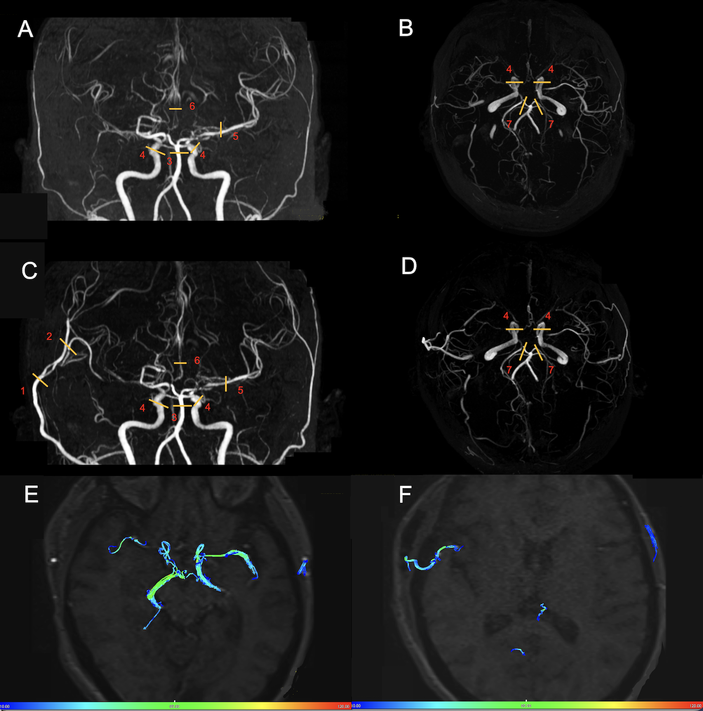
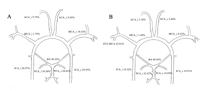

Table 1. Analysis of blood flow volume difference before and after operation of MMD patients.
Notes: a Difference between before surgery and after surgery, using a Wilcoxon Signed Rank Test. b The difference between extracranial and intracranial segments of bypass artery. * whole brain = ICA + BA. whole brain (after) = ICA + BA + STA-MCA ( Intracranial segment). Abbreviations: BFV, blood flow volume; ICA, internal carotid artery; BA, basilar artery; ACA, anterior cerebral artery; MCA, middle cerebral artery; PCA, posterior cerebral artery.