2190
Evaluating the Impact of Glymphatic Clearance Dysfunction on Cognitive Function in Cerebral Small Vessel Disease1Department of Radiology, First People's Hospital of Zigong City, Zigong, China, 2MR Research Collaboration, Siemens Healthineers, Chengdu, China
Synopsis
Keywords: Blood Vessels, Diffusion Tensor Imaging, cerebral small vessel disease; glymphatic clearance function; ALPS index; SVD score
Motivation: The pathogenesis of cerebral small vessel disease (SVD) is unclear, which may be related to glymphatic clearance.
Goal(s): To assess the association between glymphatic clearance function and the extent of cerebral damage in SVD, along with the effects on cognitive function.
Approach: We derived the ALPS index from DTI data to evaluate lymphatic function, and we utilized MR imaging to determine the SVD score as a measure of cognitive function.
Results: We found a significantly decrease in ALPS index in SVD patients compared to healthy controls, and a significantly negative correlation between the ALPS index in the left hemisphere and SVD severity.
Impact: Glymphatic clearance dysfunction may be present in patients with SVD and may be associated with decreased cognitive function.
Introduction
Cerebral small vessel disease (SVD) is an important cause of vascular diseases such as stroke and dementia. However, the pathogenesis is still poorly understood. The glymphatic clearance function is a fluid clearance pathway in the brain that flows along the perivascular space. Taoka et al developed the index for diffusion tensor image analysis along the perivascular space (ALPS index), a measurement derived from diffusion tensor imaging (DTI), to reflect glymphatic activity in the brain1. The ALPS index has since been utilized in multiple studies to evaluate lymphatic function, demonstrating its efficacy in assessing lymphatic clearance function2. The SVD score is an evaluation of a SVD imaging marker that represents the severity of brain damage in SVD patients and is associated with cognitive decline3. Therefore, this study will explore the potential link between lymphatic clearance dysfunction by employing the ALPS index and SVD score, and their relationship in cognitive function.Methods
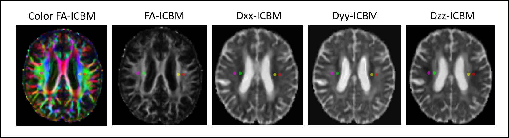
Forty-seven SVD patient and forty-seven healthy controls (HC) were recruited in the study. MRI data were acquired using 3T MR scanner (MAGNETOM VIDA, Siemens Healthineers, Erlangen, Germany), with a 64-channel head coil. DTI was acquired using echo-planar imaging sequence with the following parameters: TR/TE = 2400/79 ms, flip angle = 70°, FOV = 220×220mm2, matrix = 128×128, thickness = 4 mm, gap = 1.2 mm, slices = 66, voxel size = 1.7×1.7×4 mm3, b values = 0, 1000 s/mm2. Structural images were acquired using MP2RAGE sequence with parameters as detailed in the reference4.The ALPS index, indicative of lymphatic clearance function, was quantified employing the FSL pipeline, consistent with the reconstruction of earlier studies5. The superior corona radiata (SCR) and superior longitudinal fasciculus (SLF) were identified as the projection and association fibers, respectively, at the level of the lateral ventricle body using the JHU-ICBM-DTI-81 white matter labeled atlas. Regions of interest (ROIs) within the bilateral SCR and SLF were automatically delineated as 5mm diameter spheres on each subject’s diffusivity maps, which were depicted in Figure 1. The ALPS index was computed for both the left and right hemispheres. For each hemisphere, the index is the ratio of the mean of x-axis diffusivity in projection fibers (Dxxproj) and x-axis diffusivity in association fibers (Dxxassoc) to the mean of the y-axis diffusivity in t projection fibers (Dyyproj) and z-axis diffusivity in association fibers (Dzzassoc) as follows: ALPS index = mean (Dxxproj, Dxxassoc)/mean (Dyyproj, Dzzssoc)1. The SVD score was determined using a previously described four-point scale with neuroimaging characteristics of SVD, including white matter hyperintensities (WMH) of presumed vascular origin, deep cerebral microbleeds (CMB), lacunar infarctions (LI), and enlarged perivascular spaces (PVS).
Statistical analyses were performed by using the SPM12. Two sample T-test was used to analysis group differences. Paired t-test was used to analysis the difference between the left and right hemispheres. Pearson correlation analysis was used to analyze the association between ALPS index and SVD scores in SVD patients. The criterion for statistical significance was p < 0.05.
Result
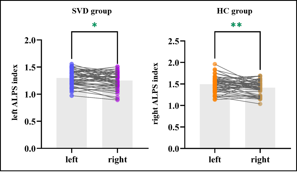
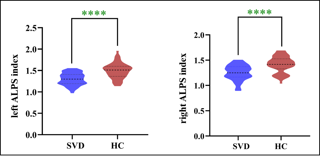
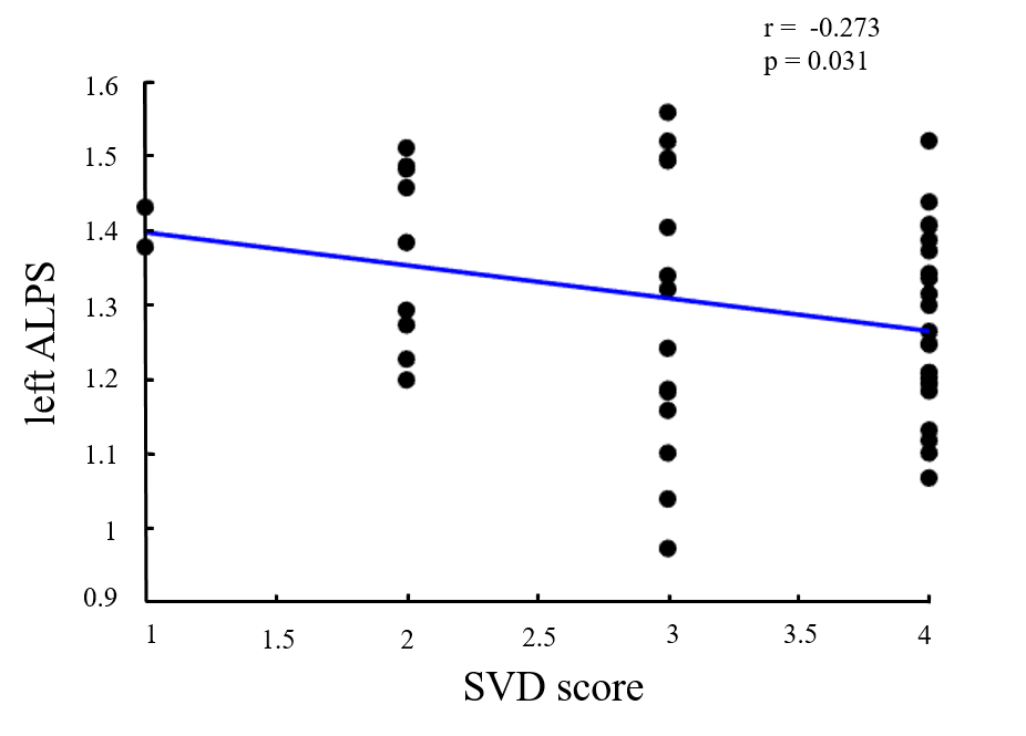
The patients and HC demographics were presented in Table 1. The ALPS index in the left and right hemispheres of the two groups were significantly different (SVD group: p = 0.021; HC group: p = 0.006) (Figure 2). Compare with HC group, the ALPS index of bilateral brain in SVD patients were significantly reduced (right: 1.251 vs 1.414, t = -4.953, p < 0.0001; left: 1.230 vs 1.496, t = -5.575, p <0.0001), as shown in Figure 3. The results of correlation analysis showed that ALPS index in the left hemisphere was negatively correlated with the SVD scores (r = -0.273, p = 0.031) (Figure 4). However, there was no correlation with ALPS index in the right hemisphere (p = 0.114).Discussion
In this study, it was found that ALPS index in SVD patients were lower than those in HC group, suggesting that SVD patients may have a wider range of pro-inflammatory factors. Glymphatic pathway disturbance can affect the clearance of inflammatory factors and increase pro-inflammatory response, thus promoting the development of SVD pathology6. Further, we found a negative correlation between APLS index and SVD score in patients, this may be due to impaired glymphatic pathways that promote cognitive decline resulting from multiple pathophysiological events in SVD.Conclusion
Our results suggest that glymphatic clearance dysfunction may be present in patients with SVD and be associated with reduced cognitive function.Acknowledgements
This work was supported by grants from the Bureau of Zigong City Science and Technology (2021ZC09).References
1. Taoka, T, Masutani Y, Kawai H, et al. Evaluation of glymphatic system activity with the diffusion MR technique: diffusion tensor image analysis along the perivascular space (DTI-ALPS) in Alzheimer’s disease cases. Jpn J Radiol. 2017; 35:172-178.
2. Zhang, W, Zhou Y, Wang J, et al. Glymphatic clearance function in patients with cerebral small vessel disease. Neuroimage. 2021; 238:118257.
3. Staals, J, Makin SD, Doubal FN, et al. Stroke subtype, vascular risk factors, and total MRI brain small-vessel disease burden. Neurology. 2014; 83(14):1228-1234.
4. Knussmann, GN, Anderson JS, Prigge MB, et al. Test-retest reliability of FreeSurfer-derived volume, area and cortical thickness from MPRAGE and MP2RAGE brain MRI images. Neuroimage: Reports. 2022; 2(2):100086.
5. Liu, X, Barisano G, Shao X, et al. Cross-Vendor Test-Retest Validation of Diffusion Tensor Image Analysis along the Perivascular Space (DTI-ALPS) for Evaluating Glymphatic System Function. Aging Dis. 2023; 6(14):325-331
6. Swardfager, W, Yu D, Ramirez J, et al. Peripheral inflammatory markers indicate microstructural damage within periventricular white matter hyperintensities in Alzheimer's disease: A preliminary report. Alzh Dement-Dadm. 2017; 7(1):56-60.
Figures