2189
MRI dynamic changes and mechanism of glymphatic system of hypertensive cerebral edema1Department of Radiology, The Affiliated Hospital of Guizhou Medical University, Guiyang, China, 2State Key Laboratory of Drug Research, Molecular Imaging Center, Shanghai Institute of Materia Medica, Chinese Academy of Sciences, Shanghai, China, 3Philips Healthcare, Shanghai, China
Synopsis
Keywords: Blood Vessels, Animals, The glymphatic system
Motivation: The pathogenesis of hypertensive cerebral edema remains elusive
Goal(s): To visualize and evaluate the transport function of the glymphatic system in the spontaneously hypertensive rats (SHR).
Approach: We used the dynamic contrast-enhanced magnetic resonance imaging (DCE-MRI) and immunofluorescence analysis
Results: And we found that there was a significant change in the transport function of the glymphatic system and a decrease in the expression of aquaporin-4 (AQP4) around blood vessels.
Impact: The combination of DCE-MRI and immunofluorescenc analysis can facilitate further exploration of mechanisms underlying brain edema in the spontaneously hypertensive animal models, thus aiding in the study of the glymphatic system.
Introduction
In 2013, Iliff et al. [1] first utilized DCE-MRI to visualize the exchange of cerebrospinal fluid (CSF) and interstitial fluid (ISF) in the rat brain, confirming the function of the glymphatic system and demonstrating the inflow of arterial CSF. The glymphatic system is a fast-acting system for exchanging fluid throughout the entire brain, driven by arterial pulsations and mediated by the perivascular space (PVS). Imaging studies in the animal models of hypertension have shown evidence of vasogenic edema and changes in cerebral hemodynamics[2]. Acute hypertension disrupts the pulsation of the vascular wall, leading to increased reflux and reduced net CSF flow in the PVS[3]. This study aims to investigate mechanisms and pathways related to the glymphatic system involved in the development of cerebral edema in spontaneous hypertension, providing a foundation for effective clinical treatment of brain edema.Methods
Subjects: We included 8 spontaneously hypertensive rats (SHR) and 8 normotensive rats (WKY) aged 18-20 weeks. All rats were housed in a controlled environment with regulated temperature and humidity. Caudal arterial blood pressure was measured using a noninvasive blood pressure meter for 15 consecutive cycles, and the data was exported for recording.MRI Data Acquisition: We used a 9.4T Biospec 94/20 USR (Bruker BioSpin, Germany) for scanning. Three-dimensional T1-weighted FLASH sequences were acquired in the sagittal plane with the following parameters: TR = 74.9ms, TE = 1.7 ms, flip angle = 15°, NA = 1, FOV = 35 × 35 × 35 mm, matrix size = 128 × 128 × 128. The scanning protocol consisted of 3 baseline scans, followed by the injection of Gd-DTPA (469 mg/mL, MW 938 Da, Bayer) into the occipital horn via a catheter at a rate of 1.6μl/min. Simultaneously, MRI data was continuously acquired for over 5 hours.
MRI Data Processing: We used Advanced Normalization Tools (ANT) to preprocess the DCE-MRI data. Six regions of interest (ROIs) in the brain were selected, namely the hippocampus, olfactory bulb, cerebellum, pons, medulla, and midbrain aqueduct. The area under the curve (AUC) was calculated for each subject's time series in each ROI.
Immunofluorescenc:Additionally, we used immunofluorescence quantitative analysis to compare the expression of AQP4 around blood vessels in the brain parenchyma.
Statistical analysis: A two-sample T-test was used to analyze the AUC values of the two groups of rats.
Results
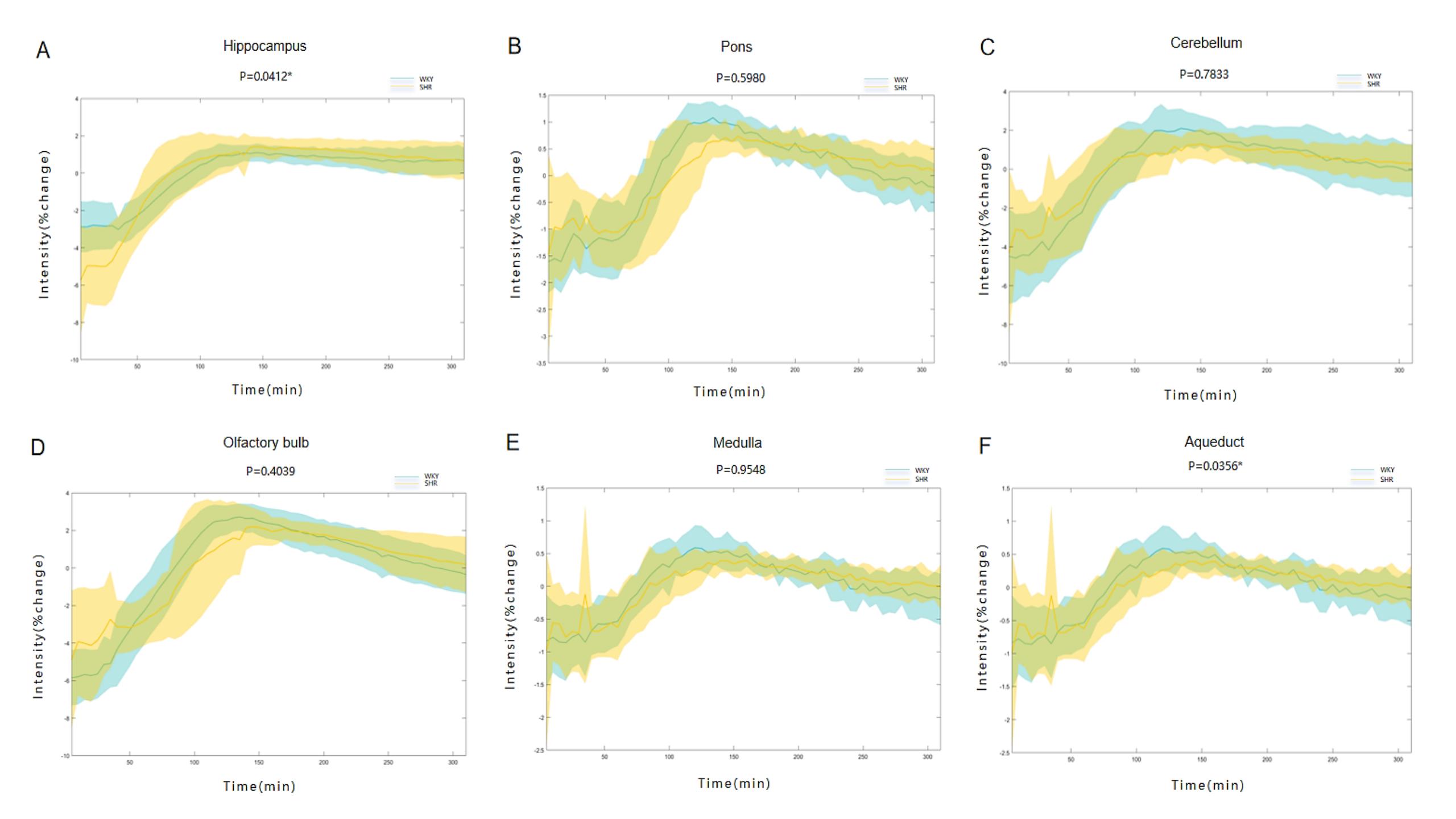
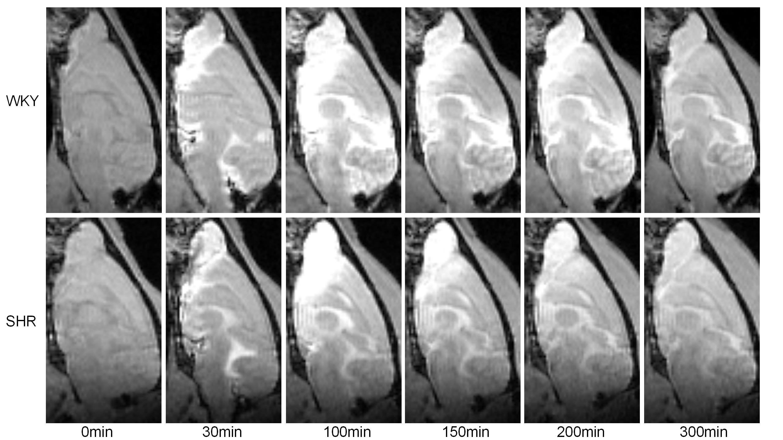
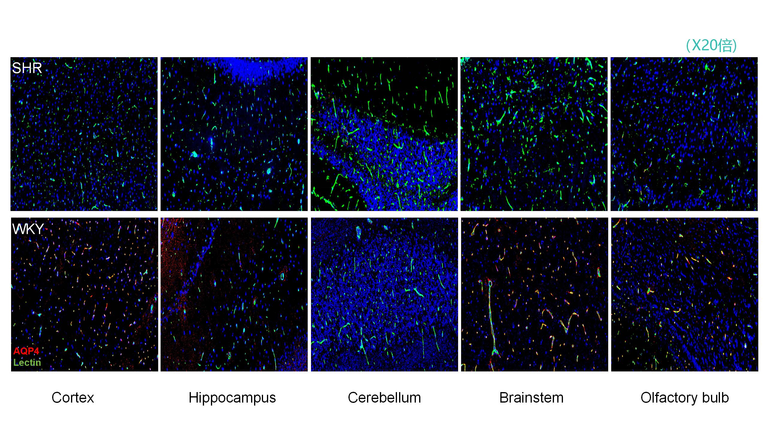
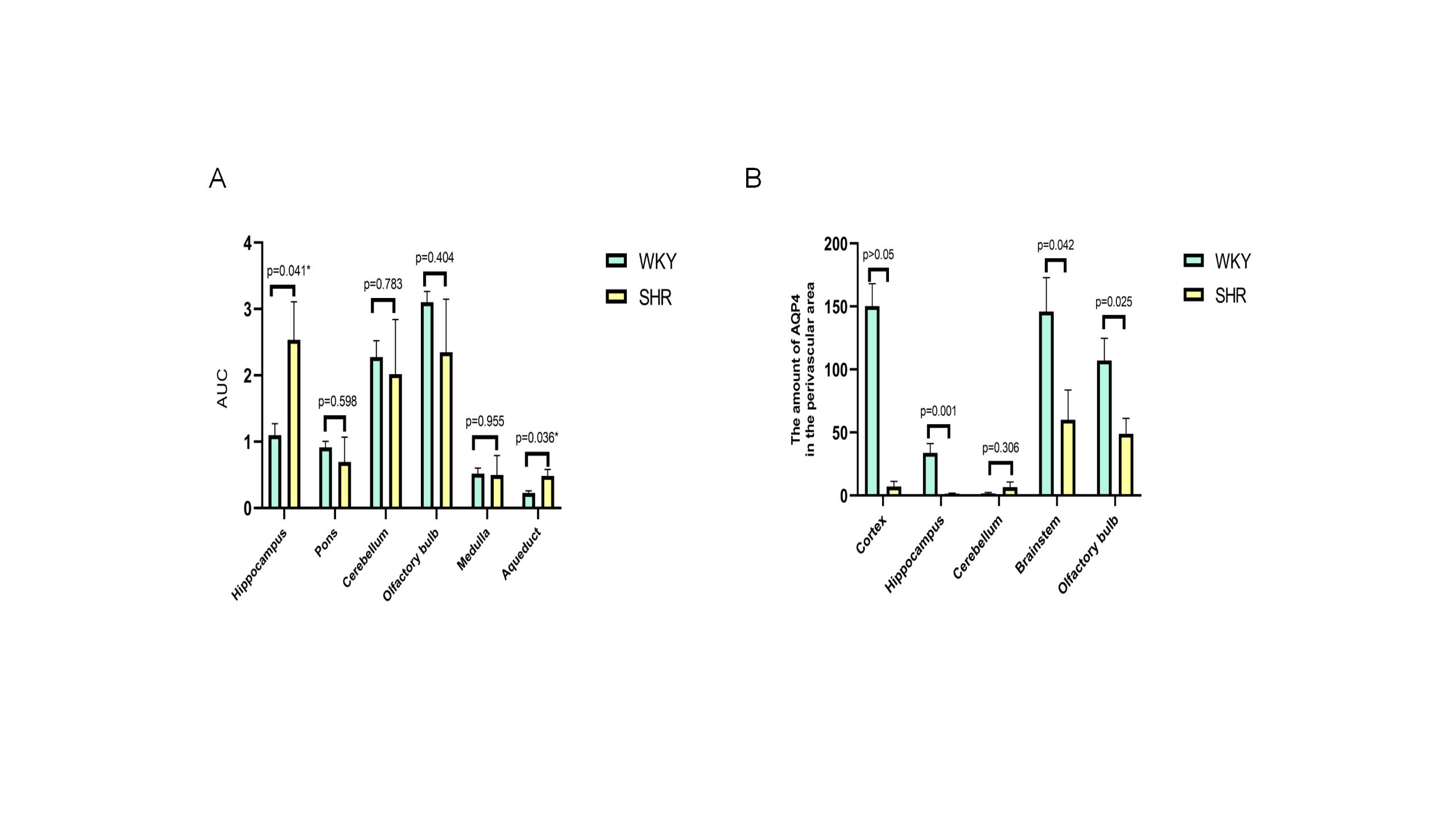
Compared to the WKY group, the SHR group exhibited significant differences in the signal intensity of the contrast agent in the hippocampus (P=0.0412) and aqueduct (P=0.0356). Additionally, we observed that the SHR group had a longer time to peak and a slower clearance rate of cerebrospinal fluid tracer agents (Figure 1-2). Furthermore, the SHR group had significantly reduced expression of AQP4 around blood vessels in the hippocampus, brainstem, and olfactory bulb compared to the WKY group (Figure 3, P<0.05).Discussion
The glymphatic system plays a vital role in transporting solutes and wastes, such as the neurotoxic soluble amyloid-β (Aβ), to the perivascular spaces[4]. And the glymphatic clearance, or perivascular CSF-interstitial fluid exchange, are mainly dependent on the astroglial water channel AQP4. Previous studies have shown that the increased CSF reflux exists in the animal models of acute hypertension[3]. However, the state does not effectively simulate the pathological condition of stable hypertension in humans. Using intra-cranial DCE-MRI, we observed significant differences in the distribution of contrast agent over time (reflecting CSF circulation) between SHR and WKY rats. Furthermore, we noted a significant reduction in the expression of AQP4, a water channel protein that plays a critical role in regulating CSF-ISF exchange, around blood vessels in SHR compared to the WKY rats[5-7]. These findings provide theoretical support and have significant implications for further exploration of the occurrence of brain edema in spontaneously hypertensive rats.Conclusion
Our findings indicate that hypertension has a significant impact on the function of the glymphatic system. This impact may directly or indirectly contribute to the development and progression of brain edema. And these findings lay the groundwork for effective clinical treatment of brain edema and improvement of glymphatic clearance function.Acknowledgements
The authors thank all staff who participated in this study.References
[1]Iliff JJ, Wang M, Liao Y, et al. A paravascular pathway facilitates CSF flow through the brain parenchyma and the clearance of interstitial solutes, including amyloid β. Sci Transl Med. 2012;4(147):147ra111.
[2]Wang Q, Huang B, Shen G, et al. Blood-Brain Barrier Disruption as a Potential Target for Therapy in Posterior Reversible Encephalopathy Syndrome: Evidence From Multimodal MRI in Rats. Front Neurol. 2019;10:1211.
[3]Mestre H, Tithof J, Du T, et al. Flow of cerebrospinal fluid is driven by arterial pulsations and is reduced in hypertension. Nat Commun. 2018;9(1):4878
[4]Verghese JP, Terry A, de Natale ER, Politis M. Research Evidence of the Role of the Glymphatic System and Its Potential Pharmacological Modulation in Neurodegenerative Diseases. J Clin Med. 2022;11(23):6964.
[5]Klostranec JM, Vucevic D, Bhatia KD, et al. Current Concepts in Intracranial Interstitial Fluid Transport and the Glymphatic System: Part I-Anatomy and Physiology. Radiology. 2021;301(3):502-514.
[6]Harrison IF, Ismail O, Machhada A, et al. Impaired glymphatic function and clearance of tau in an Alzheimer's disease model. Brain. 2020;143(8):2576-2593.
[7]Simon M, Wang MX, Ismail O, et al. Loss of perivascular aquaporin-4 localization impairs glymphatic exchange and promotes amyloid β plaque formation in mice. Alzheimers Res Ther. 2022;14(1):59.
Figures