2188
The Association of Aneurysm Wall Enhancement with Hemodynamic Relationships Between the Aneurysm Neck and Parent Artery1Department of Radiology, Zhujiang Hospital of Southern Medical University, Guangzhou, China, 2Philips Healthcare, Guangzhou, China
Synopsis
Keywords: Blood Vessels, Blood vessels, 4D Flow
Motivation: To investigate the relationship between the enhancement of intracranial cystic aneurysm walls and aneurysm morphology and hemodynamics.
Goal(s): The study aimed to identify new imaging indicators for intracranial aneurysm enhancement.
Approach: We conducted a retrospective collection of magnetic resonance images of aneurysm cases and grouped them based on aneurysm wall enhancement grades. Then the differences in morphological and hemodynamic parameters between groups were compared.
Results: The study found that the maximum flow rate at the aneurysm neck and the ratio of the aneurysm neck to the maximum flow rate of the parent artery were statistically significant.
Impact: This study investigated the hemodynamic relationship between aneurysms and the parent artery, and its potential impact on aneurysm wall enhancement. These results could offer valuable insights into the connection between aneurysm wall enhancement and hemodynamics.
Introduction
Intracranial aneurysms are characterized by localized pathological dilation of the intracranial arterial wall and have a propensity for rupture. Related epidemiological studies have revealed that the overall incidence of intracranial aneurysms in the population is approximately 3.2%[1].Research conducted by Chalouhi, N. et al.[2] has highlighted the significant role of inflammation in the initiation and progression of intracranial aneurysms. Inflammation contributes to the degradation of blood vessel walls, ultimately resulting in aneurysm expansion, development, and potential rupture. Previous studies have classified arterial wall enhancement into grades 0, 1, 2, and 3[3]. Lv, N. et al. identified a correlation between the morphological characteristics of intracranial aneurysms and the enhancement of the aneurysm wall[4]. In recent years, 4DFlow has emerged as a novel magnetic resonance imaging technique, allowing for the exploration of the interplay between hemodynamics, morphology, and aneurysm enhancement[5].Methods
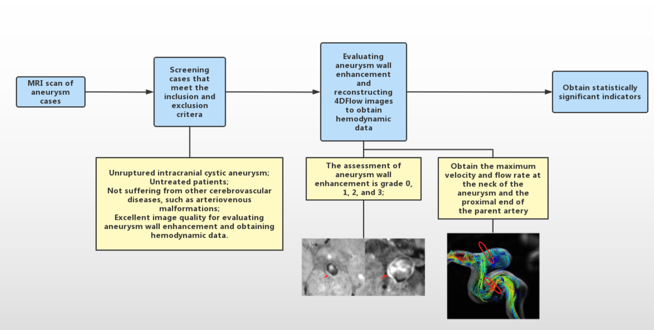
A total of 35 patients with aneurysms, collected retrospectively between September 2022 and June 2023, underwent high-resolution magnetic resonance imaging (MRI) and 4DFlow imaging. The scanning was all conducted using a Philips Ingenia ElitionX scanner, and two radiologists assessed whether there was enhancement in the aneurysm wall. The morphological characteristics of the aneurysm were measured using 3D Time-of-Flight Magnetic Resonance Angiography (3DTOF-MRA) or T1-weighted Vascular Imaging with Subtraction and Magnetization Transfer (T1WVISTA) sequences, including measurements of aspect ratio (AR), size ratio (SR), and the diameter-to-width ratio (D/W). The 4DFlow images were reconstructed using CVI42, and the host artery was measured. The maximum flow velocity and maximum flow rate at the aneurysm neck were measured. Additionally, the ratio of the aneurysm neck’s maximum flow rate to that of the parent artery was calculated. Ordered logistic regression analysis was applied to assess the influence of morphology and hemodynamics factors on arterial wall enhancement (Figure 1).Result
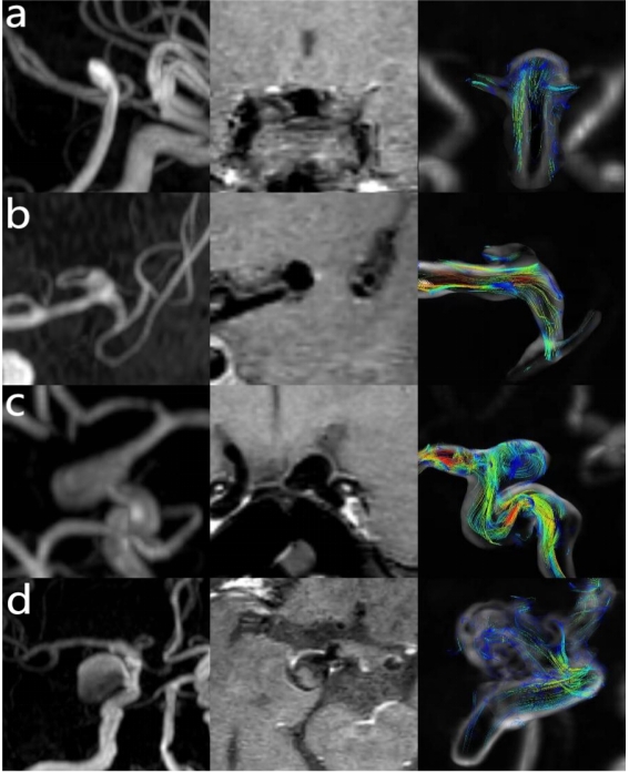
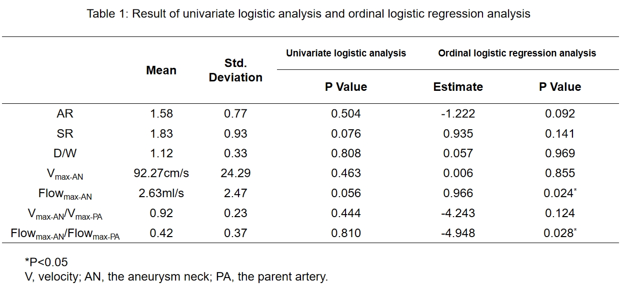
The study included data from 35 aneurysms in 33 patients, categorized into four groups based on aneurysm wall enhancement: 12 cases with grade 0 enhancement, 5 cases with grade 1 enhancement, 8 cases with grade 2 enhancement, and 9 cases with grade 3 enhancement (Figure 2). Various morphological and hemodynamic parameters were measured, including aneurysm AR, SR, D/W, the maximum flow rate at the aneurysm neck, and the ratio of the aneurysm neck’s maximum flow rate to that of the parent artery. Univariate logistic analysis indicated that the maximum flow rate at the aneurysm neck (P value=0.056) and SR (P value=0.076) showed potential associated with arterial wall enhancement. However, ordinal logistic regression analysis revealed that both the maximum flow rate at the aneurysm neck (P value=0.024) and the ratio of the maximum flow rate between the aneurysm neck and the parent artery (P value=0.028) were statistically significant in their association with arterial wall enhancement (Table 1).Discussion
The study aimed to explore the relationship between the enhancement of intracranial aneurysm walls and aneurysm morphology and hemodynamics. While previous studies have primarily focused more on wall shear stress (WSS) and its impact on enhancement[6-7], this study considered both aneurysm morphology and hemodynamics. The preliminary findings suggest that hemodynamics may play a more significant role in aneurysm wall enhancement, especially due to the small sample size and minimal differences in aneurysm morphology. Specifically, the study indicated that the maximum flow rate at the aneurysm neck may be an independent risk factor for enhancement. As the blood flow rate increases, the aneurysm may exhibit higher levels of enhancement. On the other hand, the ratio of the maximum flow rate at the aneurysm neck to the parent artery may act as a protective factor. However, it’s important to note that these conclusions are based on a small sample size and minor differences in morphology, and further research with a larger sample size and additional indicators is required for a more comprehensive understanding of this relationship.Conclusion
In conclusion, this study found that the enhancement of the intracranial aneurysm wall is associated with both morphology and hemodynamics. Specifically, the maximum flow rate at the aneurysm neck appears to be a potential independent risk factor for intracranial aneurysm wall enhancement, while the ratio of the maximum flow rate at the aneurysm neck to the parent artery may act as a protective factor. These findings provide valuable insights into the factors influencing intracranial aneurysm wall enhancement and may contribute to a better understanding of this complex phenomenon.Acknowledgements
No acknowledgement found.References
[1]van Gijn, J., Kerr, R.S. and Rinkel, G.J. (2007) ‘Subarachnoid haemorrhage’, The Lancet, 369(9558), pp. 306–318. doi:10.1016/s0140-6736(07)60153-6.
[2]Chalouhi, N. et al. (2012) ‘Biology of intracranial aneurysms: Role of inflammation’, Journal of Cerebral Blood Flow & Metabolism, 32(9), pp. 1659–1676. doi:10.1038/jcbfm.2012.84.
[3]Edjlali, M. et al. (2018) ‘Circumferential thick enhancement at vessel wall MRI has high specificity for intracranial aneurysm instability’, Radiology, 289(1), pp. 181–187. doi:10.1148/radiol.2018172879.
[4]Lv, N. et al. (2018) ‘Relationship between aneurysm wall enhancement in vessel wall magnetic resonance imaging and rupture risk of unruptured intracranial aneurysms’, Neurosurgery, 84(6). doi:10.1093/neuros/nyy310.
[5]Soulat, G., McCarthy, P. and Markl, M. (2020) ‘4d flow with MRI’, Annual Review of Biomedical Engineering, 22(1), pp. 103–126. doi:10.1146/annurev-bioeng-100219-110055.
[6]Khan, M.O. et al. (2021) ‘Association between aneurysm hemodynamics and wall enhancement on 3D vessel WALL MRI’, Journal of Neurosurgery, 134(2), pp. 565–575. doi:10.3171/2019.10.jns191251.
[7]Zhang, M. et al. (2021) ‘Associations between haemodynamics and wall enhancement of intracranial aneurysm’, Stroke and Vascular Neurology, 6(3), pp. 467–475. doi:10.1136/svn-2020-000636.
Figures