2185
Investigating the relation between cardiac-induced brain tissue strain and both global boundary conditions and local microstructure1Department of Radiology, UMC Utrecht, Utrecht, Netherlands, 2Department of Mechanical Engineering, University of Sherbrooke, Sherbrooke, QC, Canada
Synopsis
Keywords: Blood Vessels, Brain
Motivation: Recent developments enabled to measure brain tissue strain as induced by arterial pulsations in detail. This opens the opportunity to study how these strains are affected by the brain’s macroscopic environment and its local microstructure.
Goal(s): To explore to what extent the strain principal strain directions can be explained by both global boundary conditions and local tissue microstructure.
Approach: Systolic 3D strain measurements of the brain were compared with a brain model and DTI measurements.
Results: The first principal strain showed good agreement with the brain model and consistent spatial patterns were observed in comparisons between third principal strain and DTI data.
Impact: Our results help confirm previous ideas on how the brain swells during cerebral arterial pulsations while also providing a first view into the relationship between the direction of the Poisson effect and brain microstructure, opening up avenues for further research.
Introduction
Cerebral arterial pulsations are crucial for brain waste clearance, driving the convective bulk flow of cerebrospinal fluid (CSF) in perivascular spaces and facilitating CSF entry into brain tissue, enabling CSF-interstitial fluid exchange in the glymphatic system1. They are also the main drivers of the mechanical waves which are used in intrinsic magnetic resonance elastography (iMRE) to estimate the mechanical properties of the brain2. Despite this, much is unknown about to what extent local strains are determined macroscopic anatomy or the anisotropic microstructure. Previous work found that systolic forces induce displacements that are distributed in a funnel-like shape, directed from the skull down towards the foramen magnum3. Similarly, recent work which presents 3D strain maps of the brain4 has shown that the first principal strain (FPS) vector (maximum expansion) shows global, inwardly directed strains pointing towards the foramen magnum. Meanwhile, the third principal strain (TPS) vector (maximum compression) shows local heterogeneous patterns, possibly reflecting the brain microstructure. However, mechanical testing has shown no significant directional dependencies on stiffness5. This study aims to explore the relationship between cardiac-induced brain tissue strain and global boundary conditions as well as local microstructure.Method
We used data from a previously described 7T MRI study4, where Displacement Encoding with Stimulated Echoes (DENSE)6 and DTI data were acquired. The DENSE data included 3D displacement fields of the brain (3 mm isotropic resolution), time-resolved over 52.5% of the cardiac cycle (8 cardiac phases) for 9 healthy subjects, which were used to reconstruct the strain tensor of brain tissue relative to diastole. The DTI was performed axially, using spin echo with 2 mm isotropic resolution and a b-value of 800 s/mm2. The dependency of tissue strain on macro- and microstructure was investigated separately through the means of the first and third principal strains, respectively. In the first case, a simplified brain model of was constructed which takes into account the funnel-like systolic forces previously described and was voxel-wise compared with the FPS. The model consists of a weighted sum of a vector field that is directed perpendicular to the skull and a vector field that is pointing towards the foramen magnum. In the second case, the main DTI eigenvector was compared on a voxel-wise basis with the TPS by computing the angle difference for the corticospinal tract, corpus callosum, and cingulum bundle.Results
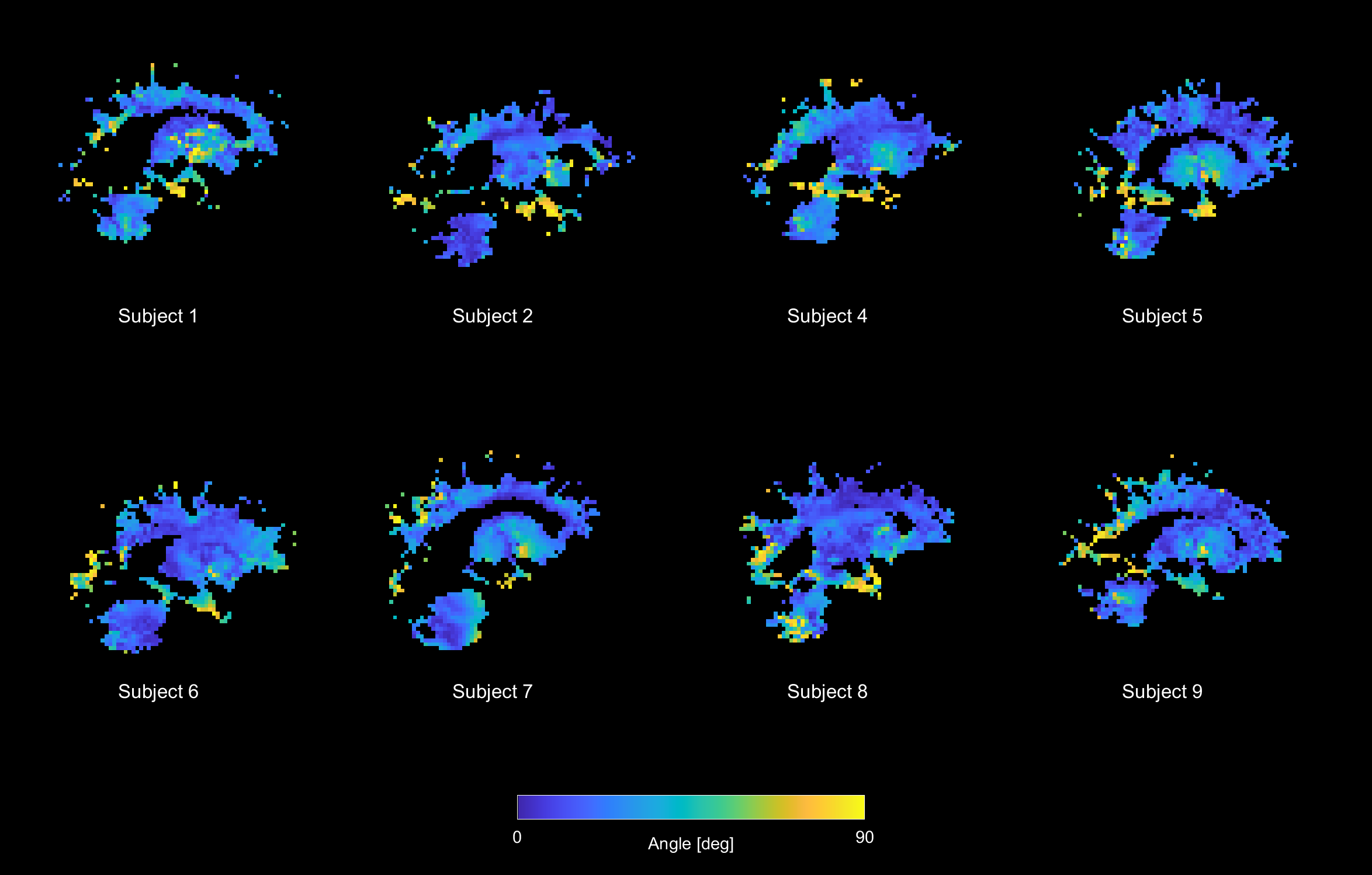
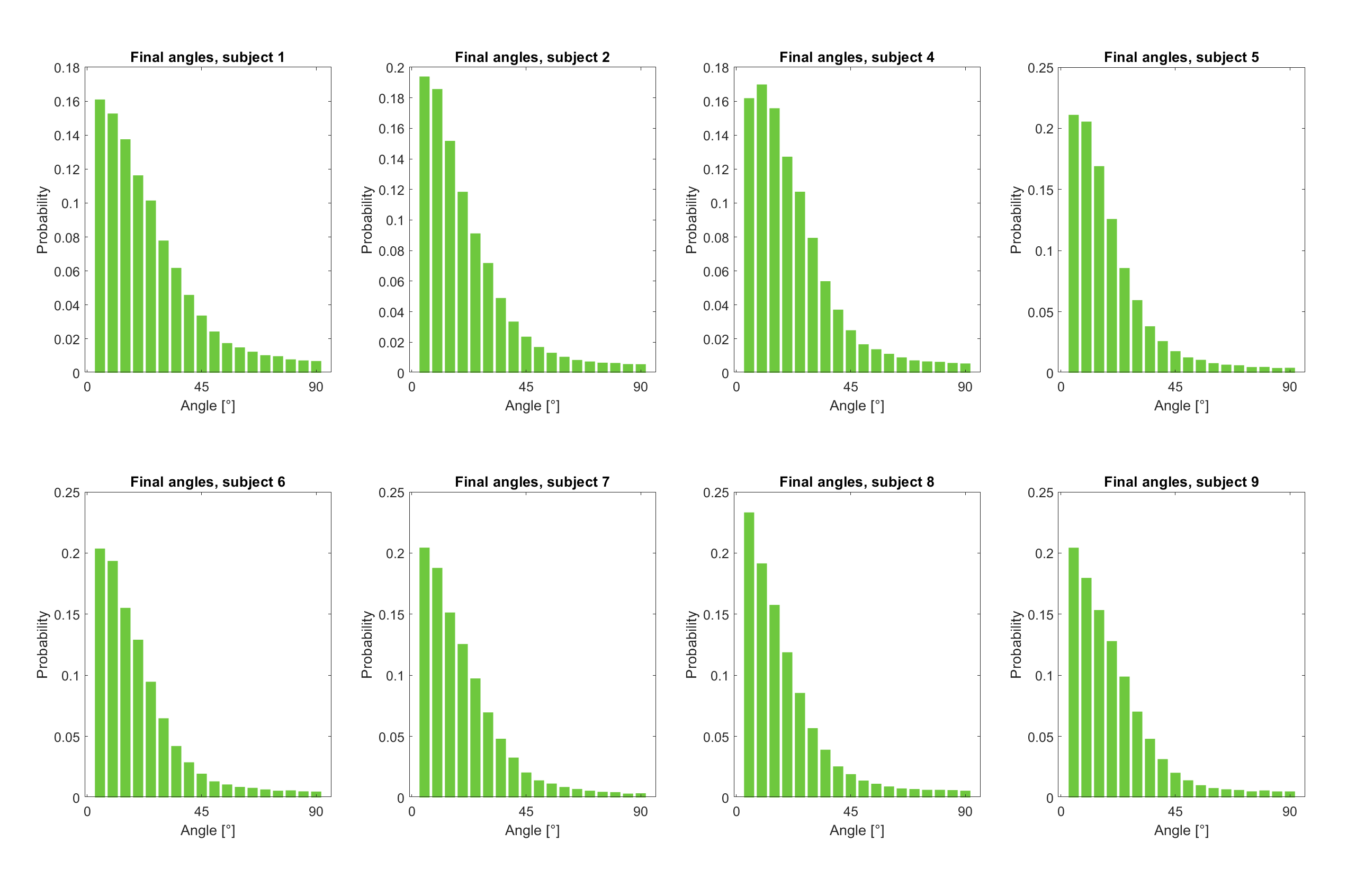
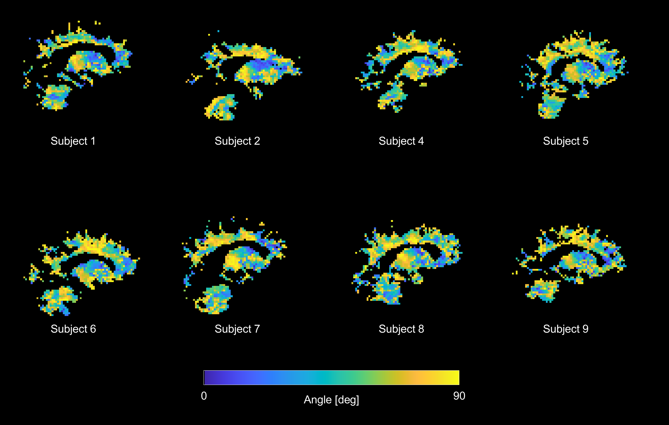
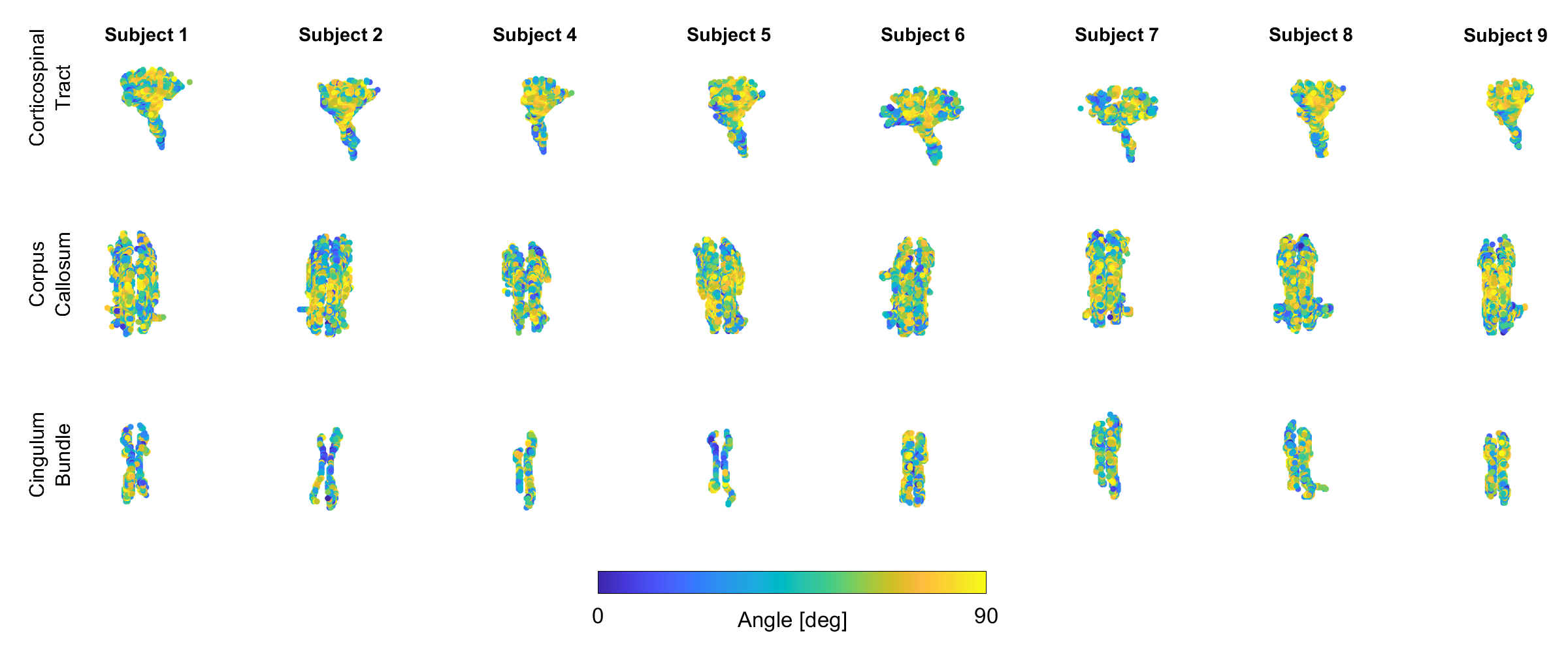
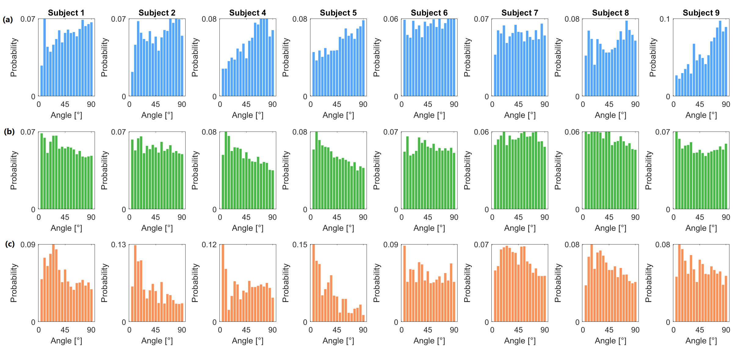
The FPS maps displayed good resemblance with the model strains (see Figure 1) with the majority of corresponding angle probability distribution centered around 0°. Due to the vectors being distributed on a sphere in 3D, the probability density distribution was corrected for the circular circumference on a sphere7. Spatial patterns can be seen in the voxel-wise angle difference between TPS and the primary DTI eigenvectors (see Figure 3). Figure 4 shows the ROIs where each voxel is color coded by the angle difference, while Figure 5 shows their respective distributions.Discussion
The brain model displays consistent behavior with FPS for the majority of the brain and similar spatial patterns are observed for all subjects. Lower angles correspond to better agreement between model and data. Areas around the temporal lobes consistently show worse agreement, possibly due to the complex shape of the skull at that region not being properly approximated in our current implementation. The back of the brain also consistently displays lower agreement, possibly due to the effects of compression in the sagittal, straight, and transverse sinuses. Results of the angle differences between the TPS and primary DTI eigenvectors show regionally dependent spatial patterns where multiple similar spatial patterns can be observed across subjects. Likewise, similar spatial patterns can be seen across subjects in the masked ROIs. The distributions over the entire ROIs show very little structure, highlighting the isotropic structure of brain tissue, which is in line with work by Budday et al.5. Future work aims to investigate ROIs with homogenous fiber orientations to further illuminate the relation between the TPS and brain tissue microstructure.Conclusion
The effects of cerebral arterial pulsations on the brain macro- and microstructure were explored through the use of a strain analysis in combination with a brain model and DTI measurements. The model was found to agree well with the FPS for a majority of the brain. Initial analysis of the angle differences between the TPS and the primary DTI eigenvector showed regionally dependent spatial patterns, however overall angle distributions imply the nature of brain microstructure being mainly isotropic. More extensive analysis is required to further illuminate the relation between cardiac-induced strain and the local microstructure.Acknowledgements
No acknowledgement found.References
1. Iliff JJ, Wang M, Zeppenfeld DM, Venkataraman A, Plog BA, Liao Y, Deane R, Nedergaard M. Cerebral arterial pulsation drives paravascular CSF-interstitial fluid exchange in the murine brain. J Neurosci. 2013 Nov 13;33(46):18190-9. doi: 10.1523/JNEUROSCI.1592-13.2013. PMID: 24227727; PMCID: PMC3866416.
2. Weaver JB, Pattison AJ, McGarry MD, Perreard IM, Swienckowski JG, Eskey CJ, Lollis SS, Paulsen KD. Brain mechanical property measurement using MRE with intrinsic activation. Phys Med Biol. 2012 Nov 21;57(22):7275-87. doi: 10.1088/0031-9155/57/22/7275. Epub 2012 Oct 18. PMID: 23079508; PMCID: PMC3797022.
3. Greitz D, Wirestam R, Franck A, Nordell B, Thomsen C, Ståhlberg F. Pulsatile brain movement and associated hydrodynamics studied by magnetic resonance phase imaging. The Monro-Kellie doctrine revisited. Neuroradiology. 1992;34(5):370-80. doi: 10.1007/BF00596493. PMID: 1407513.
4. Sloots JJ, Biessels GJ, de Luca A, Zwanenburg JJM. Strain Tensor Imaging: Cardiac-induced brain tissue deformation in humans quantified with high-field MRI. Neuroimage. 2021 Aug 1;236:118078. doi: 10.1016/j.neuroimage.2021.118078. Epub 2021 Apr 18. PMID: 33878376.
5. Budday, S., Ovaert, T.C., Holzapfel, G.A. et al. Fifty Shades of Brain: A Review on the Mechanical Testing and Modeling of Brain Tissue. Arch Computat Methods Eng 27, 1187–1230 (2020). https://doi.org/10.1007/s11831-019-09352-w
6. Aletras AH, Ding S, Balaban RS, Wen H. DENSE: displacement encoding with stimulated echoes in cardiac functional MRI. J Magn Reson. 1999 Mar;137(1):247-52. doi: 10.1006/jmre.1998.1676. PMID: 10053155; PMCID: PMC2887318.
7. Jeurissen B et al., Investigating the prevalence of complex fiber configurations in white matter tissue with diffusion magnetic resonance imaging. Hum Brain Mapp. 2013 Nov;34(11):2747-66. doi: 10.1002/hbm.22099. Epub 2012 May 19. PMID: 22611035; PMCID: PMC6870534.
Figures