2184
Feasiblity of Driven Equilibrium Single Pulse Observationof T1 (DESPOT1) at Ultra-Low Field1Pediatrics, University of Wisconsin–Madison, Madison, WI, United States, 2Medical Physics, University of Wisconsin–Madison, Madison, WI, United States, 3Waisman Center, University of Wisconsin–Madison, Madison, WI, United States, 4Neuroscience Training Program, University of Wisconsin–Madison, Madison, WI, United States, 5Hyperfine Inc, Guilford, CT, United States, 6Neuroimaging, King’s College London, London, United Kingdom, 7Medical Radiation Physics, Lund University, Lund, Sweden, 8The Bill and Melinda Gates Foundation, Seattle, WA, United States
Synopsis
Keywords: Relaxometry, Relaxometry, Low-Field MRI
Motivation: Quantitative magnetic resonance imaging can provide novel insights into the brain’s tissue microstructure, however, such methods have limited availability at ultra-low field.
Goal(s): To develop DESPOT1 approach at ultra-low field.
Approach: We acquired spoiled gradient recalled echo images across multiple flip angles at low field (64 mT) and fit the signal to estimate T1 using the DESPOT1 framework.
Results: While challenged by limited SNR at low field, our results demonstrate the feasibility to measure T1 at low field from multiple flip angle images. Through additional optimization, such methods may allow low field systems to provide sensitive measures of brain tissue microstructure.
Impact: Our results demonstrate the ability to perform T1 relaxation time mapping via DESPOT1 at ultra-low field for the first time. Improvements in the described approach could enable sensitive measurements of brain microstructure at ultra-low field.
Introduction
Quantitative measurement of the longitudinal magnetic relaxation time constant (T1) has been used to investigate brain tissue microstructure across a wide range of applications, including changes across the lifespan1,2 and clinical pathology and affords unique opportunities to understand the dynamic patterns of brain tissue microstructure1-3. Multiple methods for T1 mapping are available1,4; one common method is the Driven Equilibrium Single Pulse Observation of T1 (DESPOT1, also referred to as Variable Flip Angle)5,6 as this method allows T1 to be efficiently estimated from a series of two or more spoiled gradient recalled echo (SPGR) images with different flip angle and constant repetition time.Recent developments in low field and more portable MRI systems, such as the Hyperfine Swoop (64mT), offers the potential for a new approach to neuroimaging studies in which MRI scanner can be brought to the participant and be made more accessible to a larger range of populations. The ability to map T1 on such systems could allow for new applications of low field and portable systems, increasing enabling quantitative measurement of underlying tissue microstructure and allow low field systems to be used more readily in research applications.
Purpose
The aim of this work, therefore, was to investigate the feasibility employing DESPOT1 at 64mT to measure T1.Methods
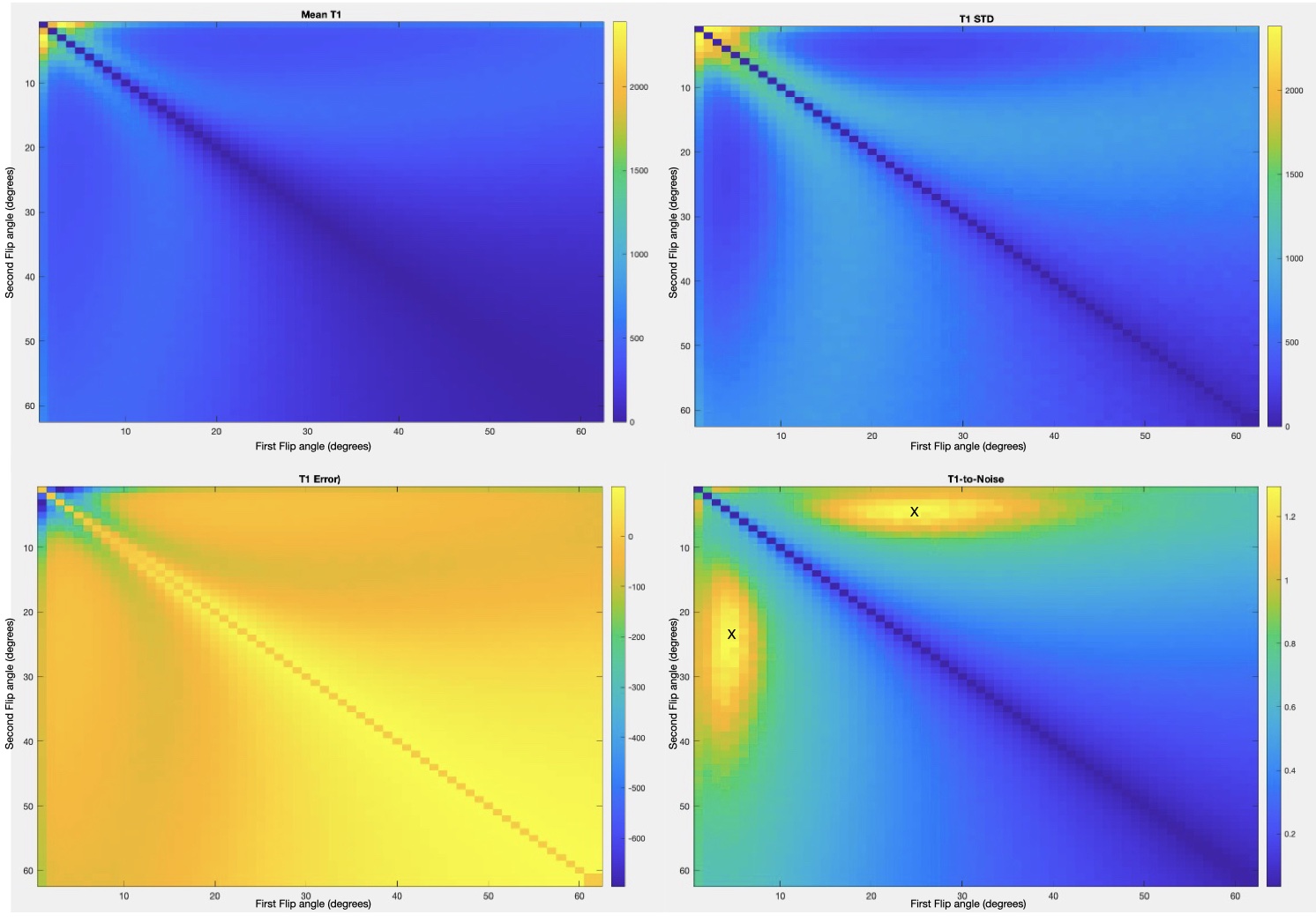
Simulations of T1 estimation at low field DESPOT1 measures T1 from a series of at least two SPGR images with differing flip angles. Flip angle choice can significantly impact the accuracy and precision of T1 estimates7,8 and therefore we initially aimed to determine the combination of flip angles that would maximize accuracy and precision. To this end, simulations that sought to maximize the T1-to-noise estimates from DESPOT1 were performed by varying two FAs between [0° 90°] in 1° increments for TR = 8 ms and T1 = 300 ms, which was assumed to reasonably represent both gray and white matter based on recent literature. Simulations were run in MATLAB utilizing 1 million iterations. Noise was added to the simulated signal for an SNR=5.To validate our approach, a series of phantom and in vivo human imaging data were acquired on a 64 mT Hyperfine Swoop system. An SPGR sequence was designed from modifying an available FISP sequence by enabling RF and gradient spoiling. Non-isotropic voxels with a larger through-plane resolution (2.8x2.8x5 mm3) were used to make scans faster while having an acceptable SNR. Additional scan parameters consisted of TR = 8 ms, TE = 3 ms, 10 averages, and approximate acquisition time of 4 minutes per flip angle.
Phantom: A series of SPGR images with varying flip angles ranging from [5° 65°] in 5° increments were acquired in the CaliberMRI Mini-Hybrid Phantom model 137. Individual flip angle images were rigidly aligned to the α= 20° image using FSL FLIRT9. T1 estimation was subsequently performed by fitting multiple flip angle SPGR images using a weighted least squares algorithm and in-house processing code.
In-vivo: A series of multiple flip angle SPGR were next acquired in a healthy adult subject with flip angles of α = [10°,25°,30°,40°,50°,60°]. Images were rigidly aligned to the α=25° image using FSL9 and T1 estimation was performed using in-house processing code.
Results
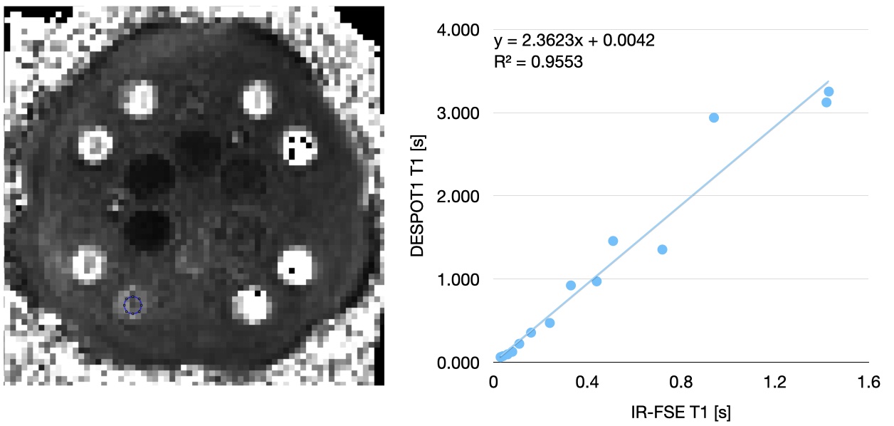
Simulation: Figure 1 shows the relative standard deviation, error, and T1-to-noise from simulations. Optimal flip angle for T1 estimation was determined to be approximately 5 degrees and 30 degrees.Phantom: Figure 2 depicts the T1 map estimated from the Caliber system phatom. Mean T1 values were extracted from the 14 T1 mimics using ImageJ and compared with estimated values using an IR-FSE based approach While a high correlation in the T1 values of the two techniques is observed (r=0.977), DESPOT1 T1 values are overestimated compared to IR-FSE based measures.
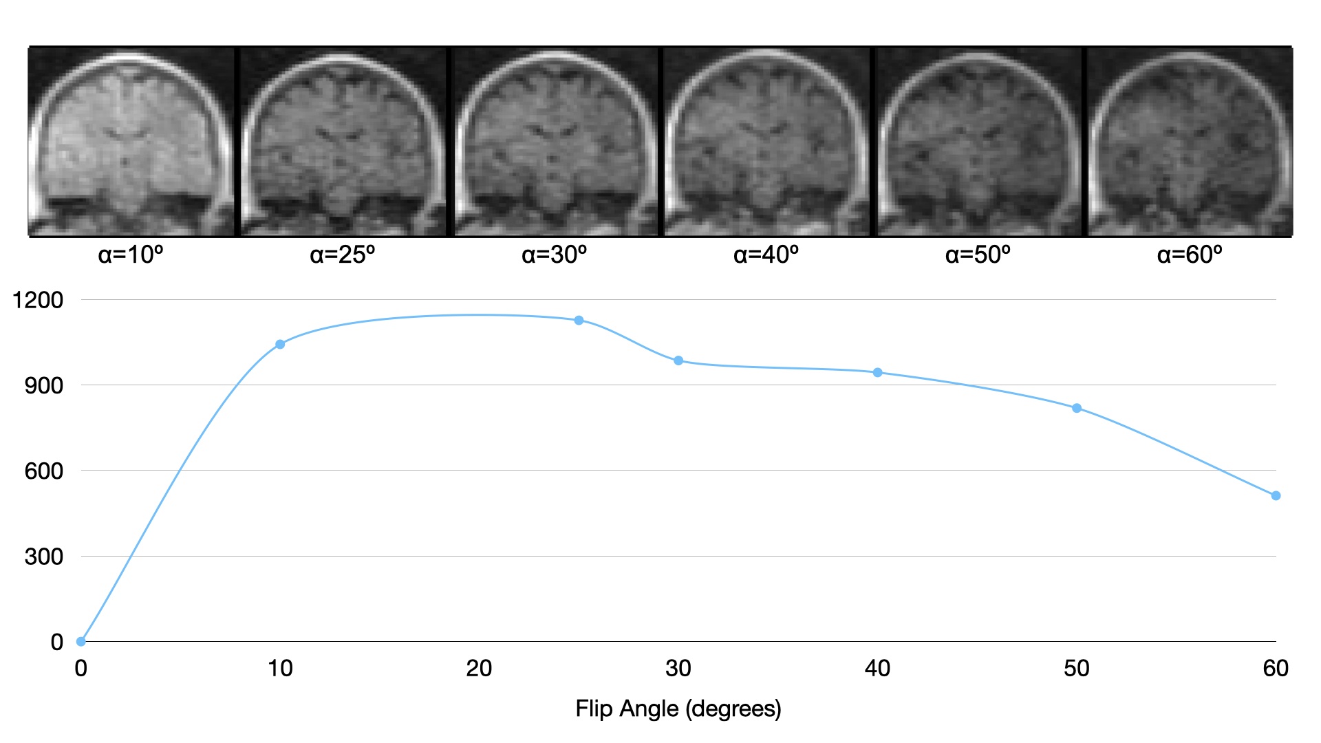
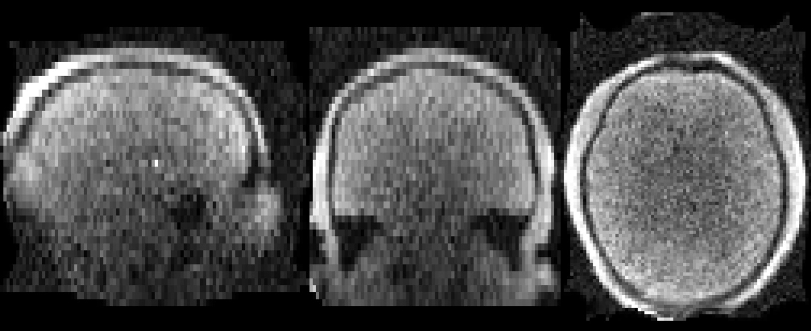
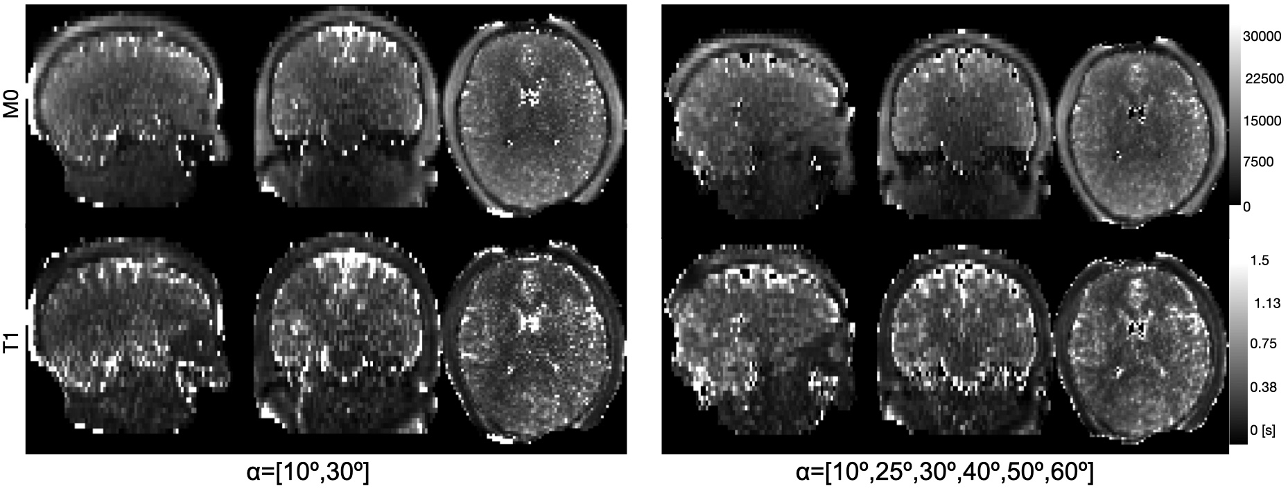
In-vivo: Figure 3 shows representative coronal slices of SPGR images acquired in healthy adult. Despite simulations indicating a lower optimal flip angle of α=5°, low SNR at this small flip angle limited its use in T1 estimation (Figure 4). Figure 5 illustrates M0 and T1 maps estimated for two flip angles [10°,30°] and across the full series of flip angles [10°,25°,30°,40°,50°,60°]. Marginal reductions in noise are observed in T1 estimates across full SPGR series.
Discussion
In this work, we have examined the ability to measure T1 from multiple flip angle SPGR images at low field (64 mT). SNR remains a challenge, particularly at low flip angles. Improvements in SNR and acquisition time may be possible through further optimization of the SPGR sequence. Nonetheless, our results demonstrate initial feasibility and suggest that T1 mapping via DESPOT1 may be achievable at ultra-low field.Acknowledgements
This work was supported by the Bill and Melinda Gates Foundation. Infrastructure support was also provided, in part, by grant U54 HD090256 from the Eunice Kennedy Shriver NICHD, National Institutes of Health (Waisman Center). We thank Hyperfine for their ongoing technical support and assistance.References
1. Deoni, S. C. Quantitative relaxometry of the brain. Top Magn Reson Imaging 21, 101-113 (2010). https://doi.org/10.1097/RMR.0b013e31821e56d8
2. Lebel, C. & Deoni, S. The development of brain white matter microstructure. Neuroimage 182, 207-218 (2018). https://doi.org/10.1016/j.neuroimage.2017.12.097
3. Alexander, A. L. et al. Characterization of cerebral white matter properties using quantitative magnetic resonance imaging stains. Brain Connect 1, 423-446 (2011). https://doi.org/10.1089/brain.2011.0071
4. Stikov, N. et al. On the accuracy of T1 mapping: Searching for common ground. Magnetic Resonance in Medicine73, 514-522 (2015). https://doi.org/https://doi.org/10.1002/mrm.25135
5. Deoni, S. C. High-resolution T1 mapping of the brain at 3T with driven equilibrium single pulse observation of T1 with high-speed incorporation of RF field inhomogeneities (DESPOT1-HIFI). J Magn Reson Imaging 26, 1106-1111 (2007). https://doi.org/10.1002/jmri.21130
6. Deoni, S. C., Rutt, B. K. & Peters, T. M. Rapid combined T1 and T2 mapping using gradient recalled acquisition in the steady state. Magn Reson Med 49, 515-526 (2003). https://doi.org/10.1002/mrm.10407
7. Wood, T. C. Improved formulas for the two optimum VFA flip-angles. Magnetic Resonance in Medicine 74, 1-3 (2015). https://doi.org/https://doi.org/10.1002/mrm.25592
8. Deoni, S. C., Peters, T. M. & Rutt, B. K. Determination of optimal angles for variable nutation proton magnetic spin-lattice, T1, and spin-spin, T2, relaxation times measurement. Magn Reson Med 51, 194-199 (2004). https://doi.org/10.1002/mrm.10661
9. Jenkinson, M., Bannister, P., Brady, M. & Smith, S. Improved optimization for the robust and accurate linear registration and motion correction of brain images. Neuroimage 17, 825-841 (2002). https://doi.org/10.1016/s1053-8119(02)91132-8
Figures