2173
Subspace modeling enables time-efficient B1-corrected volumetric water-specific relaxometry with a long echo train CASPR trajectory1School of Medicine and Health, Technical University of Munich, Munich, Germany, 2Philips GmbH Market DACH, Hamburg, Germany, 3Philips Research, Hamburg, Germany, 4Institute of Machine Learning for Biomedical Imaging, Helmholtz Munich, Neuherberg, Germany, 5School of Computation, Information and Technology, Technical University of Munich, Munich, Germany, 6Department of Computing, Imperial College London, London, United Kingdom, 7School of Biomedical Engineering and Imaging Sciences, King’s College London, London, United Kingdom, 8Department Artificial Intelligence in Biomedical Engineering, Friedrich-Alexander-Universität Erlangen-Nürnberg, Erlangen, Germany
Synopsis
Keywords: Relaxometry, Body
Motivation: The acquisition time of volumetric relaxometry techniques is constrained by the sampling efficiency and confounding factors such as fat or the B1 inhomogeneity.
Goal(s): To develop a method for volumetric T1- and T2-mapping that improves efficiency while accounting for confounding factors.
Approach: A Cartesian acquisition with spiral profile ordering is employed to increase sampling efficiency. A subspace reconstruction is proposed to perform B1- and water-specific T1- and T2-mapping considering the exact k-space sampling pattern.
Results: Simulation, phantom and in-vivo experiments were performed to evaluate the proposed method with regard to its quantification performance and its B1 sensitivity at 3T.
Impact: Subspace reconstruction in combination with a CASPR trajectory can improve the scan's efficiency and sampling flexibility while correcting for confounders such as fat or B1. The technique may be valuable to develop volumetric relaxometry in clinically acceptable scan times.
Introduction
Quantitative T1- and T2-mapping yields potential new biomarkers to complement current qualitative clinical routine imaging. While 2D relaxometry provides only limited coverage, volumetric relaxometry has shown great potential but is time-consuming and prone to motion artifacts.Highly efficient and motion-robust k-space trajectories are needed for volumetric relaxometry in clinically acceptable scan times. In addition, quantitative methods have to be designed to minimize sensitivity to confounders, such as fat or the B1 inhomogeneity, especially at 3T or higher field strengths.
Recently, Cartesian acquisitions with spiral profile ordering (CASPR) have been proposed for cardiac1,2 and abdominal applications3,4 showing a high flexibility regarding their sampling scheme, intrinsic motion robustness and can be combined with data-driven motion correction techniques. Sequence parameters such as the echo train length (ETL) directly increase the time efficiency of the sequence but simultaneously impact parameter mapping accuracy, if not considered during reconstruction.
To address these challenges, subspace reconstruction methods have shown great potential to reconstruct high-quality T2-weighted TSE images5 and improve the accuracy in volumetric quantitative imaging, e.g. in 3D-QALAS6.
The present work introduces a new method for volumetric water-specific T1- (wT1) and T2- (wT2) mapping with long ETLs and short inversion times while accounting for B1 and B0 inhomogeneity. The acquisition scheme of a previously proposed method for liver mapping7 is combined with a CASPR trajectory enabling more flexible and time-efficient sequence parameters. Subspace reconstruction combined with dictionary-matching is proposed to estimate B1 along with wT1 and wT2 maps while accounting for T1 relaxation during the acquisition.
Methods
Pulse sequenceThe pulse sequence is composed of an adiabatic modified BIR-4 preparation pulse8,7 and a two-echo bipolar gradient-echo read-out using a golden-step CASPR trajectory with center-out profile ordering (Fig.1). Four or five volumes (preps) with different contrast were acquired. Pulse durations (10.9$$$\,$$$ms and 30.9$$$\,$$$ms) and delay times between preparation and readout (200$$$\,$$$ms, 300$$$\,$$$ms and 500$$$\,$$$ms) were varied for the T2preps and T1preps, respectively.
Image reconstruction and quantification
First, a compressed-sensing reconstruction was performed with subspace operator $$$P=\mathbf{1}$$$, Fourier operator $$$F$$$ and coil sensitivity maps from a prescan $$$S$$$ using total variation (TV) regularization ($$$\alpha=0.03$$$):$$x =\arg\min_{x'}||PFSx'-y||_2^2+{\alpha}||\text{TV}(x')||_1$$A B0-map was estimated based on a dual-echo multi-resolution graph-cut algorithm9,10. B0-specific dictionary matching yielded B1, T1, and T2 maps which were forward simulated to five basis coefficients for the two echoes. The basis was computed from the Bloch-simulated dictionary ($$$\text{B0}\in[-300,300]\,$$$Hz, $$$\text{B1}\in[0.52,1.48]$$$, $$$\text{T1}\in[300,3000]\,$$$ms, $$$\text{T2}\in[10,300]\,$$$ms) using SVD with $$$\text{B0}=0\,$$$Hz.
Simulated coefficient images were used as initialization in the subsequent subspace reconstruction ($$$\alpha=0.01/0.03$$$ for phantom/in-vivo experiments), employing the subspace operator $$$P$$$ as an ETL-resolved sampling mask5 and decomposition into basis coefficients.
Virtual preparation images were generated at the sampling time of k-space center. After water-fat separation, B1-, wT1-, and wT2-mapping was performed using dictionary matching.
Simulation
A numerical body model (XCAT phantom11) was employed to generate T1, T2, PDFF and $$$R_2^*$$$ with physical realistic relaxation times at 3T (liver: $$$\text{wT1}=800\,$$$ms, $$$\text{wT2}=25\,$$$ms, $$$\text{PDFF}=10\%$$$). B1-field inhomogeneities were represented by a two-dimensional Gaussian function. Parameter maps were forward-simulated using the dictionary and the water-fat signal model. Gaussian noise was added to real and imaginary images (SNR=250). k-space acquisition was simulated using the phase encoding table of the actual scan.
Phantom and in-vivo measurements
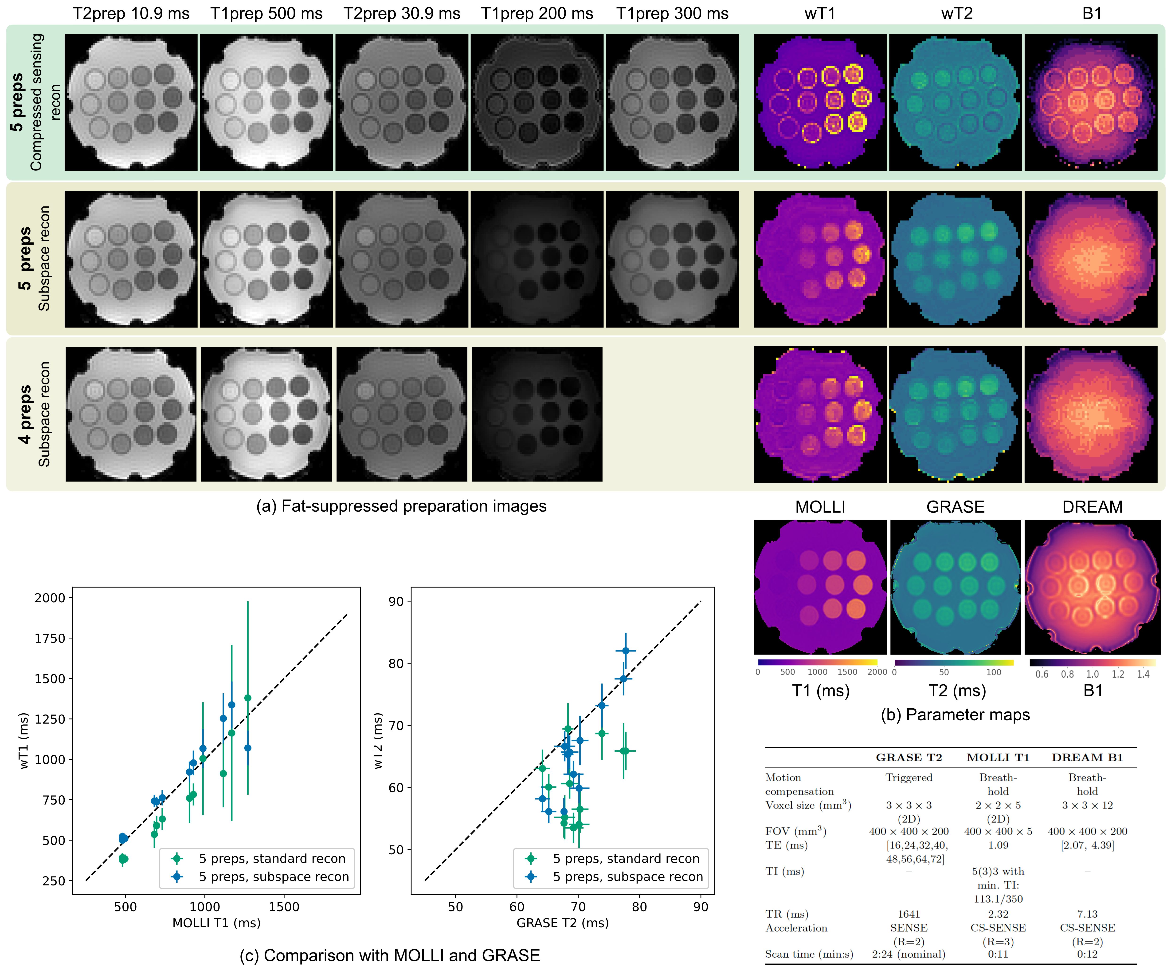
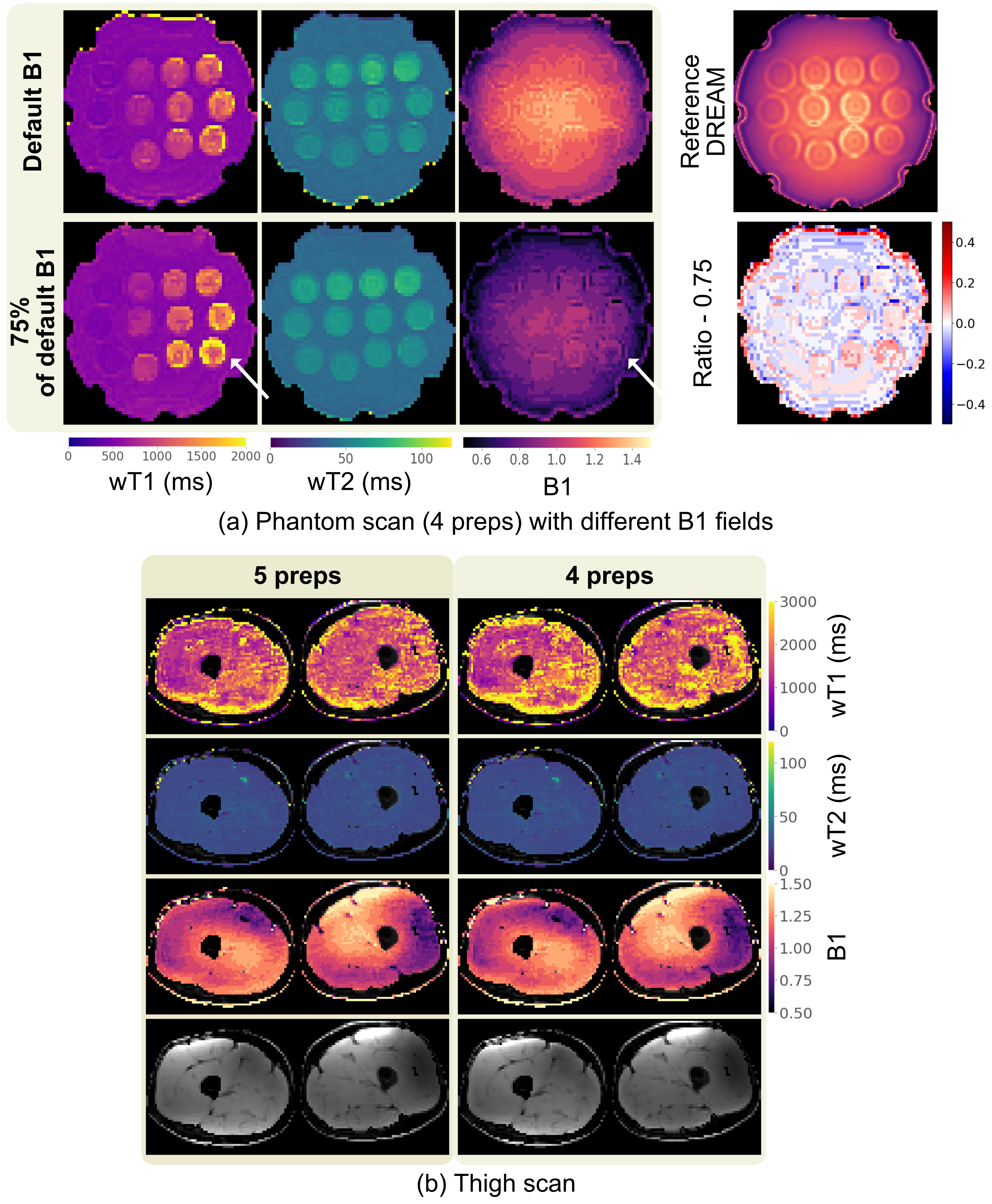
Measurements were performed at 3T (Ingenia Elition,Philips) on a water-fat T1 phantom (Calimetrix,Madison,WI,USA) and on two volunteers for the thigh and liver ($$$\text{FOV}=400\times300\times200\,$$$mm³, 3$$$\,$$$mm isotropic voxel size, $$$\text{TR/TE1/TE2}=3.4/1.0/2.1\,$$$ms, $$$\text{ETL}=130$$$, $$$N_\text{shots}=113\, N_{\text{prep}}$$$, $$$T_\text{shot}=500\,\text{ms}+T_{D}$$$, $$$\text{FA}=8^{\circ}$$$). To assess B1-mapping capabilities, a phantom scan was acquired using 3/4 of the nominal flip angle. MOLLI T1-mapping, GRASE T2-mapping, and DREAM B1-mapping were acquired as references (sequence parameters in Fig.3).
Results
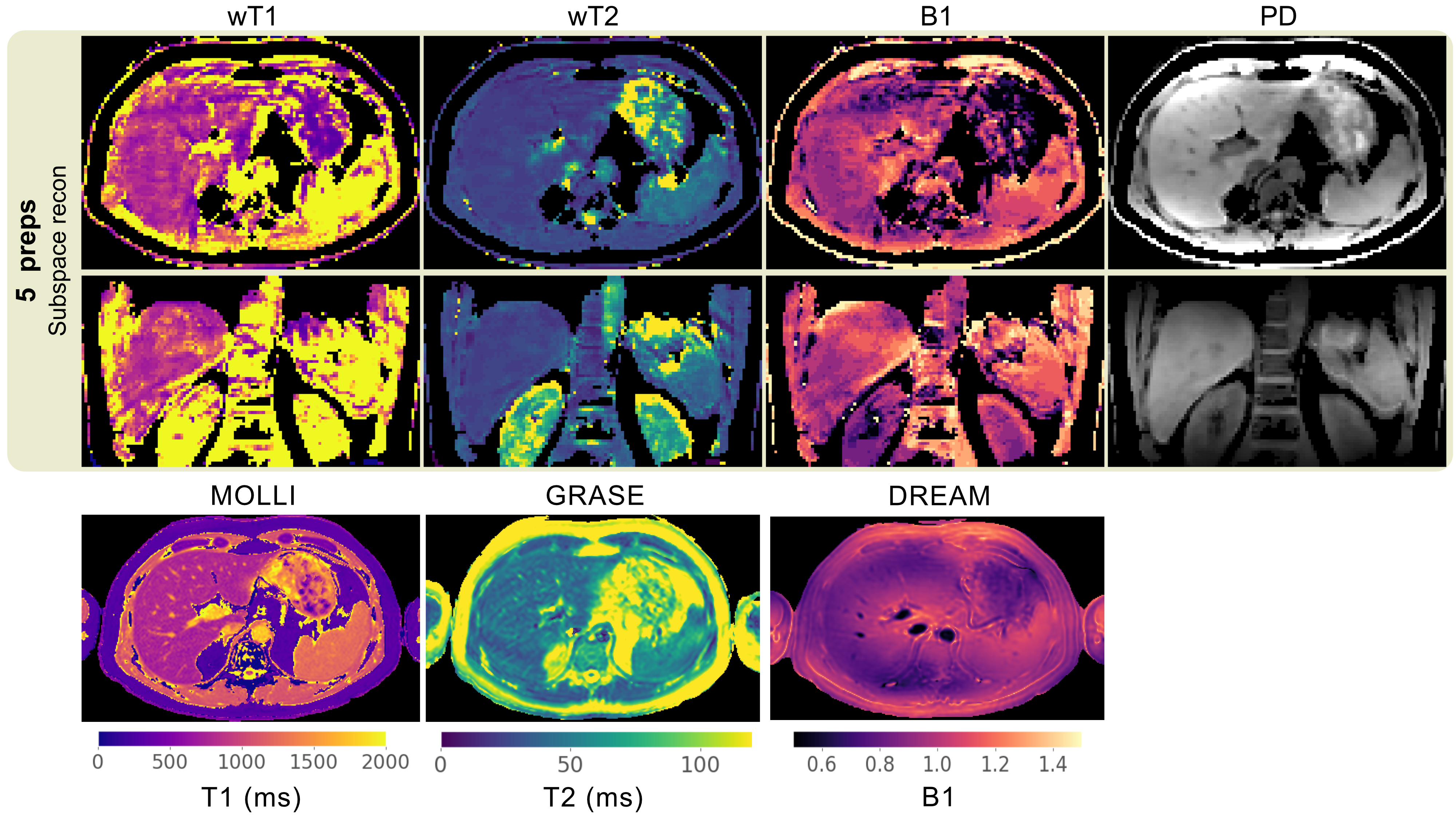
Simulation results (Fig.2) show that the proposed subspace reconstruction enables parameter mapping, even with a long ETL and short inversion time. Comparisons with reference techniques (Fig.3) show good quantification performance in the phantom, while the representation of edges is impaired compared to compressed-sensing reconstruction. The sequence performs robustly for strong B1 inhomogeneities (50-150%, Fig.4). Liver results with 5 acquired preparations are shown in Fig.5.Discussion
This work demonstrates that subspace reconstruction can be effectively employed with a CASPR trajectory using long ETL and short inversion times for combined B1-, wT1- and wT2-mapping. The resulting acquisition scheme enables greater efficiency and flexibility, e.g. compared to radial stack-of-stars trajectories7. Future work includes optimizing sequence parameters for optimal T1 and T2 sensitivity and refined regularization of the inverse subspace problem.Conclusion
A novel method was developed for wT1- and wT2-mapping using a CASPR trajectory with long ETLs and short inversion times. Preliminary results show the potential of subspace modeling for time-efficient parameter mapping with great flexibility.Acknowledgements
The present work was supported by the TUM International Graduate School of Science and Engineering (TUM-ICL Joint Academy of Doctoral Studies). The authors also acknowledge research support from Philips Healthcare.References
[1] Prieto C, Doneva M, Usman M, Henningsson M, Greil G, Schaeffter T, Botnar RM. Highly efficient respi-ratory motion compensated free-breathing coronary mra using golden-step Cartesian acquisition. Journal of Magnetic Resonance Imaging 2014; 41:738–746. 10.1002/jmri.24602.
[2] Meineke J, Schülke C, Nehrke K, Keupp J. Whole-Heart Free-Breathing 3D Cardiac Cine with Fast Respiratory Motion Compensated Reconstruction. Proc Intl Soc Mag Reson Med 30 2022; p. 1757.
[3] Milotta G, Cruz G, Neji R, Prieto C, Botnar R. 3D Isotropic-resolution Non-rigid Motion Compensated Liver T1, T2 and fat fraction mapping. Proc Intl Soc Mag Reson Med 29 2021; p. 1234.
[4] Bruijnen T, Schake T, Akdag O, Bruel CVM, Lagendijk JJW, van den Berg CAT, Tijssen RHN. Free-breathing motion compensated 4D (3D+respiration) T2-weighted turbo spin-echo MRI for body imaging,2022.
[5] Tamir JI, Uecker M, Chen W, Lai P, Alley MT, Vasanawala SS, Lustig M. T2 shuffling: Sharp,multicontrast, volumetric fast spin-echo imaging. Magnetic Resonance in Medicine 2016; 77:180-195.10.1002/mrm.26102.
[6] Jun Y, Arefeen Y, Cho J, Fujita S, Wang X, Grant PE, Gagoski B, Jaimes C, Gee MS, Bilgic B. Zero-DeepSub: Zero-Shot Deep Subspace Reconstruction for Rapid Multiparametric Quantitative MRI Using3D-QALAS, 2023.
[7] Stelter JK, Weiss K, Moquillaza EH, Harder FN, Makowski MR, Braren RF, Karampinos DC. Free-breathing Isotropic resolution self-Navigated B1-insensitive whOle liver simultaneous T1 and T2 mapping(FINO). Proc Intl Soc Mag Reson Med 31 2023; p. 0060.
[8] Weidlich D, Schlaeger S, Kooijman H, Börnert P, Kirschke JS, Rummeny EJ, Haase A, Karampinos DC. T2 mapping with magnetization-prepared 3D TSE based on a modified BIR-4 T2 preparation. NMR Biomed2017; 30:e3773. 10.1002/nbm.3773.
[9] Eggers H, Brendel B, Duijndam A, Herigault G. Dual-echo Dixon imaging with flexible choice of echo times. Magn Reson Med 2010; 65:96–107. 10.1002/mrm.22578.
[10] Stelter JK, Boehm C, Ruschke S, Weiss K, Diefenbach MN, Wu M, Borde T, Schmidt GP, Makowski MR,Fallenberg EM, Karampinos DC. Hierarchical Multi-Resolution Graph-Cuts for Water-Fat-Silicone Sepa-ration in Breast MRI. IEEE Trans Med Imag 2022; 41:3253–3265. 10.1109/tmi.2022.3180302.
[11] Segars WP, Sturgeon G, Mendonca S, Grimes J, Tsui BM. 4D XCAT phantom for multimodality imaging research. Medical physics 2010; 37:4902–4915.4
Figures
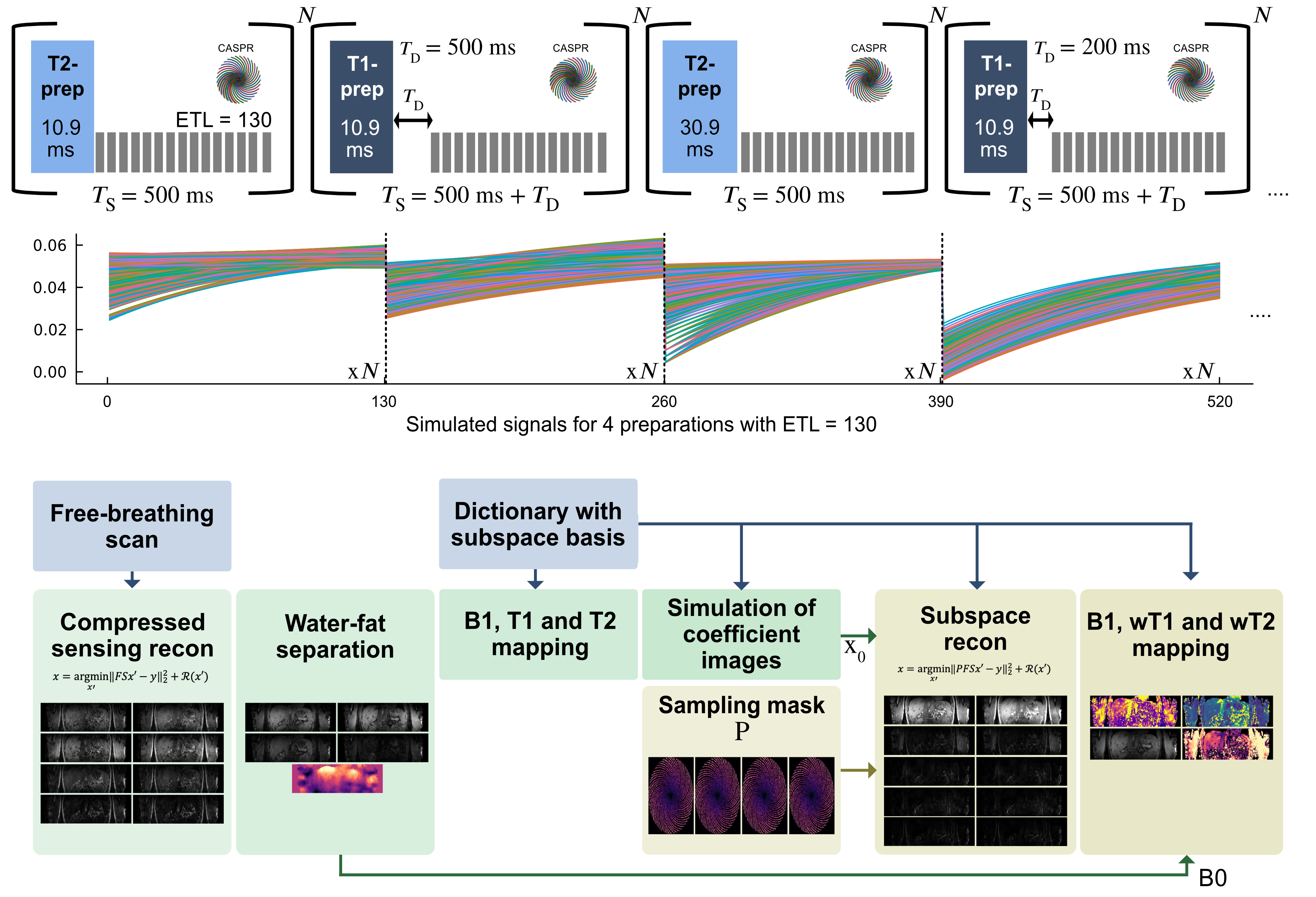
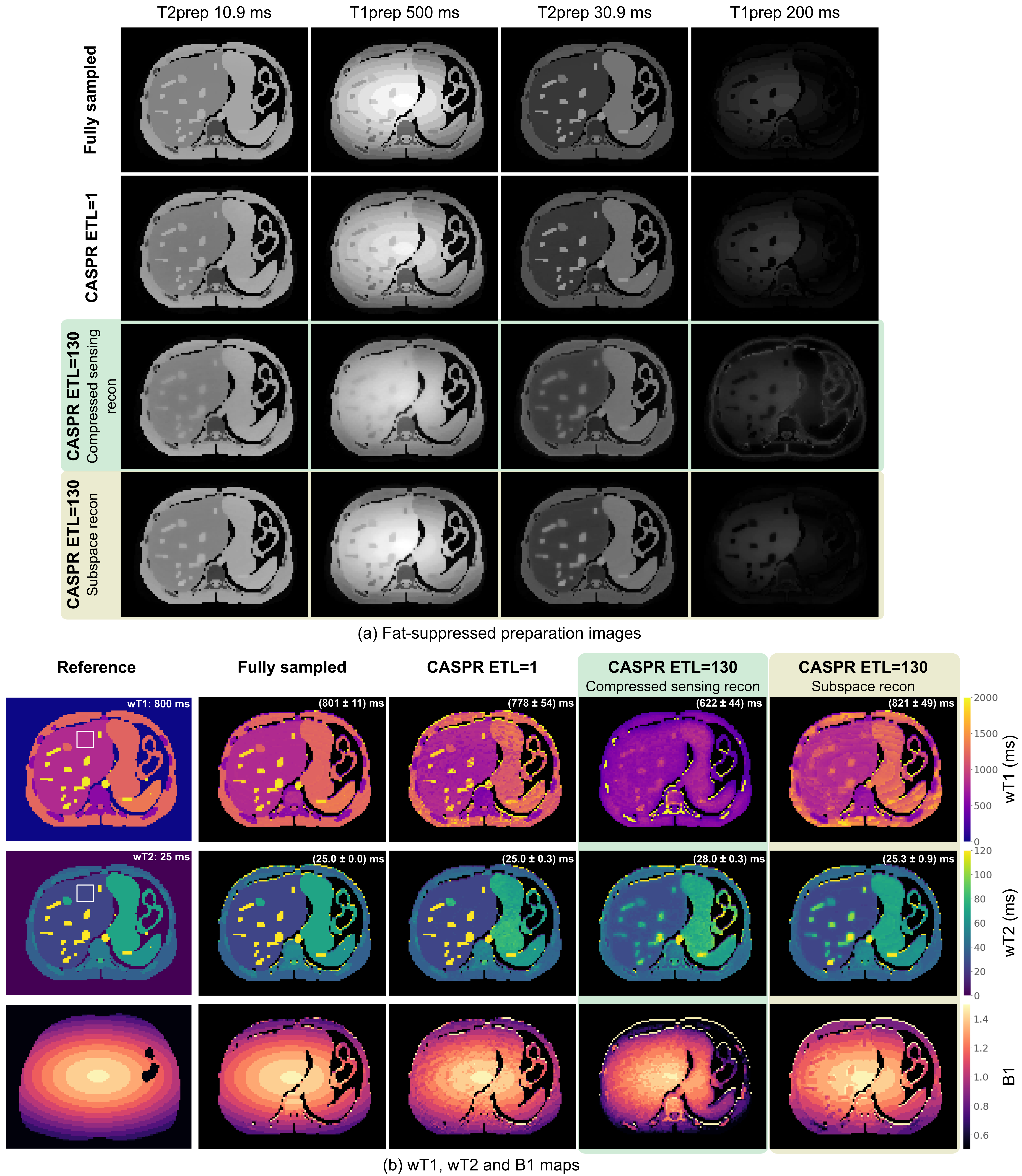
Fig.2 Simulation comparing different k-space trajectories and reconstruction techniques. Fat-suppressed preparation images are shown in (a) and parameter maps in (b). CASPR with ETL=1 refers to the dictionary as ETL=130 but only a single k-space line is acquired per shot. The ETL=130 simulation with compressed-sensing recon leads to significant bias which can be attributed to the different contrast for $$$T_D=200\,$$$ms. The subspace reconstruction can successfully improve the contrast for ETL=130. However, remaining deviations can be observed in small vessels in the wT1 map.