2167
Visualization of fine white matter bundles in the living human brain with diffusion MRI using 500 mT/m gradient strength1Athinoula A. Martinos Center for Biomedical Imaging, Department of Radiology, Massachusetts General Hospital & Harvard Medical School, Charlestown, MA, United States, 2Institute of Medical Physics and Radiation Protection, Mittelhessen University of Applied Sciences, Giessen, Germany., Giessen, Germany
Synopsis
Keywords: Tractography, Gradients, Structural Connectivity
Motivation: There is a pressing need to move beyond the brain major white matter fasciculi in our understanding and characterization of human connectional neuroanatomy.
Goal(s): To perform initial in vivo high-resolution diffusion MRI on the next-generation Connectome 2.0 scanner equipped with 500 mT/m gradient strength.
Approach: We acquired 1-mm isotropic dMRI in one healthy subject on Connectome 2.0 and evaluated its capabilities in characterizing fine fiber bundles in comparison to Connectome 1.0.
Results: The SNR boost enabled by the Connectome 2.0 stronger gradient system allows resolution of fine white matter structures in deep brain regions and near the gray-white interface.
Impact: The ability to characterize fine white matter circuitry in the living human brain within reasonable scan times opens new possibilities for investigating their role in psychiatric and neurological disorders and enables the application of clinical interventions that target these pathways.
Introduction
Data from ex vivo brain specimens and animal studies point to a pressing need to access the connectional anatomy of the human brain in vivo at higher resolution to advance our understanding of its function in health and disease. Many of the core subcortical pathways responsible for regulating vital brain functions are too small (<3 mm)1 to be imaged at the resolution that can be achieved on current state-of-the-art scanners for in vivo diffusion MRI (e.g., 1.5-2 mm)2. Validation studies have shown that spatial resolution has a greater impact than angular resolution in resolving complex fiber configurations3. While more advanced sequences4,5 can be used to image small subcortical pathways at sub-millimeter resolution with exceptional anatomical accuracy2,6, these lengthy acquisitions remain impractical for large-scale studies. The Connectome 2.0 MRI scanner was designed to image connectional anatomy in the living human brain at unprecedented resolution by pushing the maximum gradient strength (Gmax) and slew rate (SRmax) from 300 mT/m and 200 T/m/s on the original Connectome scanner (Connectome 1.0) to 500 mT/m and 600 T/m/s7. Here, we report initial results for high-resolution in vivo diffusion MRI on Connectome 2.0. We show that its ability to achieve high spatial resolution down to 1 mm isotropic with sufficient SNR is critical for capturing small fiber connections with greater accuracy than what was previously possible.Methods
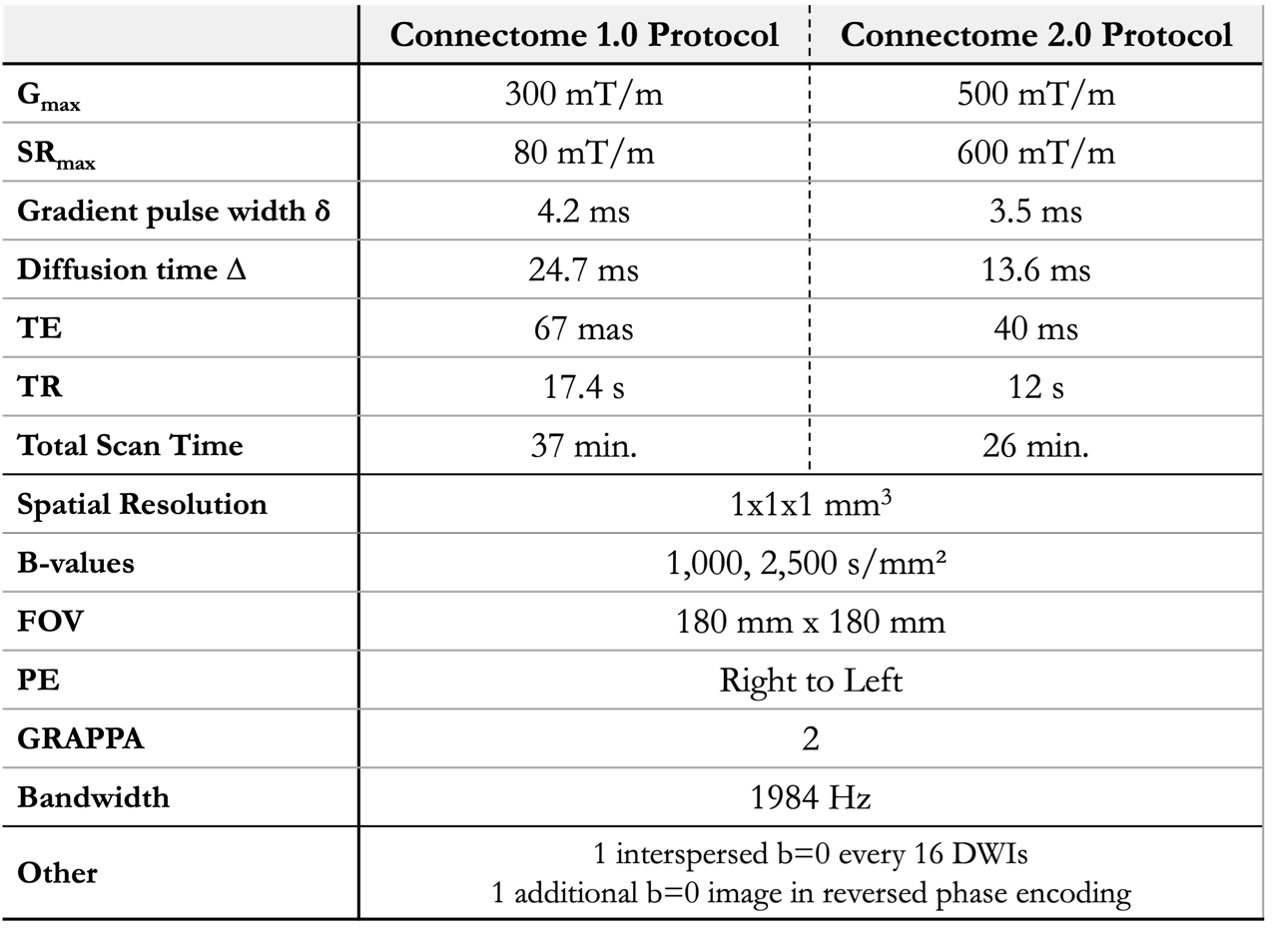
Diffusion MRI data were acquired in a healthy volunteer (34F) on the Connectome 2.0 scanner (MAGNETOM Connectom.X, Siemens Healthineers, Erlangen, Germany) using protocols with Gmax of 300 mT/m (Connectome 1.0) and 500 mT/m (Connectome 2.0) using a custom-built 72-channel head coil6. Protocol parameters for Connectome 1.0 and Connectome 2.0 acquisitions are listed in Table 1. The diffusion data were preprocessed using Gibbs ringing correction8, susceptibility and eddy current induced distortion correction9, and gradient non-linearity distortion correction10. Fiber orientation distribution functions (fODFs) were fitted to the pre-processed data using multi-shell multi-tissue constrained spherical deconvolution in MRtrix311. Whole-brain tractograms were obtained seeding local probabilistic tractography12 in every voxel within a white matter mask (5 seeds/voxel). To allow for better anatomical comparison, the datasets were co-registered13. To investigate the effects of denoising, we repeated the pre-processing adding an iterative Rician-corrected denoising14 as first step in the preprocessing pipeline.Results
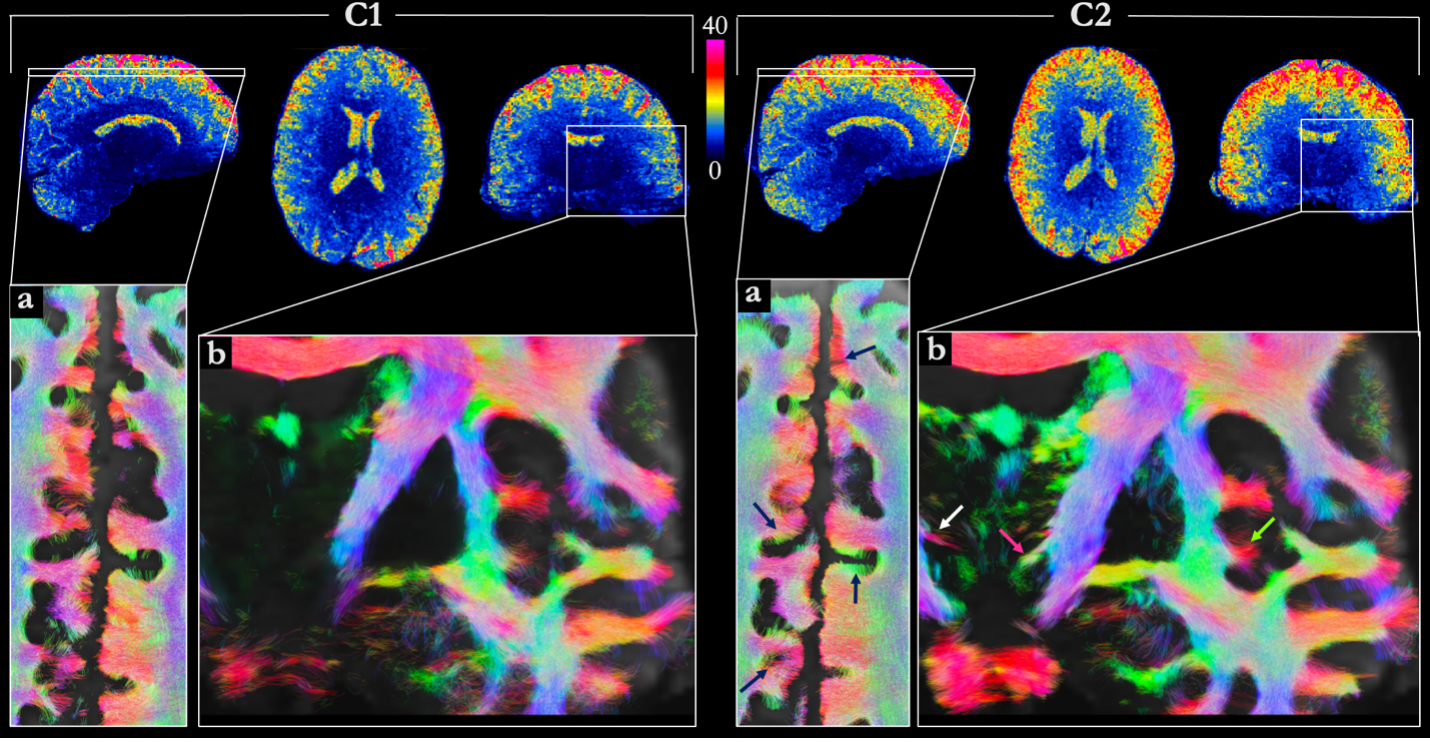
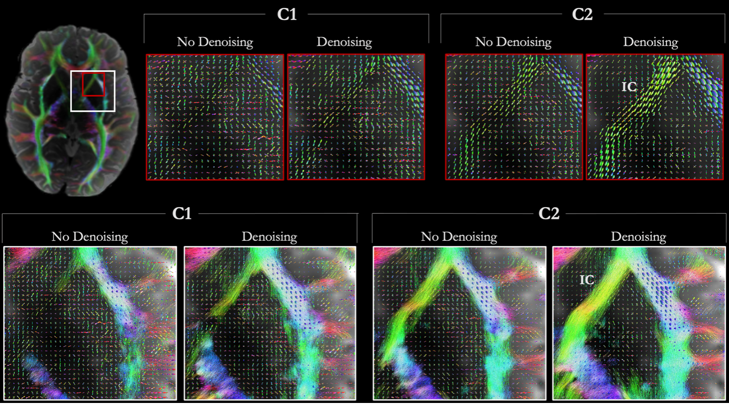
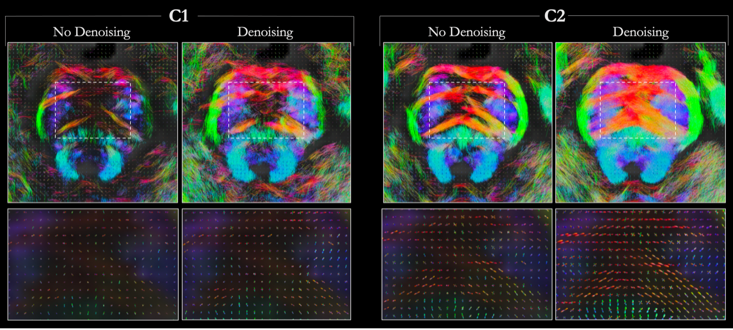
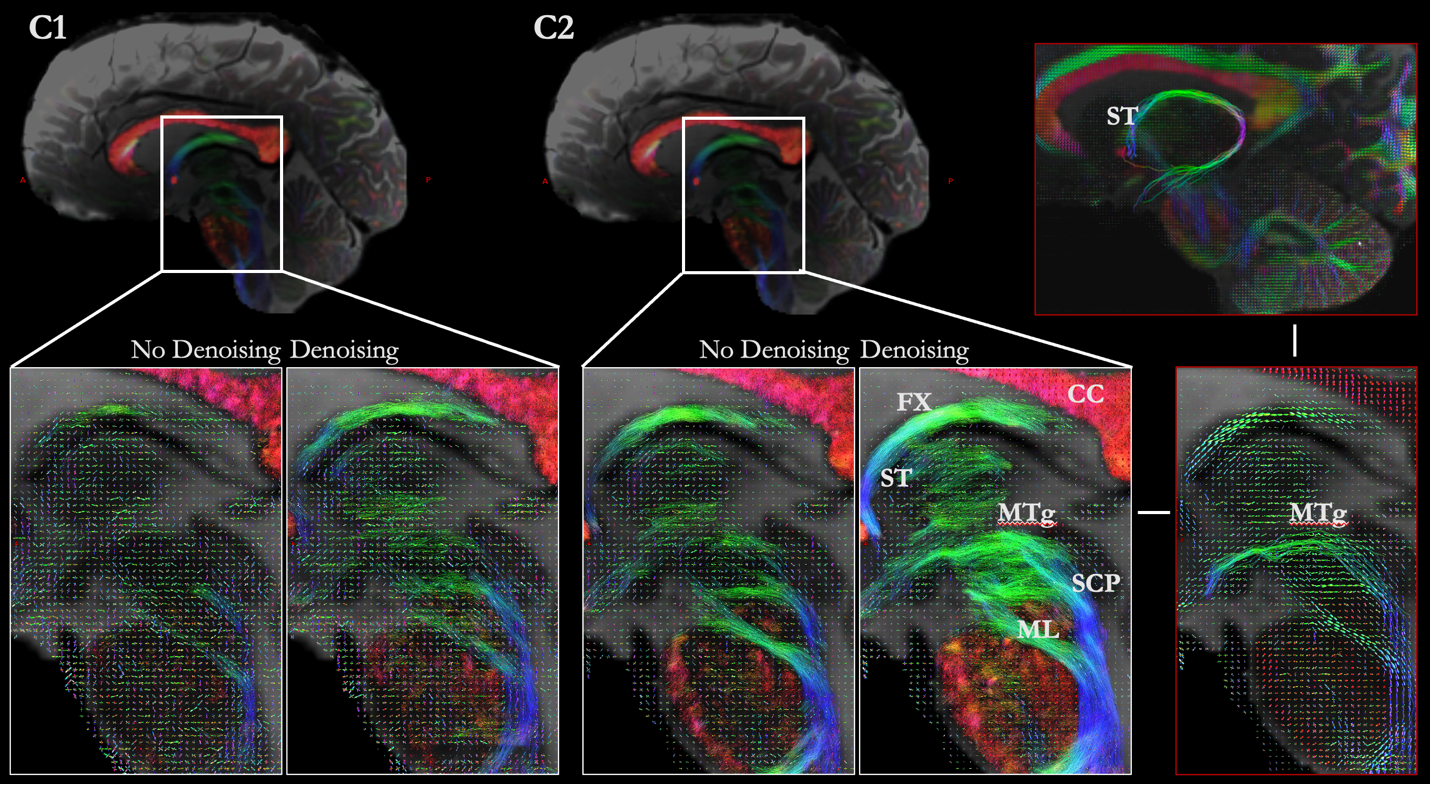
The higher-performance gradient system of Connectome 2.0 allows significant reductions in diffusion time and pulse width for the same diffusion-encoding b-value, resulting in shorter TE and higher SNR compared to Connectome 1.0 (Figure 1). This enables the resolution of smaller white-matter structures, like the radially oriented fibers around the gray and white matter boundary (Figure 1a) and the small fiber bundles leaving the internal capsule to enter subthalamic regions (Figure 1b)15. The increased SNR on Connectome 2.0 is even more noticeable in deep white matter, where more accurate fiber orientation estimates can be obtained in the anterior limb of the internal capsule (Figure 2) and in the brainstem (Figure 3,4), resulting in more accurate and complete tractography reconstructions compared to Connectome 1.0. This allows the accurate reconstruction of challenging fine fiber bundles like the mammillo-tegmental tract (MTg) and stria terminalis (ST) (Figure 4). We have previously shown that these small subcortical pathways (diameter<2 mm) could be accurately visualized in sub-millimeter diffusion MRI across 9 two-hour sessions2. Here, we show these pathways can be correctly visualized in a 30 min. scan on Connectome 2.0, while they could not be seen with the matched Connectome 1.0 protocol (Figure 4). Of note, for all the white matter structures investigated here, denoising Connectome 1.0 data does not provide the anatomical accuracy obtained with non-denoised Connectome 2.0 data.Conclusion
The SNR boost achieved on Connectome 2.0 allows the accurate visualization of small fiber configurations in vivo. This will enable mapping of the fine topographical organization in healthy subjects and individuals with psychiatric or neurological conditions, while concomitantly characterizing their underlying microstructure. It will also open new avenues for interventions that target these pathways, including deep brain stimulation16. The ability to delineate and characterize these smaller circuitries in vivo will bridge a key technology gap in the effort to advance our understanding of brain disorders.Acknowledgements
The research reported in this abstract was supported by the National Institute of Biomedical Imaging and Bioengineering of the National Institutes of Health under award number U01EB026996 and the National Institute of Neurological Disorders and Stroke of the National Institutes of Health under award number UM1NS132358.References
1. Fakhoury M. The habenula in psychiatric disorders: More than three decades of translational investigation. Neurosci Biobehav Rev. 2017;83:721-735.
2. Maffei C, Wang F, Haber SN, Yendiki A. Submillimeter dMRI protocol optimization for accurate in-vivo reconstruction of deep-brain circuitry. International Society for Magnetic Resonance in Medicine. (2022)
3. Jones R, Grisot G, Augustinack J, Magnain C, Boas DA, Fischl B, Wang H, Yendiki A. Insight into the fundamental trade-offs of diffusion MRI from polarization-sensitive optical coherence tomography in ex vivo human brain. Neuroimage. 2020 Jul 1;214:116704.
4. Setsompop, K. et al. High‐resolution in vivo diffusion imaging of the human brain with generalized slice dithered enhanced resolution: Simultaneous multislice (gSlider-SMS). Magn. Reson. Med. 79, 141–151 (2018).
5. Ramos-Llordén, G, Ning, L, Liao, C, et al. High-fidelity, accelerated whole-brain submillimeter in vivo diffusion MRI using gSlider-spherical ridgelets (gSlider-SR). Magn Reson Med. 2020; 84: 1781–1795.
6. Wang, F. et al. Motion‐robust sub‐millimeter isotropic diffusion imaging through motion corrected generalized slice dithered enhanced resolution (MC-gSlider) acquisition. Magn. Reson. Med. 80, 1891–1906 (2018).
7. Huang SY, Witzel T, Keil B, et al. Connectome 2.0: Developing the next-generation ultra-high gradient strength human MRI scanner for bridging studies of the micro-, meso- and macro-connectome. Neuroimage. 2021 Nov;243:118530.
8. Kellner, E., Dhital, B., Kiselev, V. G., & Reisert, M. (2016). Gibbs‐ringing artifact removal based on local subvoxel‐shifts. Magnetic resonance in medicine, 76(5), 1574-1581.9. Andersson, J. L., Skare, S., & Ashburner, J. (2003). How to correct susceptibility distortions in spin-echo echo-planar images: application to diffusion tensor imaging. Neuroimage, 20(2), 870-888.
10. Jovicich, J., Czanner, S., Greve, D., Haley, E., van Der Kouwe, A., Gollub, R., ... & Dale, A. (2006). Reliability in multi-site structural MRI studies: effects of gradient non-linearity correction on phantom and human data. Neuroimage, 30(2), 436-443.
11. Raffelt, D. and Connelly, A. Unsupervised 3-tissue response function estimation from single-shell or multi-shell diffusion MR data without a co-registered T1 image. ISMRM Workshop on Breaking the Barriers of Diffusion MRI, 2016.
12. Tournier, J.-D, Calamante, F. and Connelly, A. Improved probabilistic streamlines tractography by 2nd order integration over fibre orientation distributions. Proceedings of the International Society for Magnetic Resonance in Medicine, 2010, 1670
13. Reuter M, Rosas HD, Fischl B. Highly accurate inverse consistent registration: a robust approach. Neuroimage. 2010 Dec;53(4):1181-96.
14. J-Donald Tournier, Ben Jeurissen, and Daan Christiaens. (2023). Iterative model-based Rician bias correction and its application to denoising in diffusion MRI. Proc ISMRM, 31, 3795
15. Haber SN, Lehman J, Maffei C, Yendiki A. The Rostral Zona Incerta: A Subcortical Integrative Hub and Potential Deep Brain Stimulation Target for Obsessive-Compulsive Disorder. Biol Psychiatry. 2023 Jun 1;93(11):1010-1022.
16. Li, N., Baldermann, J.C., Kibleur, A. et al. A unified connectomic target for deep brain stimulation in obsessive-compulsive disorder. Nat Commun 11, 3364 (2020).
Figures