2164
Superficial White Matter Classification using Diffusion MRI Tractography with Spiking Neural Networks1Paul C. Lauterbur Research Center for Biomedical Imaging, Shenzhen Institute of Advanced Technology, Chinese Academy of Sciences, Shenzhen, China, 2University of Chinese Academy of Sciences, Beijing, China, 3University of Electronic Science and Technology of China, Chengdu, China, 4Harvard Medical School, Boston, MA, United States, 5Zhejiang University of Technology, Hangzhou, China, 6Peng Cheng Laboratory, Shenzhen, China
Synopsis
Keywords: Tractography, Brain, Diffusion MRI, Spiking Neural Network, Superficial White Matter Classification, Tractography
Motivation: The investigation of superficial white matter (SWM) poses challenges due to its small size, variability, delicate structure, high curvature, and fiber crossings in diffusion MRI tractography.
Goal(s): Our goal is to develop an innovative methodology for classifying SWM streamline clusters using diffusion MRI tractography, leveraging brain-inspired learning-based techniques.
Approach: A dual-phase method with Spiking Neural Networks (SNNs) and leaky integrate and fire (LIF) neurons is developed for the classification of 199 SWM clusters.
Results: Experiments were conducted using two open-source datasets, and our method achieves accurate SWM classification results with an accuracy of 93.73%.
Impact: The findings of this study on SWM classification hold great potential for facilitating analyses of SWM within neuroscientific research, contributing to understanding the complexities and alterations in SWM associated with various health conditions and neurological disorders.
Introduction
Diffusion magnetic resonance imaging (dMRI) tractography is an advanced method for reconstructing macro-scale brain white matter connections in vivo2,7, offering quantitative insights into structural connectivity and tissue microstructure. Whole-brain tractography generates streamlines covering deep white matter (DWM) and superficial white matter (SWM)7,9,10.The SWM, located directly beneath the cerebral cortex, predominantly consists of short association connections, such as inter-gyri short white matter fibers. SWM fibers represent a significant proportion, accounting for up to 60% of the total white matter volume11. However, classifying SWM fasciculi remains challenging due to their small size, proximity to the cortex, and issues like partial volume effects.
Numerous studies have focused on exploring SWM fasciculi1,9,10, particularly in clinical research. Most SWM tractography classification methods employ either region of interest (ROI)-guided selection or streamline-based clustering12. The streamline clustering techniques for SWM classification are automated and applicable across various health conditions9,10. However, challenges persist due to the high curvature and fiber crossings encountered during SWM tractography from dMRI.
In recent years, Spiking Neural Networks (SNNs) have gained attention as they closely mimic the behavior of biological neurons and utilize "spikes" to transmit information, resembling natural action potentials3. Given these unique characteristics, we believe SNNs are well-suited for classification of SWM clusters.
In this study, inspired by9,10, we proposed a method for the classification of 199 SWM clusters from streamline-based tractography using SNNs by utilizing a dual-phase strategy. Phase I utilizes SNNs with leaky integrate and fire neurons to extract features from tractography data, classifying the streamlines into DWM and SWM. Phase II refines the analysis by employing supervised contrastive loss (SCL), enabling precise SWM multiclass classification. This method promises a comprehensive understanding of SWM intricacies.
Method
The illustration in Figure 1 outlines the dual-phase framework developed for classifying SWM. In Phase I, a feedforward SNN with leaky integrate and fire neurons is used to extract features14 from streamlines. This network also incorporates a classifier head employing cross-entropy loss to classify the streamlines into DWM and SWM. Phase II extends the analysis by employing supervised contrastive loss in addition to cross-entropy loss to perform multiclass classification on the SWM data obtained in Phase I. In total, 199 SWM clusters are distinguished.For training and testing, data of 100 healthy individuals from the Human Connectome Project5 were utilized. The five-fold validation technique was employed. Tractography for the whole brain is performed using the Unscented Kalman Filter (UKF)6. Besides, another dataset CNP4, which includes unlabeled tractography streamline clusters from both healthy individuals and patients with various disorders, was also employed to further test the model. Training of the SNN networks used the Adam Optimizer and time steps were set to 25, executed within the PyTorch framework on NVIDIA TITAN XP GPU.
Results
The quantitative results of our proposed method on the HCP dataset are reported in Table 1, which display the evaluation metrics for the classification of SWM in both Phase I and Phase II. Notably, our approach demonstrates promising outcomes in terms of accuracy in both phases.We further test our method on subjects with different health conditions (bipolar, ADHD, and schizophrenia) for effective classification of SWM clusters using the external dataset CNP 4. We show some promising qualitative visualization results in Figure 2, demonstrating the capability of our method to accurately cluster SWM fibers even in scenarios involving out-of-distribution data.
Discussion
The challenge of SWM classification is notably complex due to various factors. In Phase I of our method, our focus was primarily on binary classification, discerning between DWM and SWM. In Phase II, the comprehensive methodology engaged a total of 199 distinct SWM categories. Dual-phase approach, which break down complex tasks into smaller subtasks, have proven to be highly effective in various medical imaging applications9,10,13. This approach allowed for a meticulous evaluation and in-depth understanding of the model's performance, offering valuable insights into its accuracy across both phases.Conclusion
We present a distinctive methodology that employs SNNs in two distinct phases to achieve precise classification of SWM. Phase I utilizes a spiking neural network with leaky integrate-and-fire (LIF) neurons, accompanied by a classifier to discern between DWM and SWM. Phase II focuses on the accurate implementation of multiclass classification techniques to classify various classes within SWM effectively. The findings of this study hold great potential for facilitating analyses of SWM within neuroscientific research.Acknowledgements
This research was partly supported by the National Natural Science Foundation of China (62222118, U22A2040), Guangdong Provincial Key Laboratory of Artificial Intelligence in Medical Image Analysis and Application (2022B1212010011), Shenzhen Science and Technology Program (RCYX20210706092104034, JCYJ20220531100213029), and Key Laboratory for Magnetic Resonance and Multimodality Imaging of Guangdong Province (2023B1212060052).
References
1. Shinohara H et al. Pyramid-Shape Crossings and Intercrossing Fibers Are Key Elements for Construction of the Neural Network in the Superficial White Matter of the Human Cerebrum. Cereb Cortex 2020;30(10):5218–28.
2. Guevara, Miguel, et al. "Superficial white matter: A review on the dMRI analysis methods and applications." Neuroimage 212 (2020): 116673.
3. Eshraghian, Jason K., et al. "Training spiking neural networks using lessons from deep learning." Proceedings of the IEEE (2023).
4. Poldrack, Russell A., et al. "A phenome-wide examination of neural and cognitive function." Scientific data 3.1 (2016): 1-12.
5. Van Essen, David C et al. “The WU-Minn Human Connectome Project: an overview.” NeuroImage vol. 80 (2013): 62-79. doi:10.1016/j.neuroimage.2013.05.041
6. Zhang, Fan, et al. "An anatomically curated fiber clustering white matter atlas for consistent white matter tract parcellation across the lifespan." NeuroImage 179 (2018): 429-447.
7. Zhang, Fan, et al. "Quantitative mapping of the brain’s structural connectivity using diffusion MRI tractography: A review." Neuroimage 249 (2022): 118870.
8. Shastin, Dmitri, et al. "Surface-based tracking for short association fiber tractography." Neuroimage 260 (2022): 119423.
9. Xue, Tengfei, et al. "Superficial white matter analysis: An efficient point-cloud-based deep learning framework with supervised contrastive learning for consistent tractography parcellation across populations and dMRI acquisitions." Medical Image Analysis 85 (2023): 102759.
10.Xue, Tengfei, et al. "Supwma: consistent and efficient tractography parcellation of superficial white matter with deep learning." 2022 IEEE 19th International Symposium on Biomedical Imaging (ISBI). IEEE, 2022.
11.Schüz A, Braitenberg V, Miller R, The Human Cortical White Matter: Quantitative Aspects of Cortico-Cortical Long-Range Connectivity. Schüz, A.; Miller, R.: In Cortical Areas: Unity and Diversity, 377- 385 (2002), 2002.
12.Schilling, Kurt G et al. “Short superficial white matter and aging: a longitudinal multi-site study of 1293 subjects and 2711 sessions.” Aging brain vol. 3 (2023): 100067. doi:10.1016/j.nbas.2023.100067.
13.Panda, Ananya et al. “Two-stage deep learning model for fully automated pancreas segmentation on computed tomography: Comparison with intra-reader and inter-reader reliability at full and reduced radiation dose on an external dataset.” Medical physics vol. 48,5 (2021): 2468-2481. doi:10.1002/mp.14782.
14.Zhang, Fan, et al. "Deep white matter analysis (DeepWMA): fast and consistent tractography segmentation." Medical Image Analysis 65 (2020): 101761.
Figures
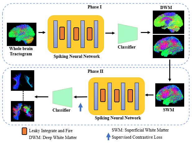
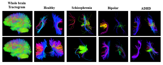
Figure 2. Qualitative visualization results of superficial white matter clusters on CNP dataset of healthy individuals and patients with disorders.
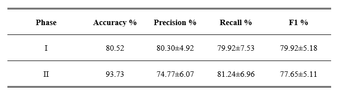
Table 1. Quantitative results of dual-phase SNNs-based method on the HCP dataset.