2162
Resolving complex white matter architecture in the human brain at 64 mT1CUBRIC, School of Psychology, Cardiff University, Cardiff, United Kingdom, 2Universidad de Valladolid, Valladolid, Spain, 3Hyperfine Inc., Guilford, CT, United States, 4School of Physics and Astronomy, Cardiff University, Cardiff, United Kingdom
Synopsis
Keywords: Tractography, Low-Field MRI
Motivation: Facilitating white matter mapping in traditionally inaccessible low income settings.
Goal(s): Demonstration of a viable protocol to resolve complex white matter architecture at low field.
Approach: A multi-modality protocol was devised to allow feasible acquisition of multi-direction diffusion weighted imaging (DWI) datasets at 64mT. A modified DWI protocol is shown, employing voxel-wise encoding non-uniformity calibration, extended readout, and optimal sampling density. Machine learning based denoising was employed to significantly reduce scan duration. T1 weighted scans were super-resolved to guide tractography.
Results: Anatomically-constrained tractography was shown to be viable at low field, and fine white matter microstructure was successfully recovered.
Impact: This work demonstrates that a 64mT system is capable of resolving microstructural detail even with lowered SNR, and gradient nonuniformity. This may have relevance in studies examining white matter neurodevelopment in historically underrepresented populations in low-income settings.
Introduction
Low-field (LF) MRI is becoming increasingly available, facilitating non-invasive imaging for both diagnosis and research in underserved populations. Diffusion weighted imaging (DWI) is a powerful neuroscience research tool, providing unique insights into tissue microstructure and brain connectivity. However, pulsed field gradients (PFGs) used for DWI induce eddy currents, causing temporal and spatial deviations from the desired gradient amplitude. Such effects are more pronounced in permanent magnet LF systems, due to magnet or yoke conductivity. These effects may be further exacerbated in low cost MRI systems, where gradient spatio-temporal uniformity is often sacrificed, resulting in nonuniform diffusion encoding and biased estimates of diffusivity and its directionality. DWI at LF is challenging due to SNR limitations, and has so far been limited to estimation of the diffusion tensor (DT)1. Here, for the first time, we demonstrate recovery of fibre orientation density function (ODF) in each voxel at 64 mT, through constrained spherical harmonic deconvolution2 (CSD). We show retrieval of complex fibre architecture, including dispersing and crossing fibres, providing more realistic tract reconstruction than for the DT alone.Methods
A 15-direction DWI scheme was implemented to investigate the utility of low field systems in fibre tracking. The scanner was first calibrated with the vendor's B1 and B0 mapping sequences. One T1, 15 b=900 s/mm2 isotropically-distributed diffusion-weighted and one b=0 volumes were collected using the vendor's 3D fast-spin echo DWI sequence (TE=76ms, TR=1000ms, ETL=44; FoV=180x216x198mm)3, modified to allow control of diffusion-encoding gradients, and oversampled by 1.44x the Nyquist limit, about 2.5x fewer samples than the product sequence. Data were reconstructed to 3mm isotropic voxels with the default reconstruction pipeline, and subsequently denoised with DDM2 4. Total acquisition time was 86 minutes.Diffusion measurements were calibrated with a voxel-wise technique5 against an isotropic 40/60% polyvinylpyrrolidone (PVP) / water solution phantom, characterised by the US National Insitute of Standards and Technology, whose temperature was tracked throughout the scan with a fibre optic thermometer to give accurate ADC estimates. Four complete repeats of the CSD protocol were collected, alongside a fifth trial dataset. Both diffusion encoded and b=0 scans were smoothed with a Gaussian filter with standard deviation of 9mm, as was a mask containing the phantom. Computation of ADC on the smoothed data, followed by dividing by the smoothed mask gave a smooth ADC map without edge artifacts. This was calibrated against the known value of the diffusion coefficient for PVP at each temperature to give a spatially uniform image of diffusion-encoding error. The inverse of this was applied to measurements for each corresponding volume to the trial dataset, to validate that it reduced error in ADC measurements, and then subsequently applied in-vivo.
An additional T1-weighted scan was collected (resolution=1.6x1.6x5mm) and superresolved to 1mm isotropic with synthSR6,7,8 , and FSL's FAST used to create grey / white matter interface masks. The super-resolved T1 was registered to the DW-volumes using ANTs, and MRTrix used for constrained spherical deconvolution (CSD) to order 4. Tissue-specific fibre population response functions were estimated from the FAST tissue masks, and CSD used to estimate ODFs in white matter voxels. Anatomically-constrained tractography9 was performed with the ODF maps, and hand-drawn regions of interest used to separate specific tracts.
Results & Discussion
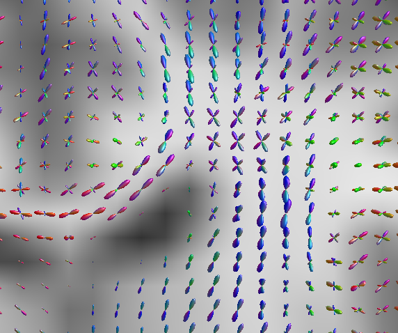
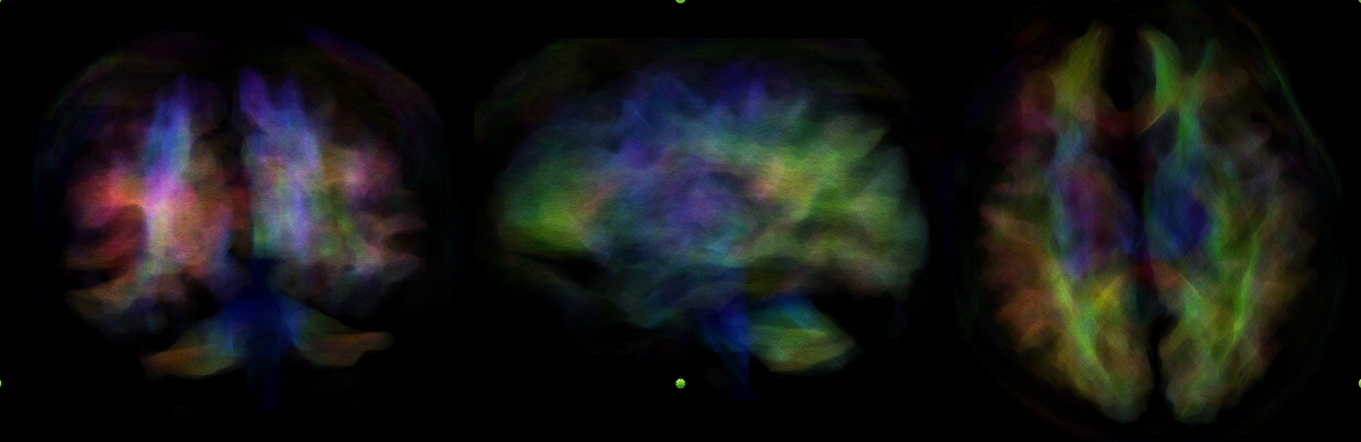
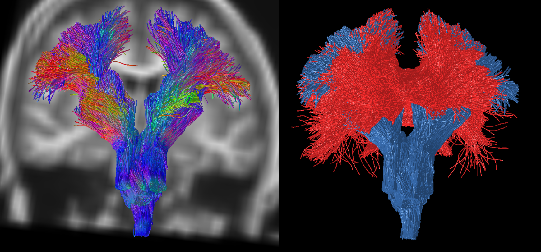
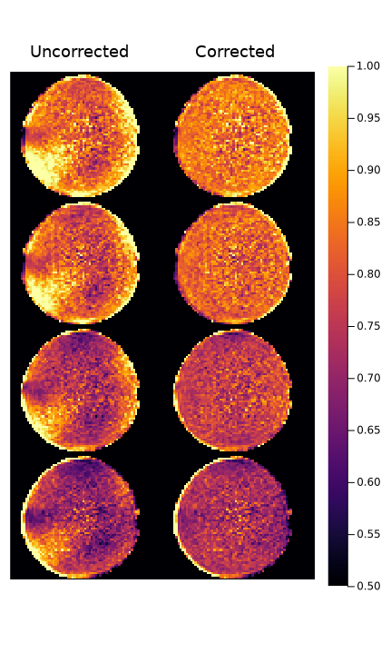
Multipeak ODFs were observed in voxels where crossing fibres were expected, including in the intersection of the corpus callosum and corticospinal tracts (Figure 1). Detail could be resolved in whole-brain tractography of major fibre bundles (Figure 2), with tracts displaying familiar form and directionality. Fibre dispersion was highly visible in the corticospinal tract (Figure 3), improving on prior work. Validation of the isotropic phantom calibration is shown to be repeatable in a test dataset (Figure 4), demonstrating that simple calibration procedures may be used to improve ADC quantification in LF DWI.Significant bias was observed in colour maps, suggesting errors in quantification of diffusion. Direct measurement of gradient fields on a per-coil basis, either through probing or phantom based measurements may offer improved ADC estimation and resolve some of the bias still observed even in corrected ADC maps. Additionally, the ML-assisted reconstruction used in the product sequence was not employed here as it produced biased estimates; with further training a direction agnostic ML reconstruction scheme may further improve SNR.
Conclusion
This work demonstrates the feasibility of performing CSD on low cost, low field (64mT) systems, showing that major tracts may be readily resolved even in regions of complex fibre architecture (Figure 3), that are otherwise non-amenable to DT-MRI. This extends the reach of white matter connectivity research to low resource settings.Acknowledgements
This work was made possible by generous support from the Bill and Melinda Gates Foundation through the UNITY project.References
[1] Plumley A, Padormo F, Cercignani M, et al. Tensors and Tracts at 64 mT. In: Proceedings of the 2023 ISMRM & ISMRT Annual Meeting & Exhibition. Toronto, ON, Canada, 2023, p. 5080.
[2] Tournier, J.-D., Calamante, F., & Connelly, A. (2007). Robust determination of the fibre orientation distribution in diffusion MRI: Non-negativity constrained super-resolved spherical deconvolution. NeuroImage, 35(4), 1459–1472. https://doi.org/https://doi.org/10.1016/j.neuroimage.2007.02.016
[3] Rafael O'Halloran, Hadrien Dyvorne, Laura Sacolick, Jo Schlemper, Michal Sofka, Sadegh Salehi, Samantha By, Riana Schleicher, Edmond Knopp, Kevin Sheth, and W. Taylor Kimberly. Diffusion-Weighted Imaging at 0.064 T
[4] Xiang T, Yurt M, Syed AB, et al. DDM2: Self-Supervised Diffusion MRI Denoising with Generative Diffusion Models. In: The Eleventh International Conference on Learning Representations. 2023.
[5] Lee, Y., Kettinger, A. O., Wilm, B. J., Deichmann, R., Weiskopf, N., Lambert, C., Pruessmann, K. P., & Nagy, Z. (2020). A comprehensive approach for correcting voxel-wise b-value errors in diffusion MRI. Magnetic Resonance in Medicine, 83(6), 2173–2184. https://doi.org/10.1002/mrm.28078
[6] Iglesias, J. E., Billot, B., Balbastre, Y., Tabari, A., Conklin, J., Gilberto González, R., Alexander, D. C., Golland, P., Edlow, B. L., & Fischl, B. (2021). Joint super-resolution and synthesis of 1 mm isotropic MP-RAGE volumes from clinical MRI exams with scans of different orientation, resolution and contrast. NeuroImage, 237, 118206. https://doi.org/https://doi.org/10.1016/j.neuroimage.2021.118206
[7] Iglesias, J. E., Schleicher, R., Laguna, S., Billot, B., Schaefer, P., McKaig, B., Goldstein, J. N., Sheth, K. N., Rosen, M. S., & Kimberly, W. T. (2022). Quantitative Brain Morphometry of Portable Low-Field-Strength MRI Using Super-Resolution Machine Learning. Radiology, 306(3), e220522. https://doi.org/10.1148/radiol.220522
[8] Iglesias, J. E., Billot, B., Balbastre, Y., Magdamo, C., Arnold, S. E., Das, S., Edlow, B. L., Alexander, D. C., Golland, P., & Fischl, B. (2023). SynthSR: A public AI tool to turn heterogeneous clinical brain scans into high-resolution T1-weighted images for 3D morphometry. Science Advances, 9(5), eadd3607. https://doi.org/10.1126/sciadv.add3607
[9] Smith RE, Tournier J-D, Calamante F, et al. Anatomically-constrained tractography: Improved diffusion MRI streamlines tractography through effective use of anatomical information. Neuroimage 2012; 62: 1924–1938.
Figures