2160
HARP: Hierarchical Anatomical Refinement of Pathways in Tractography1University of Eastern Finland, Kuopio, Finland, 2University of Helsinki, Helsinki, Finland, 3A.I. Virtanen Institute for Molecular Sciences, University of Eastern Finland, Kuopio, Finland, 4Aalto University School of Science, Espoo, Finland
Synopsis
Keywords: Tractography, Tractography & Fibre Modelling
Motivation: Whole brain tractography is highly challenging, and it is prone to false positive streamlines despite the use of Anatomically Constraint Tractography (ACT).
Goal(s): This study aims to reduce implausible streamlines in tractograms by introducing a novel refinement approach, thereby enhancing the accuracy of white matter connectivity analyses.
Approach: We developed Hierarchical Anatomical Refinement of Pathways (HARP), an advanced modification of ACT. HARP integrates increasingly detailed anatomical priors in a hierarchical fashion to improve accuracy of tractograms.
Results: Amount of implausible streamlines in ACT-based tractograms are relatively low but they are systematic. HARP is effective in further reducing false positive connections in tractograms.
Impact: Hierarchical Anatomical Refinement of Pathways (HARP) enhances tractography, offering neuroscientists and clinicians a new level of precision in brain connectivity analysis, which could impact our understanding of the brain and its disorders.
Introduction
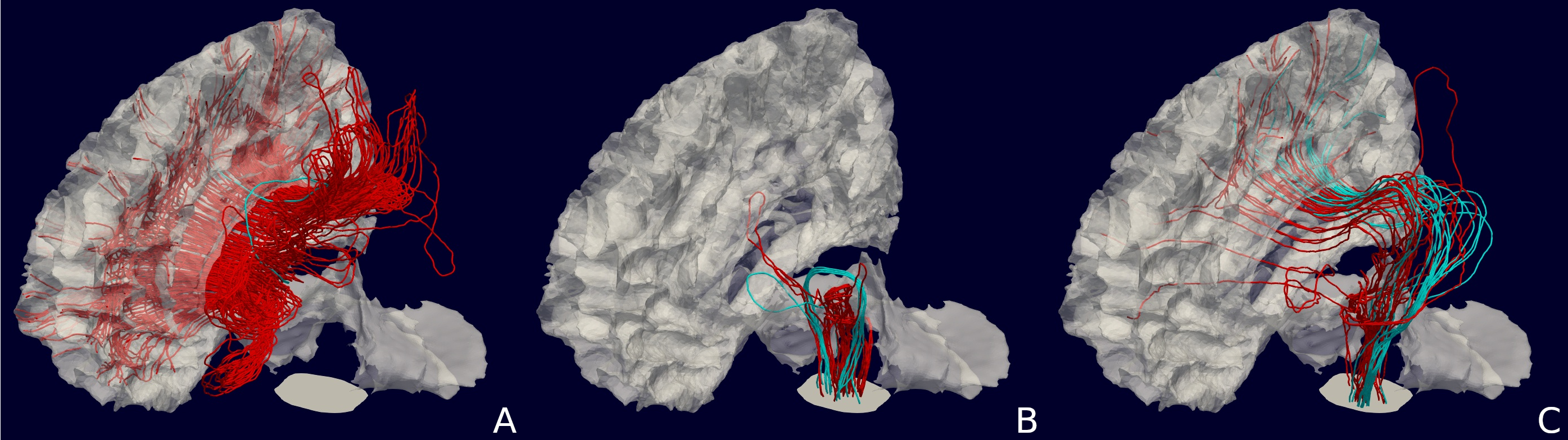
Tractography has paved the way for the in-vivo study of the brain's white matter. A well-known limitation of tractography is the presence of false positive connections—reconstructed streamlines that do not have a counterpart in the actual brain1,2. This limitation imposes significant doubts on the validity of any subsequent analysis performed on tractograms3,4.Anatomically Constrained Tractography (ACT) has set the standard for addressing this issue by filtering streamlines based on anatomical priors5. These priors primarily include the white-matter (WM), gray-matter (GM), subcortex (SUB) and cerebrospinal-fluid (CSF). By constraining the streamline termination points, ACT has been shown to remove implausible streamlines—those terminating within the WM, or cross CSF. However, ACT still allows for implausible loops like those shown in Figure 1, whose extents are largely unknown to researchers.
In this study, we propose a hierarchical refinement to ACT, named Hierarchical Anatomical Refinement of Pathways (HARP), which augments anatomical precision by leveraging subdivisions of well-known brain regions, thereby enhancing the neuroanatomical classification of streamlines and concurrently diminishing the false positives that are present in ACT.
Methods
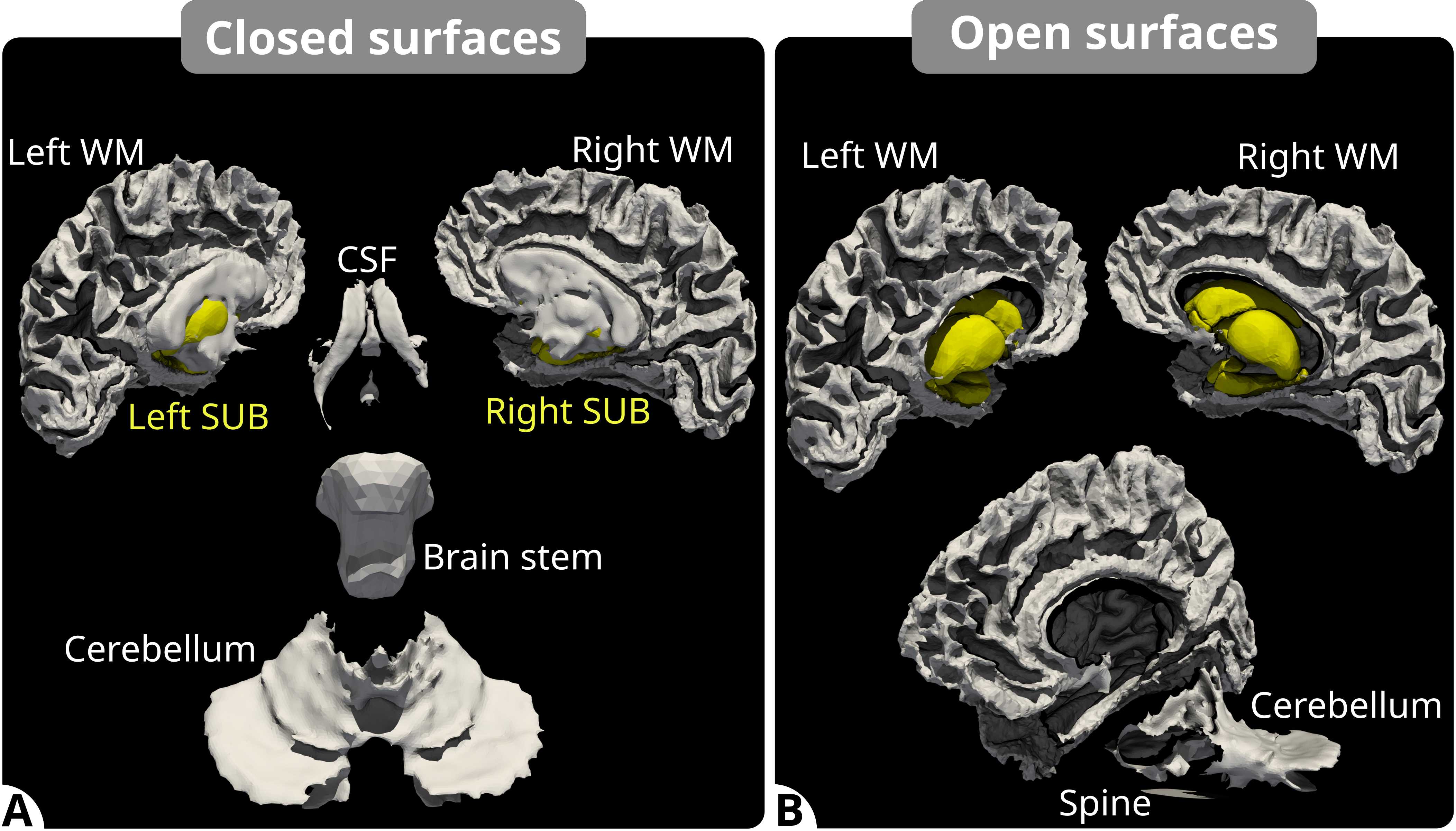
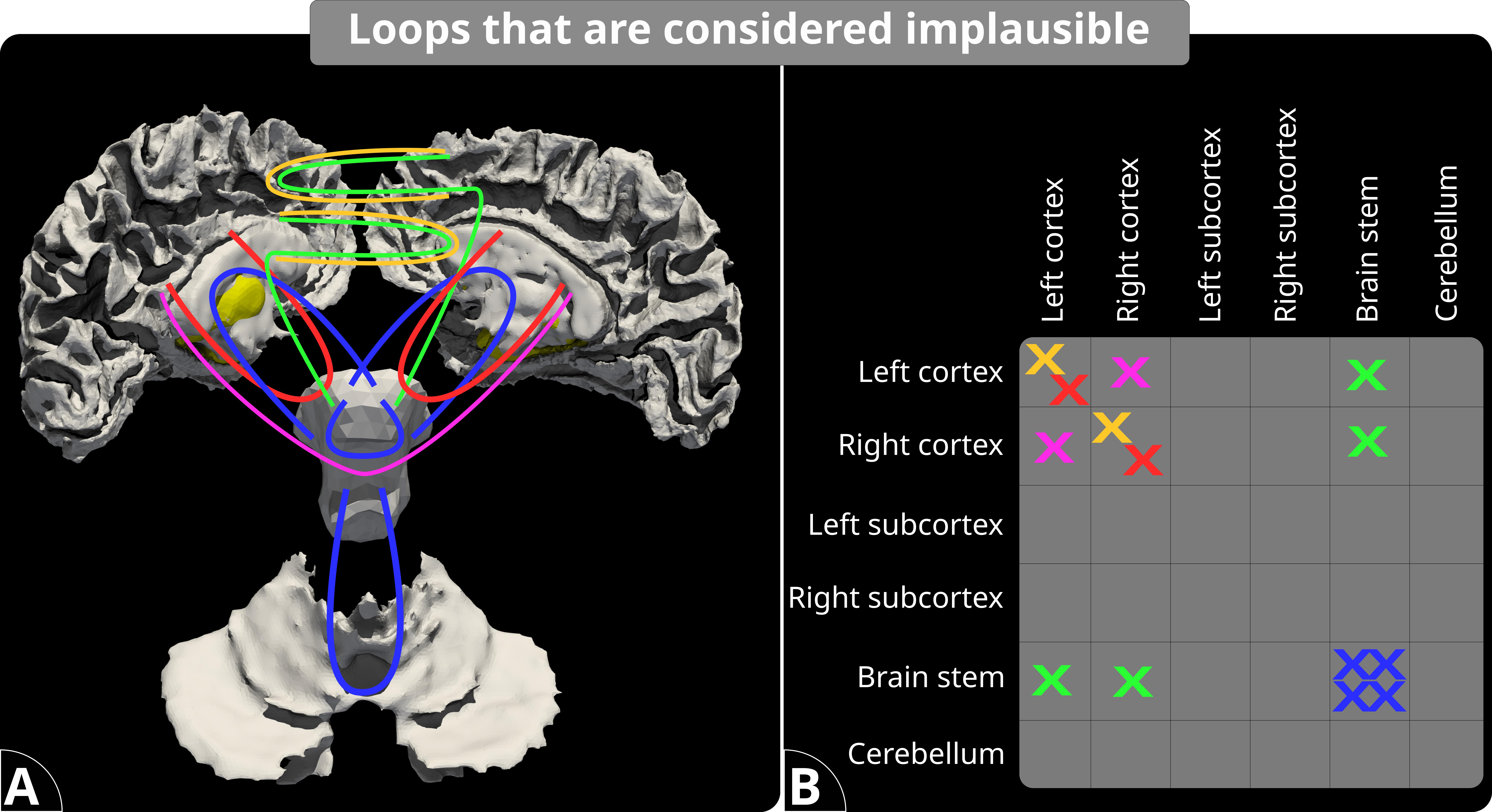
To improve the anatomical plausibility of streamlines, with HARP we implement a layered set of anatomical rules that extend the current ACT framework. To that end, we subdivide the brain into more granular regions, enabling distinction not only between the hemispheres for WM and SUB but also segmenting the CSF, cerebellum, brainstem, and spinal cord. The use of a refined segmentation not only mitigates the false positives present in ACT by eliminating anatomically implausible loops, but it also enables a more accurate neuroanatomical categorization of streamlines, splitting them into well-known association, projection, and commissural fibers.At the top layer of our hierarchy, like ACT, we make sure that streamlines do not end inside WM, and they do not enter CSF. On the second layer, using anatomical segmentation of brain regions shown in Figure 2, we classify streamlines according to their trajectory as described in Figure 3. Such categorization is exclusive, meaning that streamlines cannot be assigned to two categories simultaneously. The classification is done in an order. At the end, a streamline is either, (i) among a know class in the table, (ii) implausible, or (iii) unclassified.
Experiments
We tested HARP against standard ACT protocols using 100 million streamlines generated from the MRI data of 10 subjects from the Human Connectome Project (HCP)6. Two tractography algorithms, MRtrix3's iFOD27 and Trekker's parallel transport tractography (PTT)8, were employed to study the efficacy of HARP. Both tracking pipelines utilized ACT; however, they differed in their anatomical constraints: MRtrix3's implementation relied on partial volume fraction images, whereas Trekker's approach utilized surface mesh representations, as shown in Figure 2, to guide streamline propagation and termination, which is akin to the approach in9.Results
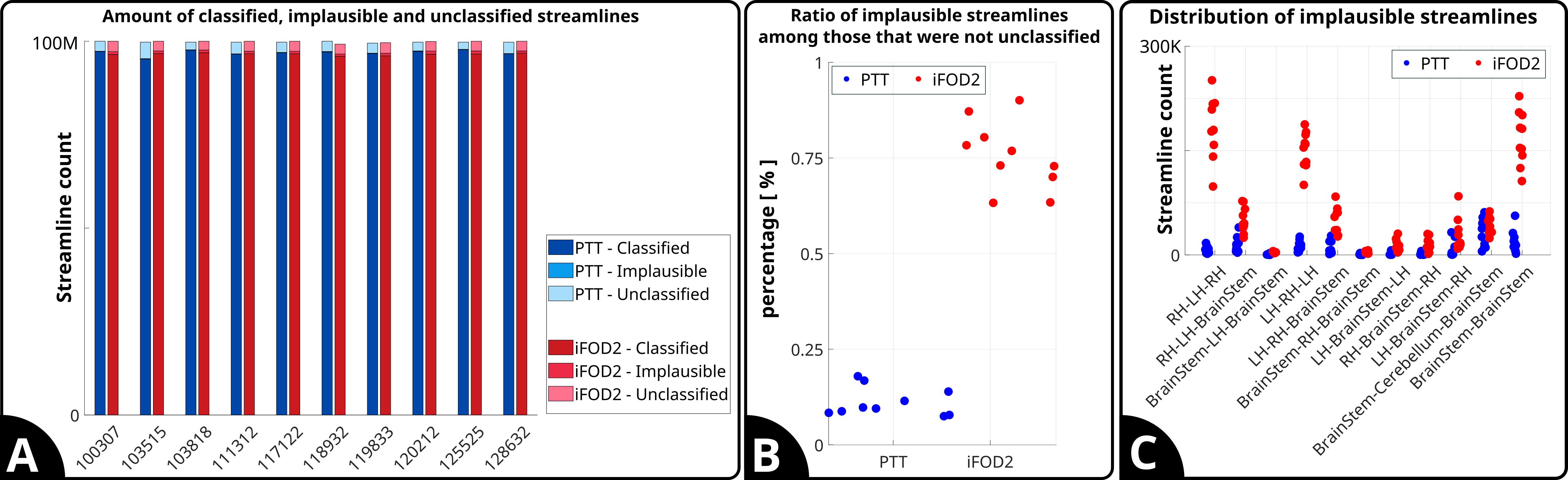
Figure 4A depicts the total counts of streamlines classified as "Classified," "Implausible," and "Unclassified" for each subject when using PTT and iFOD2. For both algorithms, the majority of streamlines are classified, with a relatively small proportion being implausible or unclassified. Panel B shows the ratio of implausible streamlines to the total number of streamlines that were not unclassified. It can be seen that PTT consistently results in a lower ratio of implausible streamlines compared to iFOD2. Panel C illustrates the distribution of implausible streamlines across different brain regions. We observe that iFOD2 yields a higher number of implausible streamlines in the cerebellum to brain stem region, whereas PTT shows a more even distribution of implausible streamlines across different regions.Discussion
Based on the loops we investigated, our results show that ACT-based whole brain tractograms contain relatively low amount implausible streamlines; however their presence is systematic, and they are accumulated in certain areas, such as in the brain stem. Taking into account the intricate and densely interconnected nature of brain's wiring, we highlight that 1% of implausible streamlines could have a disproportionate effect on the overall connectivity pattern of the brain. Therefore, there is a need to quantify the impact of these errors and explore the benefits of applying the HARP approach to remove them.Conclusion
Overall, our results demonstrate the potential of HARP to improve the anatomical plausibility of tractography by reducing false positives. However, they also underscore the need for further refinement of the hierarchical constraints and their application, particularly in complex brain regions where fiber pathways are dense and crossing, such as the cerebellum and brain stem. Future work should focus on going deeper into HARP levels and defining more refined rules to ensure the most accurate reconstruction of the brain's white matter pathways.Acknowledgements
This project has received funding from the Research Council of Finland through grants #348631and #353798. Data were provided by the Human Connectome Project, WU-Minn Consortium (Principal Investigators: David Van Essen and Kamil Ugurbil; 1U54MH091657) funded by the 16 NIH Institutes and Centers that support the NIH Blueprint for Neuroscience Research; and by the McDonnell Center for Systems Neuroscience at Washington University.References
- Thomas, C., Ye, F.Q., Irfanoglu, M.O., Modi, P., Saleem, K.S., Leopold, D.A. and Pierpaoli, C., 2014. Anatomical accuracy of brain connections derived from diffusion MRI tractography is inherently limited. Proceedings of the National Academy of Sciences, 111(46), pp.16574-16579.
- Schilling KG, Nath V, Hansen C, Parvathaneni P, Blaber J, Gao Y, Neher P, Aydogan DB, Shi Y, Ocampo-Pineda M, Schiavi S. Limits to anatomical accuracy of diffusion tractography using modern approaches. Neuroimage. 2019 Jan 15;185:1-1.
- Jbabdi, S. and Johansen-Berg, H., 2011. Tractography: where do we go from here?. Brain connectivity, 1(3), pp.169-183.
- Maffei C, Girard G, Schilling KG, Aydogan DB, Adluru N, Zhylka A, Wu Y, Mancini M, Hamamci A, Sarica A, Teillac A. Insights from the IronTract challenge: Optimal methods for mapping brain pathways from multi-shell diffusion MRI. NeuroImage. 2022 Aug 15;257:119327.
- Smith RE, Tournier JD, Calamante F, Connelly A. Anatomically-constrained tractography: improved diffusion MRI streamlines tractography through effective use of anatomical information. Neuroimage. 2012 Sep 1;62(3):1924-38.
- Van Essen DC, Smith SM, Barch DM, Behrens TE, Yacoub E, Ugurbil K, Wu-Minn HCP Consortium. The WU-Minn human connectome project: an overview. Neuroimage. 2013 Oct 15;80:62-79.
- Tournier JD, Calamante F, Connelly A. Improved probabilistic streamlines tractography by 2nd order integration over fibre orientation distributions. InProceedings of the international society for magnetic resonance in medicine 2010 May 1 (Vol. 1670). John Wiley & Sons, Inc, New Jersey.
- Aydogan DB, Shi Y. Parallel transport tractography. IEEE transactions on medical imaging. 2020 Oct 26;40(2):635-47.
- Yeh CH, Smith RE, Dhollander T, Connelly A. Mesh-based anatomically-constrained tractography for effective tracking termination and structural connectome construction. InProc ISMRM 2017 (Vol. 58).
Figures