2156
Multi-modal multi-scale imaging reveals that long-association cortico-cortical systems are composed of short-range relay fibers1Athinoula A. Martinos Center for Biomedical Imaging, Massachusetts General Hospital and Harvard Medical School, Charlestown, MA, United States, 2Department of Pharmacology & Physiology, University of Rochester School of Medicine and Dentistry, Rochester, New York; Department of Psychiatry, McLean Hospital, Harvard Medical School, Belmont, MA, United States
Synopsis
Keywords: Tractography, White Matter, Structural Connectivity, Fiber Pathways
Motivation: Obtaining accurate anatomical information of connectional neuroanatomy across scales is crucial to improve in-vivodiffusion MRI techniques and advance our understanding of the brain white matter circuitries.
Goal(s): To reveal the mesoscopic organization of the SLF-I, a major cortico-cortical fiber association system of the human brain.
Approach: We combine multi-scale, multi-species, multi-modality connectional data from humans and macaques to investigate the structural connectivity of medial fronto-parietal cortical regions.
Results: We provide preliminary novel evidence that the SLF-I is composed of a succession of shorter relay fibers, which, in lower-resolution dMRI tractography result erroneously in a long, direct association bundle.
Impact: The mesoscopic anatomical validation of major white matter pathways in the human brain will increase the accuracy of their reconstruction in vivo, and open new avenues for our understanding of the functional substrates of these different connections.
Introduction
The current gap in spatial resolution between diffusion MRI (mm) and axons (μm) results in errors of dMRI tractography in regions with challenging fiber configurations1. Ex-vivo imaging allows us to obtain accurate anatomical information at higher resolution that can inform in vivo dMRI tractography2. Here we combine data across multiple modalities, scales, and species to clarify the connectivity of the dorsal branch of the superior longitudinal fasciculus (SLF-I), a major fiber association system running within the white matter of the superior frontal gyrus (SFG). Anatomic tracing studies in monkeys described the SLF-I as connecting the postero-medial parietal regions (PGm, PE, PEc) to different regions of the superior frontal gyrus (SFG) (6D, 8B, 9)3. Due to the complexity of SFG, where multiple projection systems merge with superficial U-fibers and frontal projections of the cingulum bundle, the morphology of the human SLF-I remains controversial. Tractography and post-mortem dissections have yielded conflicting results, some supporting direct, long connections from the parietal to the more frontal regions, and others supporting shorter or no SLF-I fibers4,5. Here, we combine multi-scale, multi-species, multi-modality data to investigate the mesoscopic organization within the SLF-I fiber system.Methods
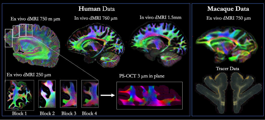
The different datasets used in this work are presented in Figure 1 and acquisition details are specified below.Human data:
1) In vivo 1.5 mm dMRI: 3T MGH Connectom 1.0 Scanner, 2D EPI, 552 dMRI volumes (40 b=0, 64 b = 1000, 64 b = 3000; 128 b = 5000; 256 b = 10000 s/mm2)6.
2) In vivo 760 μm dMRI: 3T MGH Connectom 1.0 Scanner, gSlider-SMS, 2808 dMRI volumes (144 b=0, 420 b=1000, 840 b=2500 s/mm2, paired reversed PE volumes)7.
3) Ex vivo 750 μm dMRI: 3T Siemens Trio scanner, 3D diffusion-weighted SSFP, 68 dMRI volumes (TR=30.21 ms, TE=25.12 ms, 8 b=0, 60 b=4,000 s/mm2).
4) Ex vivo 250 μm dMRI: 4 small blocks (roughly 2x2x1cm) were cut from dataset 3 and scanned on a 9.4T Bruker Biospec System (3D EPI, TR=75 ms, TE=43 ms, GRAPPA = 2, 515 dMRI volumes, maximum b = 40,000 s/mm2, 48 h).
5) PS-OCT 3 μm in plane: polarization-sensitive optical coherence tomography (PS-OCT) data were acquired on a system developed in-house [8].
Macaque data:
1) Ex vivo 700 μm dMRI NHP data: Small-bore 4.7T Bruker BioSpin scanner, 3D EPI sequence, 48h. 514 dMRI volumes (maximum b = 40, 000 s/mm2)1.
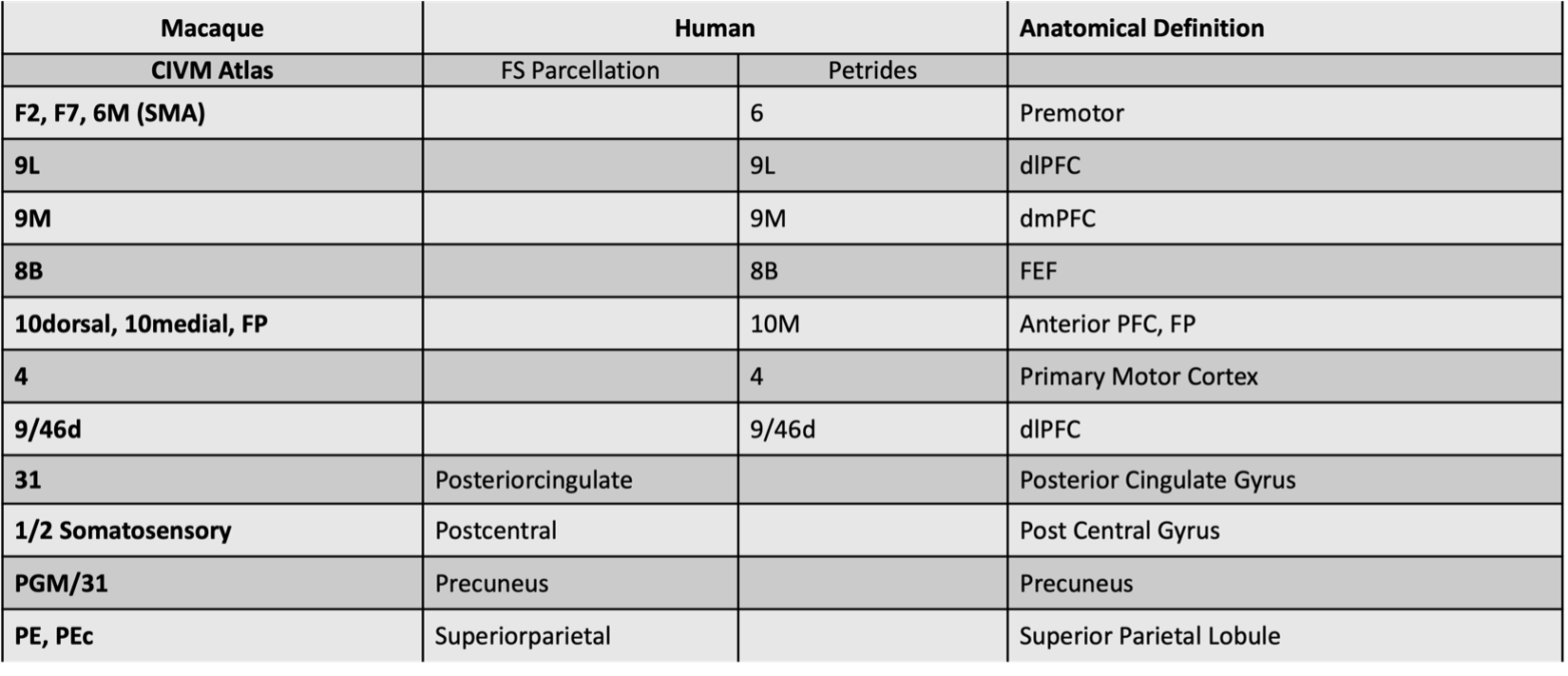
2) Tracer data: one male rhesus macaque received an injection in the frontal pole (10M), as described previously9. Pre-processing: All dMRI data were denoised using MP-PCA in MRtrix310 and corrected for motion/eddy current distortions in FSL10. We created a common parcellation scheme for macaque and human data to maximize anatomical comparison (Table 1). For each dataset, fiber orientation distributions were estimated using constrained spherical deconvolution11 and connectivity matrices were generated (angle threshold:45º, step-size: 0.5 x voxel size) in MRtrix312. PS-OCT data were processed as previously described13 and tractography was performed using an in-house algorithm developed in Julia.
Results
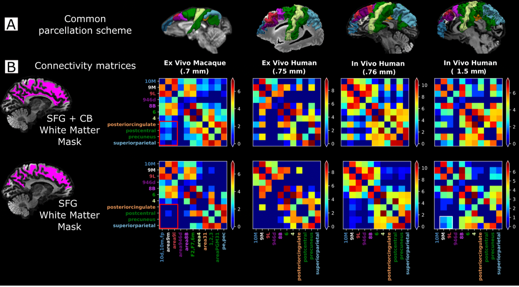
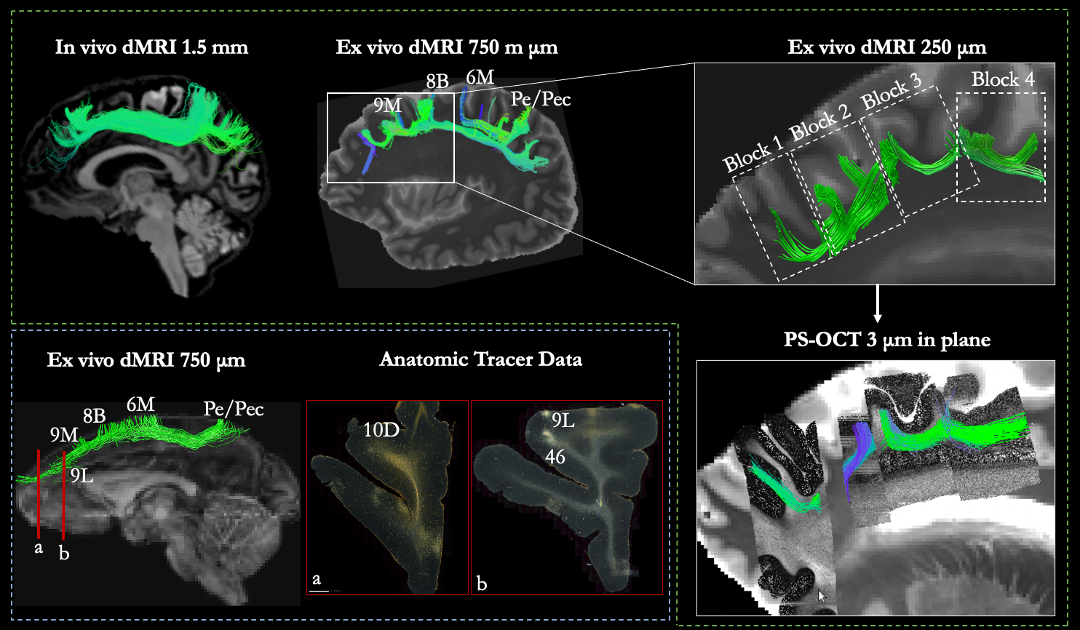
Results show that most of the long, direct connections between parietal and frontal regions course within the cingulum bundle (white rectangles in figure 2), in accordance with the literature14. White-matter fibers originating in the parietal regions and coursing withing the SFG white matter mainly terminate in areas 4, 6, and 8B, as previously reported2. The number of direct, long connections coursing within the SFG white matter increases in lower resolution data (white square, figure 1). We qualitatively compared these long-range tractography reconstructions obtained from in vivo lower resolution dMRI data with four higher-resolution datasets: 1) virtual dissections of fibers coursing between the same areas obtained from high-resolution ex vivo dMRI data (750 and 250 μm); virtual dissections of fibers obtained from PS-OCT orientation data; virtual dissections of fibers in the ex vivo dMRI macaque data; anatomic tracer data in macaque (Figure 3). We found that all the higher-resolution datasets provide complementary evidence to support that the medial SFG has mainly short-range connections that come in and out the cortex, rather than long-range direct connections.Conclusion
By comparing data across multiple modalities, scales, and species, we provide preliminary novel evidence that the SLF-I is composed of a succession of shorter relay fibers, which, in lower-resolution dMRI tractography result erroneously in a long, direct association bundle. These preliminary results point to the need to move beyond the conceptualization of white matter bundles as monolithic structures connecting wide portions of cortex and investigate the connectivity of the human brain at a finer scale. The delineation and characterization of these pathways will advance our understanding of the functional substrates of the brain connectome.Acknowledgements
The research conducted in this work was supported by the National Institute of Biomedical Imaging and Bioengineering (R01-EB021265) and the National Institute of Neurological Disorders and Stroke (R01-NS119911, UM1NS132358-01). Additional support was provided by the National Institute of Mental Health (R01-MH045573, P50-MH106435).References
1. Maffei C, Girard G, Schilling KG, […], Yendiki A. Insights from the IronTract challenge: Optimal methods for mapping brain pathways from multi-shell diffusion MRI. Neuroimage. 2022 Aug 15;257:119327.
2. Yendiki A, Aggarwal M, Axer M, Howard AFD, van Walsum AVC, Haber SN. Post mortem mapping of connectional anatomy for the validation of diffusion MRI. Neuroimage. 2022 Aug 1;256:119146.
3. Petrides M, Pandya DN. Projections to the frontal cortex from the posterior parietal region in the rhesus monkey. J Comp Neurol. 1984 Sep 1;228(1):105-16.
4. Kamali A, Flanders AE, Brody J, Hunter JV, Hasan KM. Tracing superior longitudinal fasciculus connectivity in the human brain using high resolution diffusion tensor tractography. Brain Struct Funct. 2014 Jan;219(1):269-81.
5. Wang X, Pathak S, Stefaneanu L, Yeh FC, Li S, Fernandez-Miranda JC. Subcomponents and connectivity of the superior longitudinal fasciculus in the human brain. Brain Struct Funct. 2016 May;221(4):2075-92.
6. Fan Q, Witzel T, Nummenmaa A, Van Dijk KRA, Van Horn JD, Drews MK, Somerville LH, Sheridan MA, Santillana RM, Snyder J, Hedden T, Shaw EE, Hollinshead MO, Renvall V, Zanzonico R, Keil B, Cauley S, Polimeni JR, Tisdall D, Buckner RL, Wedeen VJ, Wald LL, Toga AW, Rosen BR. MGH-USC Human Connectome Project datasets with ultra-high b-value diffusion MRI. Neuroimage. 2016 Jan 1;124(Pt B):1108-1114.
7. Wang, F. et al. Motion‐robust sub‐millimeter isotropic diffusion imaging through motion corrected generalized slice dithered enhanced resolution (MC-gSlider) acquisition. Magn. Reson. Med. 80, 1891–1906 (2018).
8. Wang H, Akkin T, Magnain C, Wang R, Dubb J, Kostis WJ, Yaseen MA, Cramer A, Sakad ž i ć S, Boas D, 2016. Polarization sensitive optical coherence microscopy for brain imaging. Opt. Lett 41 (10), 2213–2216.
9. Lehman JF, Greenberg BD, McIntyre CC, Rasmussen SA, Haber SN, 2011. Rules ventral prefrontal cortical axons use to reach their targets: implications for diffusion tensor imaging tractography and deep brain stimulation for psychiatric illness. J. Neurosci. 31, 10392–10402.
10. Andersson, J. L., Skare, S., & Ashburner, J. (2003). How to correct susceptibility distortions in spin-echo echo-planar images: application to diffusion tensor imaging. Neuroimage, 20(2), 870-888.
11. Daan Christiaens, Ben Jeurissen, Chun-Hung Yeh, Alan Connelly. MRtrix3: A fast, flexible and open software framework for medical image processing and visualisation, NeuroImage,2019, 202; 116137.
12. Tournier, J.-D, Calamante, F. and Connelly, A. Improved probabilistic streamlines tractography by 2nd order integration over fibre orientation distributions. Proceedings of the International Society for Magnetic Resonance in Medicine, 2010, 1670.
13. Jones R, Maffei C, Augustinack J, Fischl B, Wang H, Bilgic B, Yendiki A. High-fidelity approximation of grid- and shell-based sampling schemes from undersampled DSI using compressed sensing: Post mortem validation. Neuroimage. 2021 Dec 1;244:118621.
14. Heilbronner SR, Haber SN. Frontal cortical and subcortical projections provide a basis for segmenting the cingulum bundle: implications for neuroimaging and psychiatric disorders. J Neurosci. 2014 Jul 23;34(30):10041-54.
15. Calabrese E, Badea A, Coe CL, Lubach GR, Shi Y, Styner MA, Johnson A (2015) "A diffusion tensor MRI atlas of the postmortem rhesus macaque brain" Neuroimage 117:408–416[16 ] Tang W, Jbabdi S, Zhu Z, Cottaar M, Grisot G, Lehman JF, Yendiki A, Haber SN, 2019. A connectional hub in the rostral anterior cingulate cortex links areas of emotion and cognitive control.
Figures