2155
Creation of a cross-population white matter atlas for concurrent mapping of brain connections between Eastern and Western populations1University of Electronic Science and Technology of China, Chengdu, China, 2Nanjing University of Science and Technology, Nanjing, China, 3West China Hospital, Sichuan University, Chengdu, China, 4Harvard Medical School, Boston, MA, United States, 5Boston University, Boston, MA, United States
Synopsis
Keywords: Tractography, White Matter, diffusion MRI, fiber clustering, tractography parcellation
Motivation: Existing white matter atlases are usually created based on a certain population, which may omit subtle differences across populations from different cultures.
Goal(s): This study presents a fine-scale white matter atlas that is created concurrently using high-quality diffusion MRI data from both Eastern and Western populations.
Approach: The curated atlas includes a cluster-level parcellation of 800 fiber clusters from the entire brain and an anatomical tract parcellation of 53 major long-range white matter connections.
Results: Comparative assessment between the two populations within the atlas shows highly visually similar white matter geometry but significant differences when measuring streamline counts in the fiber parcels.
Impact: We propose a diffusion MRI tractography atlas that enables concurrent white matter parcellation across Eastern and Western populations. While the white matter geometry is visually similar, the number of streamlines in the fiber parcels differs significantly between the two populations.
INTRODUCTION
The study of brain differences across Eastern and Western populations using neuroimaging provides important insights for understanding potential cultural and genetic influences on the process of cognition and mental health[1–3]. Brain MRI atlases serve as essential tools for standardizing anatomical references in the assessment of brain structure and function across different populations[4–6], and have been used to measure variations in brain morphology, cortical thickness, and functional connectivity between Eastern and Western people[1–3,7–11]. Yet, there has been no comprehensive investigation into the comparison of white matter (WM) tracts between Eastern and Western populations.This study presents a diffusion MRI (dMRI) tractography atlas that enables concurrent mapping of brain WM connections across Eastern and Western people. The atlas is created concurrently using the high-quality dMRI data provided in the Human Connectome Project (HCP)[12] and the Chinese Human Connectome Project (CHCP)[13], where each parcel includes streamlines from both populations. We assess population-wise differences between these two cohorts, leveraging the HCP and CHCP components within the curated atlas.
METHODS
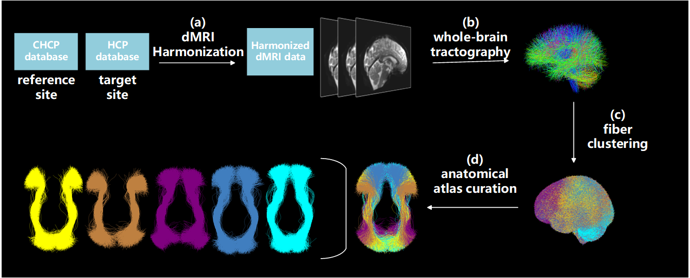
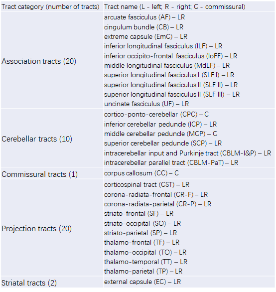
Dataset: The HCP dataset includes dMRI data acquired from healthy adults living in the USA, and the CHCP dataset includes data from healthy adults living in China. We selected 153 subjects from each dataset with matched age and sex distributions (HCP: 24.2±1.4 years, 72 females and 81 males; CHCP: 23.9±2.4 years, 68 females and 85 males) to create the atlas.Atlas creation: Figure-1 gives the method overview. First, dMRI Harmonization is performed to remove the inter-site variability resulting from scanner differences while preserving inter-subject variability. We use our novel machine learning algorithm[14] to reconcile the raw dMRI signals across the two datasets. Second, whole-brain tractography is performed to reconstruct WM fibers from the entire brain. We use our advanced two-tensor Unscented Kalman filter (UKF) algorithm[15,16], which accounts for crossing fibers and offers sensitive and reliable fiber tracking within a wide range of populations[17–19]. Third, fiber clustering is performed to subdivide the tractography into fine-scale parcels. We use our well-established spectral clustering pipeline for simultaneous tractography streamline clustering across all HCP and CHCP subjects[20,21]. This generates a fiber clustering atlas including 800 fiber clusters, with a total of 2.4 million streamlines. Last, anatomical atlas curation is performed to annotate each fiber cluster with an anatomical label. We use our ORG atlas built only using the HCP data as a reference[18,22]. We calculate the distances between the clusters in the new atlas and those in the ORG atlas and then assign each new atlas cluster with the label of the closest ORG cluster. In total, our curated atlas contains an anatomical tract parcellation including 53 major anatomical WM tracts (Table-1).
Comparison between HCP and CHCP: Our atlas is created using the HCP and CHCP datasets concurrently so that each cluster includes streamlines from both populations. This enables an unbiased comparison of the corresponding WM structures between the populations. We separate each cluster and each tract in the atlas into two components according to the origin of the streamlines (HCP or CHCP). For each cluster and tract, we measure the number of streamlines and perform t-tests (with an FDR correction) to assess between-population differences.
RESULTS
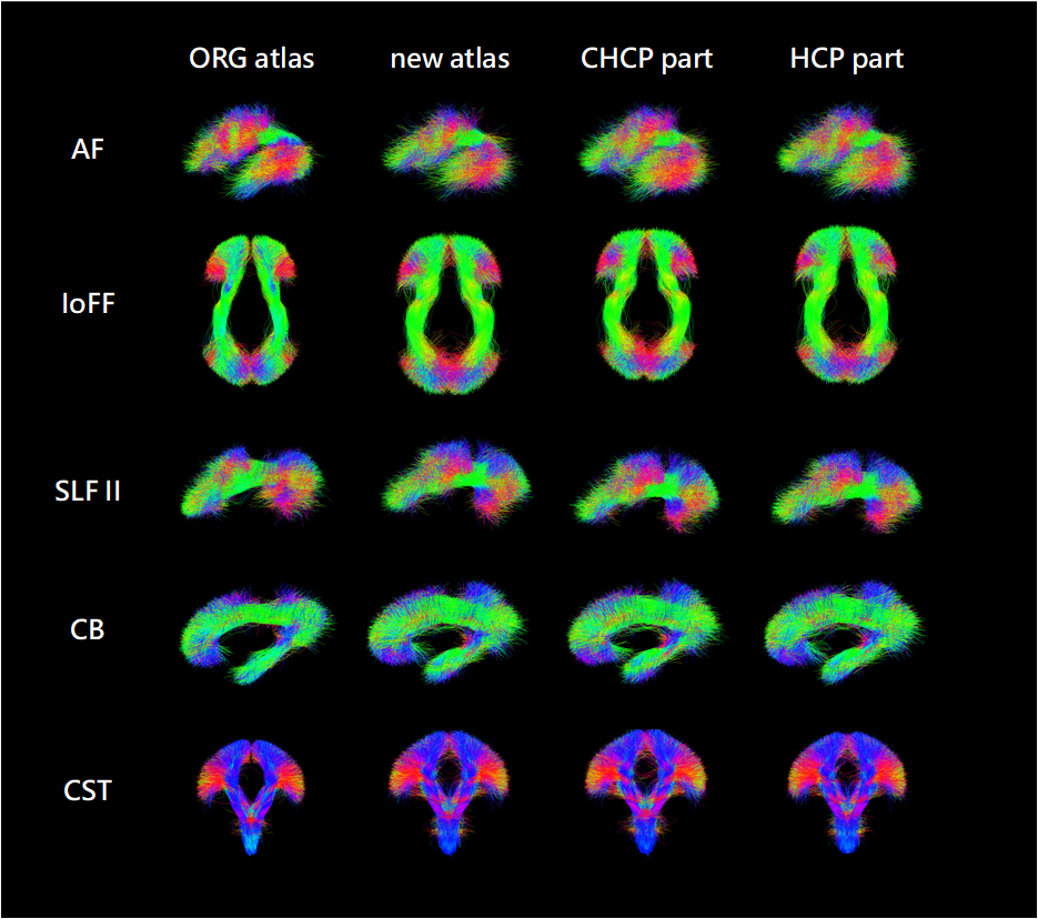
Figure-2 gives a visualization of example anatomical tracts in the proposed atlas, as well as those belonging to the HCP and CHCP components, with a comparison to the ORG atlas. Overall, the shape of these anatomical tracts across the new atlas, the ORG atlas, the HCP component, and the CHP component are highly visually similar. In addition, we quantify the existence of streamlines belonging to a certain population and find that 100% of fiber clusters comprised streamlines from both populations. These results show a generally comparable WM parcellation between the HCP and CHCP populations.For the cluster-level and tract-level comparison results, we find a large number of clusters and tracts with significant differences between the two populations. Across the 800 fiber clusters, 454 clusters (56.75%) have a significantly different number of streamlines, where 201 clusters in HCP have more streamlines and 253 clusters in CHCP have more streamlines. Among the 53 fiber tracts, 30 tracts (56.60%) have a significantly different number of streamlines, where 19 tracts in HCP have more streamlines and 11 tracts in CHCP have more streamlines.
DISCUSSION & CONCLUSION
This study presents a novel cross-population WM atlas for concurrent mapping of brain connections between Eastern and Western people. We perform a comparative assessment of the WM connections and identify widespread differences between the two populations in terms of streamline counts.Acknowledgements
This work is in part supported by the National Natural Science Foundation of China (No. 62371107) and the National Institutes of Health (R01MH125860, R01MH119222, R01MH132610, R01NS125781).References
1. Han, S. & Ma, Y. Cultural differences in human brain activity: a quantitative meta-analysis. Neuroimage 99, 293–300 (2014).2. Ge, J. et al. Cross-language differences in the brain network subserving intelligible speech. Proc. Natl. Acad. Sci. U. S. A. 112, 2972–2977 (2015).
3. Gao, T., Han, X., Bang, D. & Han, S. Cultural differences in neurocognitive mechanisms underlying believing. Neuroimage 250, 118954 (2022).
4. Laird, A. R. et al. ALE Meta-Analysis Workflows Via the Brainmap Database: Progress Towards A Probabilistic Functional Brain Atlas. Front. Neuroinform. 3, 23 (2009).
5. Mazziotta, J. et al. A probabilistic atlas and reference system for the human brain: International Consortium for Brain Mapping (ICBM). Philos. Trans. R. Soc. Lond. B Biol. Sci. 356, 1293–1322 (2001).
6. Collins, D. L., Neelin, P., Peters, T. M. & Evans, A. C. Automatic 3D intersubject registration of MR volumetric data in standardized Talairach space. J. Comput. Assist. Tomogr. 18, 192–205 (1994).
7. Tang, Y. et al. The construction of a Chinese MRI brain atlas: A morphometric comparison study between Chinese and Caucasian cohorts. Neuroimage 51, 33–41 (2010).
8. Yang, G. et al. Sample sizes and population differences in brain template construction. Neuroimage 206, 116318 (2020).
9. Kochunov, P. et al. Localized morphological brain differences between English-speaking Caucasians and Chinese-speaking Asians: new evidence of anatomical plasticity. Neuroreport 14, 961–964 (2003).
10. Kang, D. W. et al. Differences in cortical structure between cognitively normal East Asian and Caucasian older adults: a surface-based morphometry study. Sci. Rep. 10, 20905 (2020).
11. Wei, X. et al. Native language differences in the structural connectome of the human brain. Neuroimage 270, 119955 (2023).
12. Elam, J. S. et al. The Human Connectome Project: A retrospective. Neuroimage 244, 118543 (2021).
13. Ge, J. et al. Increasing diversity in connectomics with the Chinese Human Connectome Project. Nat. Neurosci. 26, 163–172 (2023).
14. Cetin Karayumak, S. et al. Retrospective harmonization of multi-site diffusion MRI data acquired with different acquisition parameters. Neuroimage 184, 180–200 (2019).
15. Malcolm, J. G., Shenton, M. E. & Rathi, Y. Filtered multitensor tractography. IEEE Trans. Med. Imaging 29, 1664–1675 (2010).
16. Reddy, C. P. & Rathi, Y. Joint Multi-Fiber NODDI Parameter Estimation and Tractography Using the Unscented Information Filter. Front. Neurosci. 10, 166 (2016).
17. Gong, S. et al. Free water modeling of peritumoral edema using multi-fiber tractography: Application to tracking the arcuate fasciculus for neurosurgical planning. PLoS One 13, e0197056 (2018).
18. Zhang, F. et al. An anatomically curated fiber clustering white matter atlas for consistent white matter tract parcellation across the lifespan. Neuroimage 179, 429–447 (2018).
19. Zhang, F. et al. Deep white matter analysis (DeepWMA): Fast and consistent tractography segmentation. Med. Image Anal. 65, 101761 (2020).
20. O’Donnell, L. J. & Westin, C.-F. Automatic tractography segmentation using a high-dimensional white matter atlas. IEEE Trans. Med. Imaging 26, 1562–1575 (2007).
21. O’Donnell, L. J., Wells, W. M., III, Golby, A. J. & Westin, C.-F. Unbiased Groupwise Registration of White Matter Tractography. in Medical Image Computing and Computer-Assisted Intervention – MICCAI 2012 (eds. Ayache, N., Delingette, H., Golland, P. & Mori, K.) 123–130 (Springer Berlin Heidelberg, 2012).
22. Zhang, F. et al. Test-retest reproducibility of white matter parcellation using diffusion MRI tractography fiber clustering. Hum. Brain Mapp. 40, 3041–3057 (2019).
Figures