2154
Evaluation of diffusion MRI methods and fiber tracking algorithms for optimal imaging of the anterior visual pathway1Montreal Neurological Institute, McGill University, Montreal, QC, Canada, 2Department of Ophthalmology, McGill University, Montreal, QC, Canada
Synopsis
Keywords: Diffusion Acquisition, Diffusion/other diffusion imaging techniques, Anterior Visual Pathway
Motivation: Diffusion-weighted MRI of the anterior-visual-pathway (AVP) is challenging due to the small diameter of the optic nerve and susceptibility artifacts.
Goal(s): To compare the performance of diffusion MRI acquisition, tractography, reconstruction, and fiber-tracking methods in reconstructing the AVP.
Approach: The overlap between a mask based on track-density and the T1-weighted imaged AVP was used to evaluate the accuracy of the reconstruction.
Results: Readout segmented EPI (RESOLVE) and the combined use of Constant Solid Angle (CSA) reconstruction with Parallel Transport Tractography (PTT) scored best.
Impact: Our findings support an optimal selection of methods for imaging the AVP in clinical research and radiology clinics.
Introduction
The anterior visual pathway (AVP) plays a crucial role in transmitting visual information from the retina to the lateral geniculate nucleus. It consists of the optic nerve, optic chiasm, and optic tract. However, performing diffusion-weighted MRI (dMRI) for the AVP presents complexities. These challenges arise from factors such as the narrow dimensions of the optic nerve and optic tract, subject motion artifacts, the impact of surrounding tissues and Cerebro-Spinal-Fluid, causing partial volume effects, and magnetic susceptibility at the optic canal. This study's primary objective is to assess six different dMRI protocols, aiming to pinpoint the most effective sequence for precise diffusion data related to the AVP. Alongside comparing diffusion sequences, we systematically evaluate various tractography reconstruction and fiber tracking methods, considering parameters like angle and step size. Our goal is to identify the parameter combination that aligns best with anatomical data. This analysis identifies which dMRI protocol and associated tractography parameters are most proficient in reconstructing the AVP.Methods
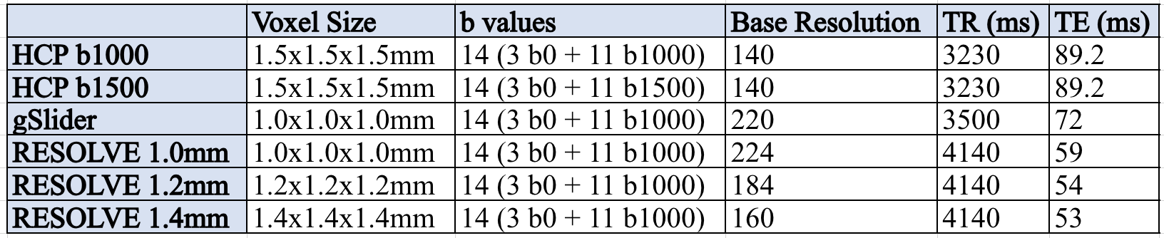
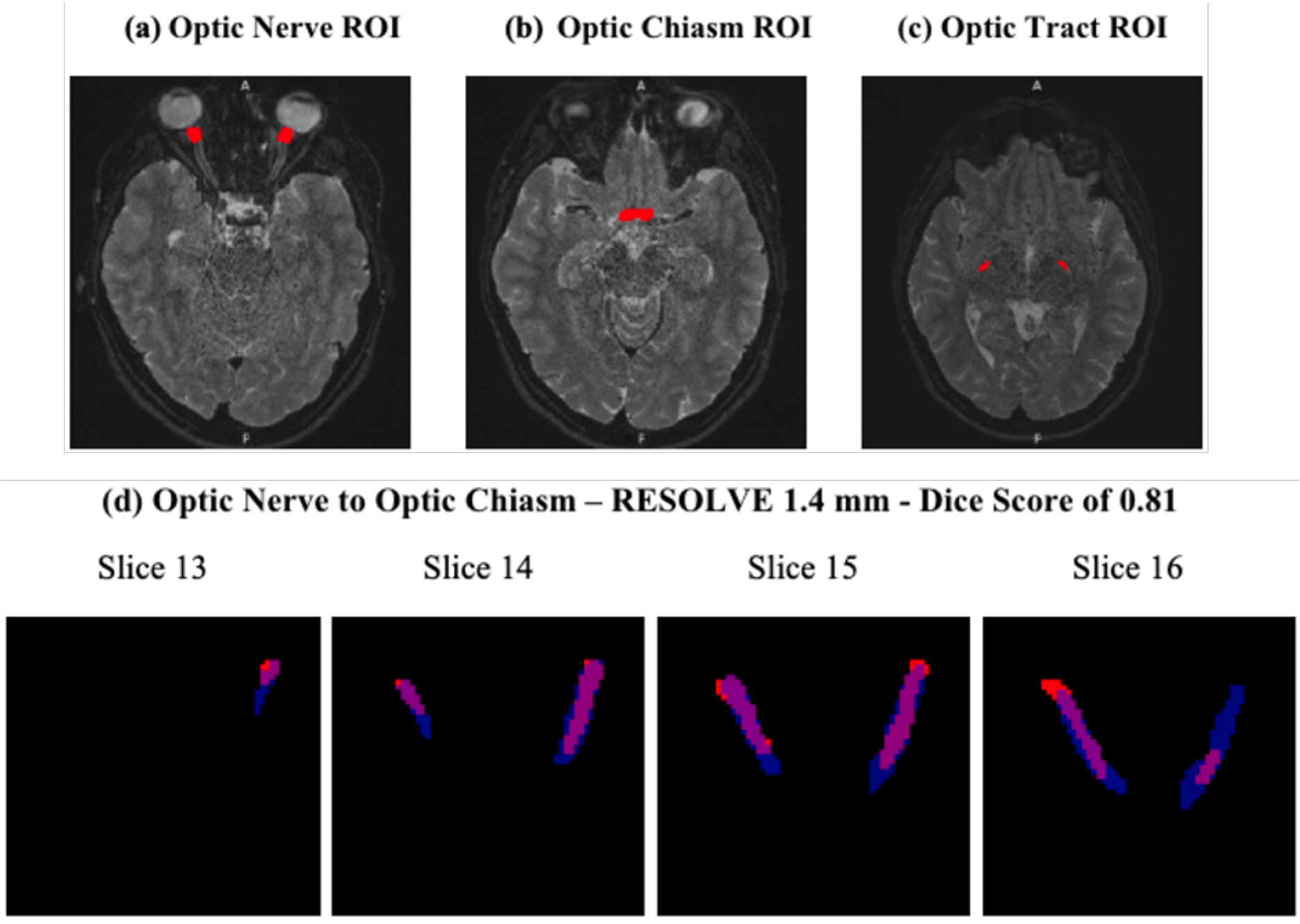
Data from 14 participants were collected. The study includes two dMRI sequences resembling those used in the Human Connectome Project (HCP) with b-values of 1000 and 1500, a high-resolution gSlider sequence, and three readout segmented EPI (RESOLVE) sequences with voxel sizes of 1.0 mm, 1.2 mm, and 1.4 mm. Additionally, each participant underwent a structural T1-weighted scan.For localized tractography of the AVP, we manually delineated specific ROI points at anatomical locations, such as the optic nerves (ON), optic chiasm (OC), and optic tracts (OT), for each participant. We also manually created ground truth masks encompassing the optic nerves and optic tracts.
To evaluate the efficacy of diffusion sequences, we compared the AVP tracks generated from these sequences with anatomical ground truth data, utilizing Dice scores for this comparison. We aimed to optimize tractography parameters for AVP reconstruction by varying the angle and step size of the streamlines. Angle sets the maximum deviation of each streamline in its direction of propagation, and step size determines the maximum propagation length of each streamline. Furthermore, various reconstruction methods for estimating the Fiber Orientation Distribution Function (fODF) from a single fiber white matter population signal estimate were compared. These methods included Constant Solid Angle (CSA), Constrained Spherical Deconvolution (CSD), Robust and Unbiased Model-Based Spherical Deconvolution (RUMBA), and Sparse Fascicle Model (SFM). To perform tractography, we employed an array of fiber tracking methods such as EuDX, Probabilistic Tracking (PT), Deterministic Tracking (DT), Particle Filter Tracking (PFT), and Parallel Transport Tractography (PTT). By utilizing these tracking methods along with different seed points and endpoints, we conducted tractography for distinct AVP regions.
Results
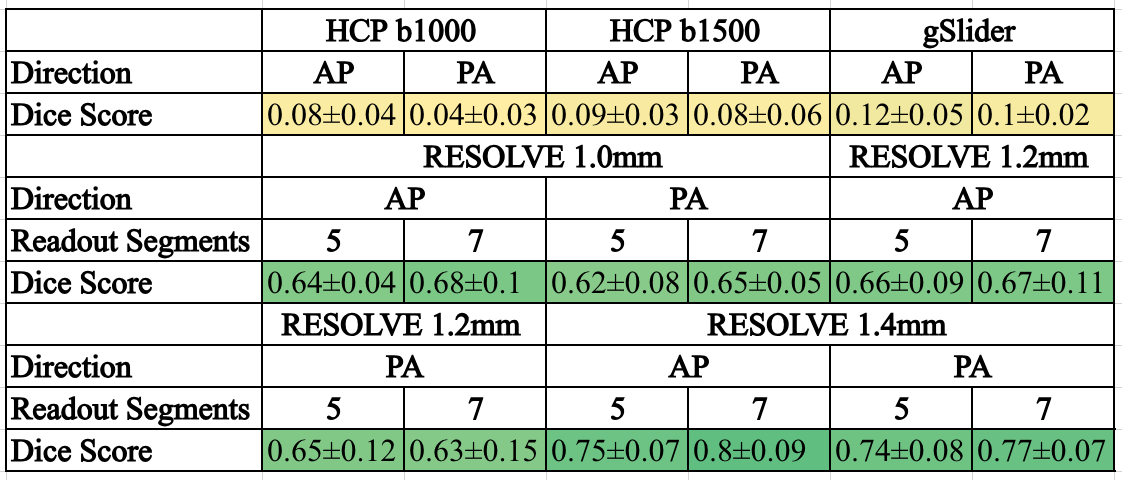
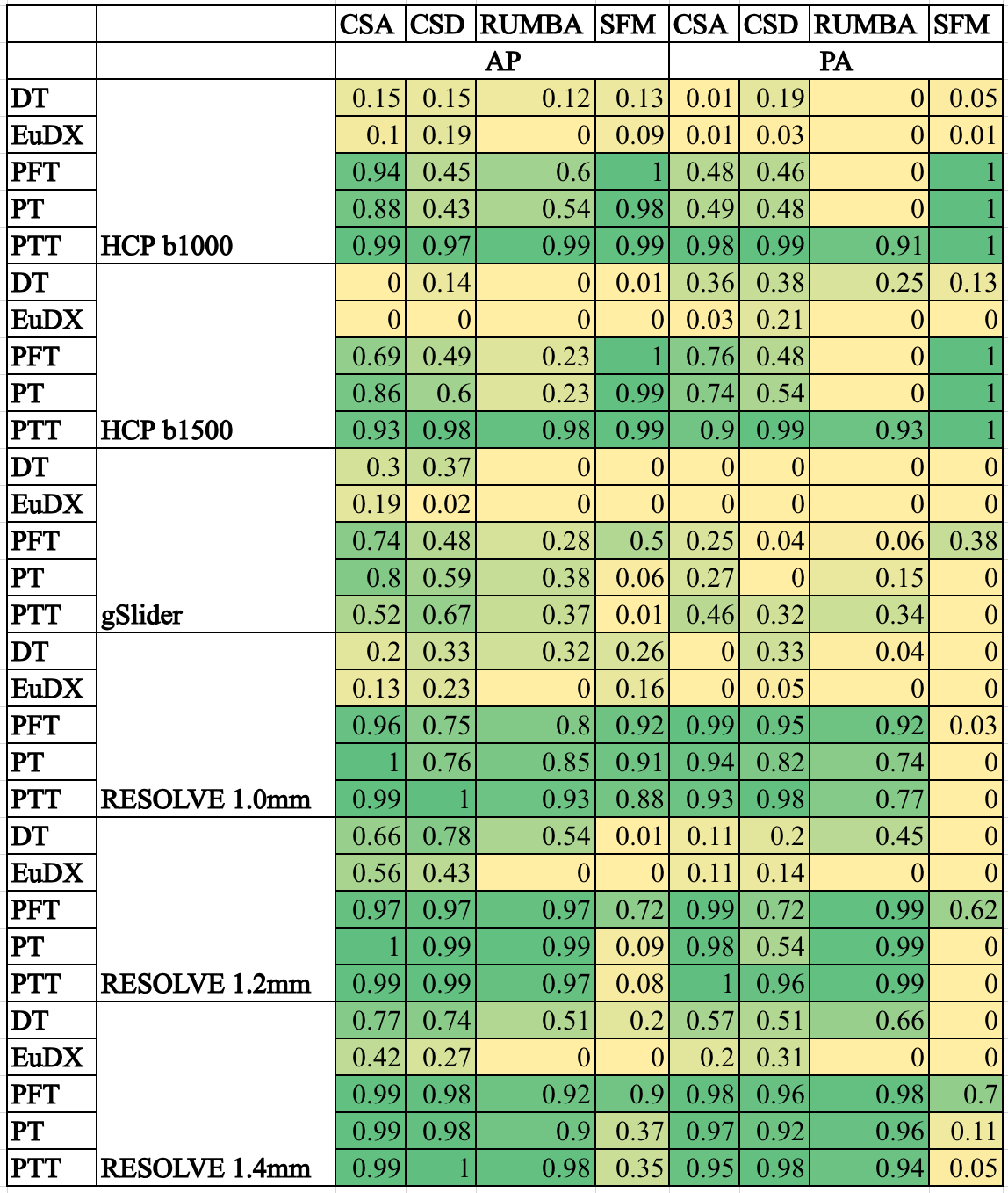
We individually assessed reconstruction of the optic nerve and optic tract segments, along with the entire AVP segment. Dice scores were calculated to compare track density maps with structural anatomy masks. This analysis encompassed all six diffusion sequences and their respective parameters.The results presented in Table 2 highlighted the consistent and robust performance of the three RESOLVE acquisitions. They demonstrated reliable tract density overlap. Conversely, the gSlider sequence and the HCP-like diffusion sequences with b-values of 1000 and 1500 failed to generate the expected tracts, underscoring the superiority of the RESOLVE sequences. Notably, the AP direction with 7 readout segments consistently yielded higher track density overlap compared to the PA direction for all three RESOLVE sequences.
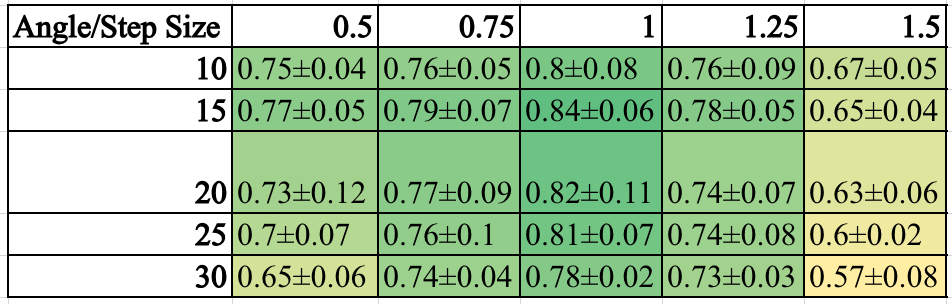
Furthermore, Table 3 established that the optimal parameters for reconstructing the AVP are a maximum angle of 15 degrees and a step size of 1.0 mm. Lastly, our final analysis involved comparing Dice scores for different reconstruction algorithms and fiber tracking methods across all six dMRI sequences within the context of the AVP segment.
Discussion
Our findings indicate that among the methods we tested, the RESOLVE sequence, with 1.4 mm isotropic voxels acquired in the AP direction with seven readout segments, stands out as the most effective choice for AVP diffusion MRI. This configuration offers the optimal track overlap with the anatomical data, making it the preferred option for AVP reconstruction. Additionally, a maximum angle of 15 degrees and a step size of 1.0 mm demonstrate superior performance among the tractography parameters, yielding relatively higher Dice scores.In terms of reconstruction methods, CSA and CSD display high Dice scores, with CSA consistently achieving higher Dice scores. Particle filter tracking (PFT), which is a probabilistic tractography method and parallel transport tractography (PTT) outperform their counterparts, with PTT proving more consistent and superior in terms of dice scores.
In future studies, we plan to use these methods to compare diffusion parameters in the AVP between patients with optic neuropathy and healthy controls.
Acknowledgements
No acknowledgement found.References
- He, J et. al. (2021). Comparison of multiple tractography methods for reconstruction of the retinogeniculate visual pathway using diffusion MRI. Human brain mapping, 42(12), 3887-3904.
- Seeger, A. et. al. (2018). Advanced diffusion-weighted imaging in patients with optic neuritis deficit–value of reduced field of view DWI and readout-segmented DWI. The Neuroradiology Journal, 31(2), 126-132.
- Zhou, F et. al. (2022). Reproducibility and feasibility of optic nerve diffusion MRI techniques: single-shot echoplanar imaging (EPI), readout-segmented EPI, and reduced field-of-view diffusion-weighted imaging. BMC Medical Imaging, 22(1), 1-11.
Figures