2150
Exploring the precision of robust modelling methods for diffusion-weighted MRI1Radiology, Hämeenlinna Central Hospital, Hämeenlinna, Finland, 2Baby Brain Activity Center, Children’s Hospital, Helsinki University Hospital and University of Helsinki, Helsinki, Finland, 3Wellcome Centre for Integrative Neuroimaging, FMRIB, Nuffield Department of Clinical Neurosciences, University of Oxford, United Kingdom, Oxford, United Kingdom
Synopsis
Keywords: Diffusion Modeling, Diffusion/other diffusion imaging techniques, DTI, DKI, robust modeling, outliers, motion correction
Motivation: Clinical research with infants, subject motion can cause many subjects being excluded from analyses due to large parts of their data is corrupted by outliers. While robust modelling methods can mitigate this problem, how they affect dMRI model estimate precision is not well known.
Goal(s): We demonstrate how dMRI model precision can be evaluated with two robust modelling strategies.
Approach: We used white-matter simulation to compare multi-tensor model precision between 1) Gaussian Process outlier replacements and ordinary model estimator to 2) robustly weighted model estimation.
Results: Model precision estimation is possible with both robust approaches, but outlier replacement can cause inflated precision estimates.
Impact: Our aim is to enable larger sample sizes for clinical dMRI research by decreasing the need to exclude subject due to subject motion. Additionally, we provide new robust tools to evaluate the precision of dMRI model estimates.
Introduction
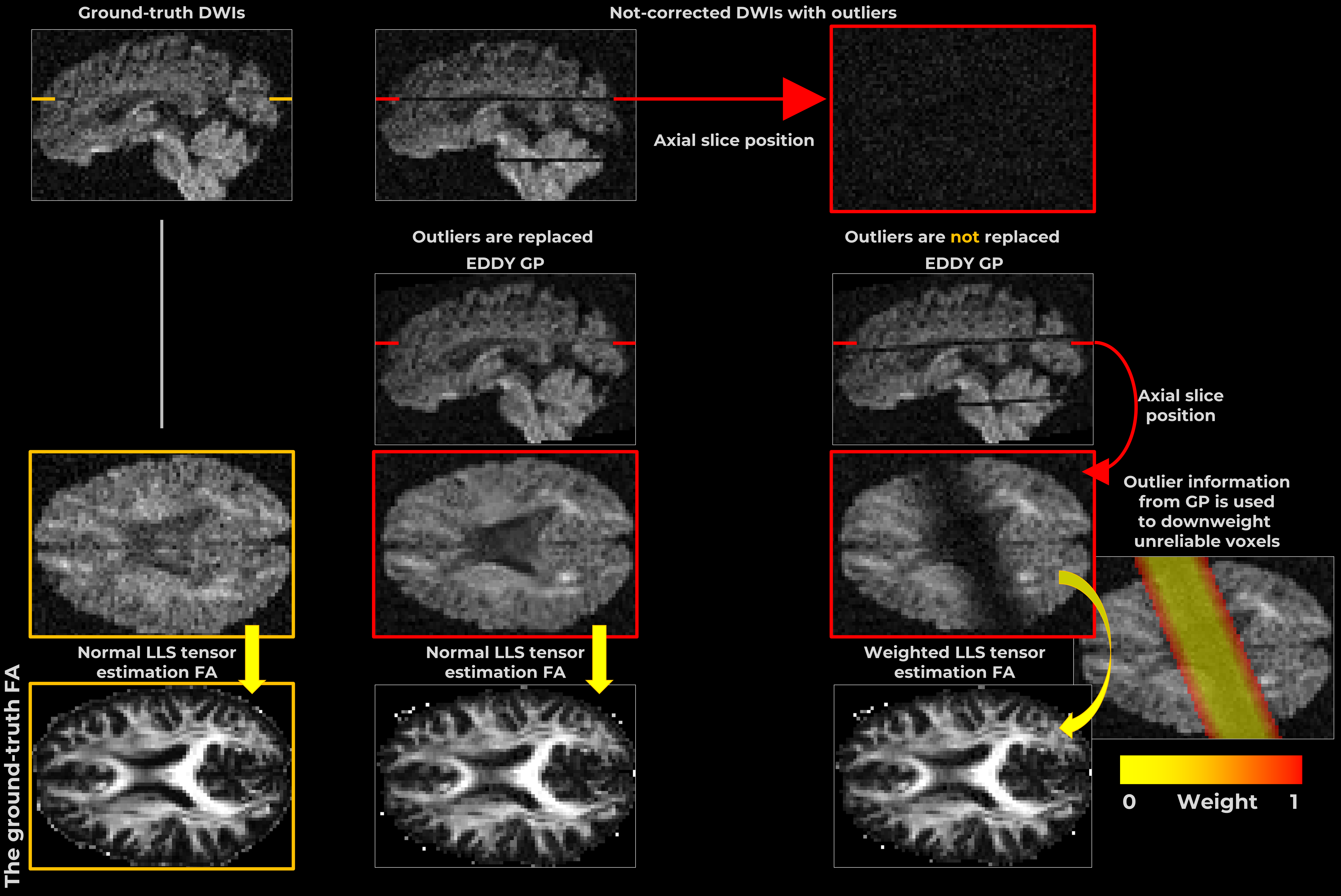
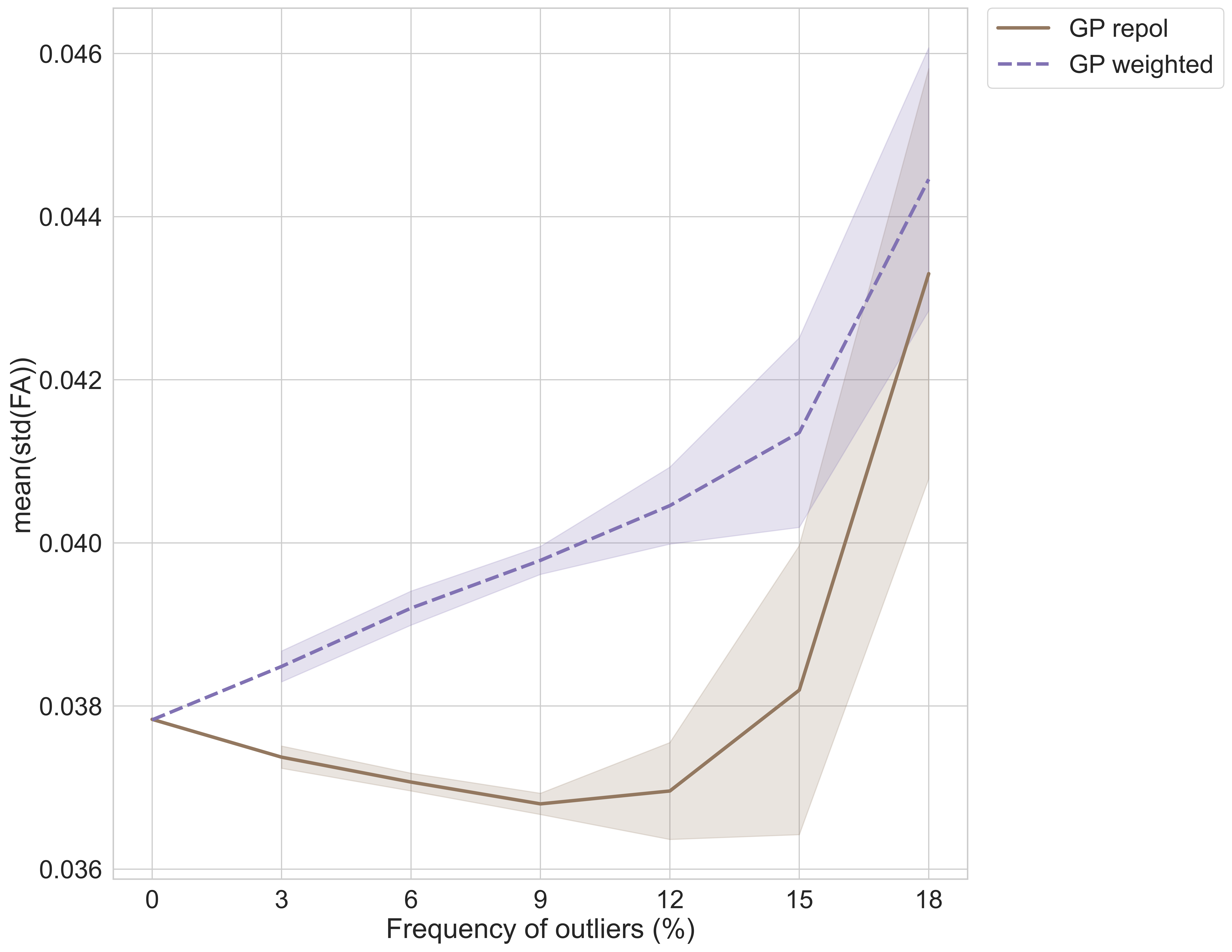
Diffusion-weighted MRI (dMRI) applications such as tractography and microstructural modelling require fitting a mathematical model to the measurements. An accurate model estimation can be impeded by signal dropout outliers that originate from subject motion during the acquisition1,2. While the origin of these outliers and different mitigation strategies such as robust modelling (e.g., Figure 1) have been explored in previous studies3, effects on the precision of the estimated model parameters are not well known. For example, if one subject has many outliers (more missing data), the precision of the fitted model should be lower than for a subject with no outliers. This could result in differences probabilistic tractography even if there are no real structural differences between the subject’s brains.Robust modelling can be done with two different strategies, the first is to replace outliers and use normal model estimator whereas the second strategy is to downweight or exclude the outliers during model estimation1,2,4,5. The outlier replacement is a convenient approach as it does not require altering any software used in downstream data processing. However, a recent study pointed out that outlier replacement can lead to inflated precision for the estimated model parameters3. We reproduced this finding successfully (Figure 2) and extended the analysis with a simulation to consider a multi-tensor model. Our aim is to investigate how the precision of parameters obtained from the multi-tensor model are affected by the increasing number of outliers in data.
Methods
We made a Python adaptation from FSL’s EDDY[1] Gaussian Process (GP) predictor that and used for outlier replacements so we could focus solely on the voxel-wise model fitting and its precision estimates.We generated a ground-truth (GT) dMRI signal mimicking a signal from a white-matter voxel with two crossing fibers using diffusion imaging in Python (DIPY) library’s multi_tensor_dki function with b=1000 and b=2000 shells with 64 gradient directions each. Details for volume fractions and tensor coefficients were drawn from a previous work6. GT was used to create 1000 noisy samples with b0 signal-to-noise ratio of 40 which were used to estimate GP hyperparameters.
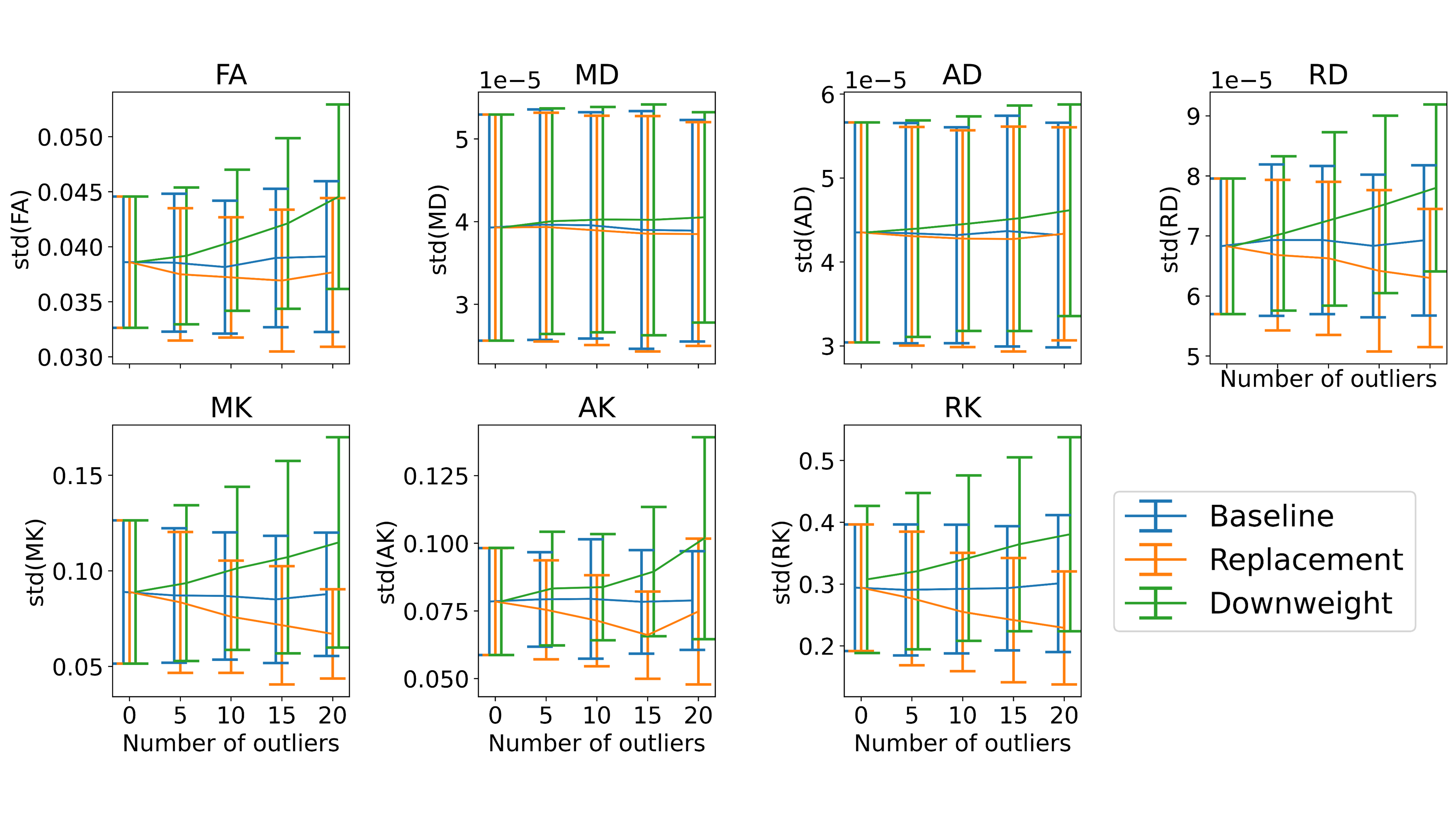
We selected a random subset of 50 noisy samples for random outlier placements. We varied the number of outliers, so each shell had 0, 5, 10, 15, or 20 outliers. The multi-tensor model was fitted to these samples with the two robust strategies outlier replacement and downweighting. Dataset with zero outliers gave us a baseline on model parameter precision when only noise is considered.
We calculated fractional anisotropy (FA), mean diffusivity (MD), radial diffusivity (RD), axial diffusivity (AD), mean kurtosis (MK), axial kurtosis (AK), and radial kurtosis (RK) using 100 bootstrap samples for each outlier setup. We used ordinary wild residual bootstrap for the baseline and replacement cases whereas for the weighted modelling case we used robust wild residual bootstrap7.
Results
Figure 2 shows a comparison between outlier replacement and weighted modelling as function of incremental outlier frequency in data. This result is based on whole brain simulations that have undergone the full EDDY-pipeline. Details of this simulation can be found from the previous study3.Figure 3 shows a toy example where we isolated the robust model estimation from the rest of the EDDY-pipeline to evaluate how the model precision is related to the incremental number of outliers in data. Please, note that the x-axis is different from Figure 2 on purpose as we wanted to explore higher outlier frequencies.
Discussion
Results in figures 2 and 3 support each other: outlier replacement in all cases seem to result in higher precision for the parameter estimates than weighted modelling. The precision of FA, RD, and kurtosis tensor derived parameters seem to be more sensitive to outliers than MD and AD. However, this could be due to random chance as parameter like AD is likely sensitive to outliers that would occur in the gradient direction that is closely aligned with the axon. With the random outlier placements, it is possible that such outlier positions were not evaluated.Conclusion
Our preliminary results indicate that decision how outliers in data are handled can influence the dMRI model parameter estimates, especially their precision. This in turn could, in theory, affect applications such as probabilistic tractography and therefore confound connectivity analyses. In future, this kind of precision analysis could be added to dMRI quality control protocols to help clinical researchers who might deal with datasets containing large numbers of outliers.Acknowledgements
V.S. was supported by the Orion Research Foundation sr, Finland and Instrumentarium Science Foundation sr, Finland. The authors wish to thank the Finnish Computing Competence Infrastructure (FCCI) for supporting this project with computational and data storage resources.References
1. J. L. R. Andersson, M. S. Graham, E. Zsoldos, and S. N. Sotiropoulos, “Incorporating outlier detection and replacement into a non-parametric framework for movement and distortion correction of diffusion MR images,” NeuroImage, vol. 141, pp. 556–572, 2016, doi: 10.1016/j.neuroimage.2016.06.058.
2. V. Sairanen, A. Leemans, and C. M. W. Tax, “Fast and accurate Slicewise OutLIer Detection (SOLID) with informed model estimation for diffusion MRI data,” NeuroImage, vol. 181, pp. 331–346, Nov. 2018, doi: 10.1016/j.neuroimage.2018.07.003.
3. V. Sairanen and J. Andersson, “Outliers in diffusion-weighted MRI: Exploring detection models and mitigation strategies,” NeuroImage, vol. 283, p. 120397, Dec. 2023, doi: 10.1016/j.neuroimage.2023.120397.
4. J. L. Schafer, “Multiple imputation: a primer,” Stat. Methods Med. Res., vol. 8, no. 1, pp. 3–15, Feb. 1999, doi: 10.1177/096228029900800102.
5. J. F. Mangin, C. Poupon, C. Clark, D. Le Bihan, and I. Bloch, “Distortion correction and robust tensor estimation for MR diffusion imaging,” Med. Image Anal., vol. 6, pp. 191–198, 2002, doi: 10.1016/S1361-8415(02)00079-8.
6. R. Neto Henriques, M. M. Correia, R. G. Nunes, and H. A. Ferreira, “Exploring the 3D geometry of the diffusion kurtosis tensor—Impact on the development of robust tractography procedures and novel biomarkers,” NeuroImage, vol. 111, pp. 85–99, May 2015, doi: 10.1016/j.neuroimage.2015.02.004.
7. V. Sairanen, A. Leemans, D. K. Jones, and C. M. W. Tax, “Rebooting diffusion MRI uncertainty distributions in the presence of outliers with ROBOOT,” 2018.
Figures