2145
Atlas-based analysis of diffusion imaging may predict efficacy of constraint-induced movement therapy in stroke rats1Ren Ji Hospital, School of Medicine, Shanghai Jiao Tong University, Shanghai, China, 2Huashan Hospital, School of Medicine, Fudan University, Shanghai, China
Synopsis
Keywords: Diffusion Analysis & Visualization, Data Analysis
Motivation: Atlas-based diffusion imaging of whole brain white matter analysis might be helpful in evaluating obscure motor function recovery in chronic stroke.
Goal(s): Our goal was to evaluate the potential of diffusion imaging atlas-based in predicting motor function recovery in stroke rats with constraint-induced movement therapy.
Approach: We used atlas-based analysis of diffusion imaging to conduct multi-parameter measurements of white matter fibers in the entire brain, combined with catwalk-automated gait analysis to test quantificationally evaluated the motor function.
Results: Atlas-based analysis of diffusion observations indicated that treatment with constraint-induced movement therapy improved microstructural integrity of axon and myelin.
Impact: Atlas-based analysis of diffusion imaging were intended to advance non-invasive imaging approaches for understanding microstructural changes of axon and myelin in chronic stroke.
Introduction
Constraint-Induced Movement therapy (CIMT) has recently shown potentiality for chronic stroke as a method of behavior-based neurorehabilitation1. Atlas-based analysis (ABA) of diffusion imaging may be helpful in evaluating obscure lesions in brain injury of stroke rats by analyzing the whole brain white matter based on an anatomical atlas2. The aim of this study was to predict efficacy of CIMT for motor function recovery in stroke rats using ABA of diffusion imaging.Methods
Thirty-six male Sprague-Dawley (SD) rats were randomly divided into three groups: a middle cerebral artery occlusion group (MCAO n=6), a therapeutic group (MCAO + CIMT n=24), and a sham-operated group (sham n=6). The therapeutic group was further divided into four groups according to the brain damage with unilateral or bilateral CIMT rehabilitation training as follows: moderate group (unilateral/bilateral n=6/6), severe group (unilateral/bilateral n=6/6). Catwalk and diffusion MRI scans were conducted on the groups of rats at 6 (as the baseline before rehabilitation training), 14 and 28 days after surgery. We used the ABA method to conduct multi-parameter measurements of white matter fibers in the entire brain. Regions of interest (ROIs) analyses were conducted on 29 regions, independent anatomically, on the basis of the DTI atlas of the P72 rat brain. Statistical analyses of fractional anisotropy (FA), radial diffusivity (RD), axial diffusivity (AD) values were performed with a two-sample t test. DTI was used to measure the microstructural integrity changes of axon and myelin sheath. The Catwalk-automated gait analysis test quantificationally evaluated the motor function of MCAO rats after CIMT treatment.Results
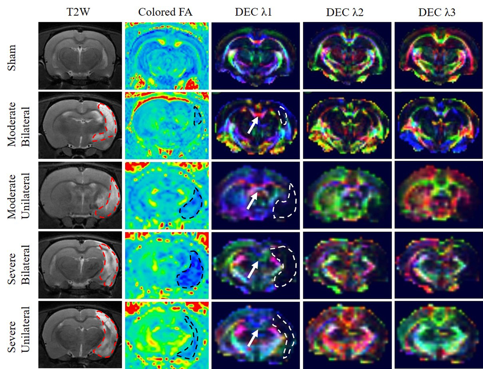
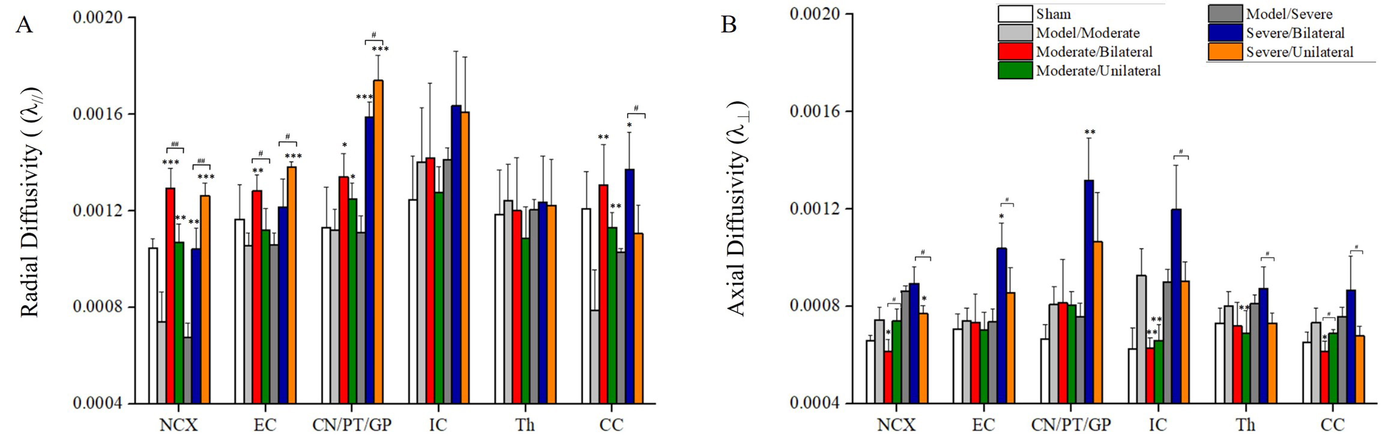
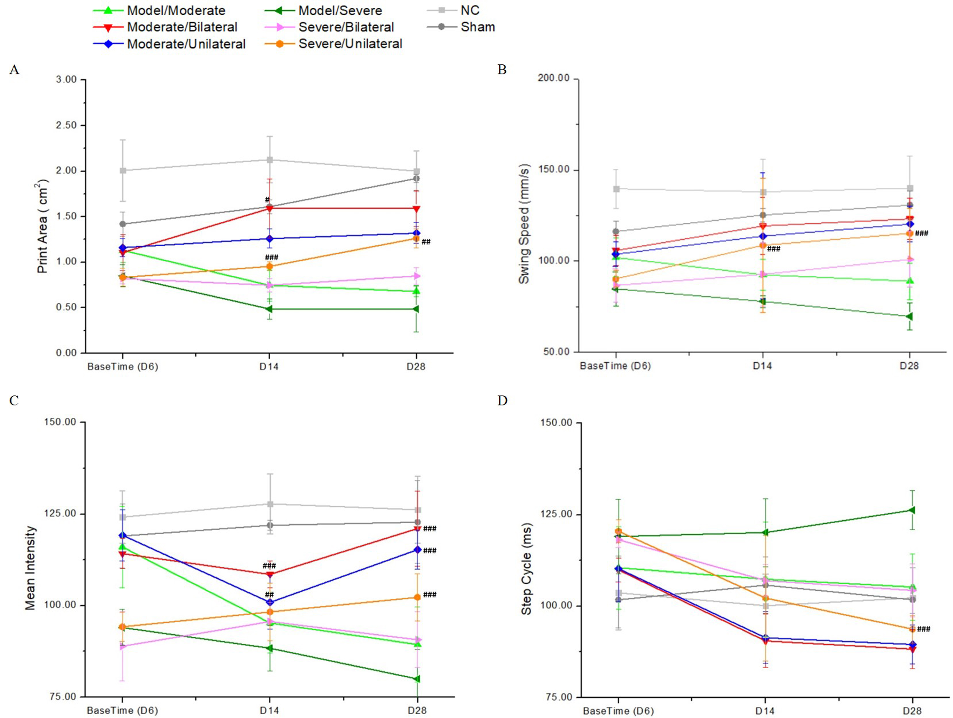
Compared to baseline, FA and RD were increased significantly in the moderate group in the neocortex, external capsule, caudate/putamen/globus pallidus, internal capsule, thalamus, corpus callosum (Fig 1). It's notable that rats treated with moderate/bilateral CIMT significantly increased RD in the neocortex and corpus callosum as compared to model groups (Fig 2). In addition, for the severe stroke rats, the RD of corpus callosum in unilateral group increased significantly compared with bilateral group. These DTI observations indicated that treatment with CIMT improved microstructural integrity of axon and myelin. Our catwalk data indicated that CIMT treatment alleviated the gait deficit of ischemic stroke rats. The paw print area and mean intensity prominently increased in moderate/bilateral and severe/unilateral groups as compared to corresponding model MCAO rats on 28th day after MCAO (Fig 3A & C). The swing speed significantly increased in CIMT groups (both moderate and severe groups) rats as compared to model group (Fig 3B). In addition, CIMT (both moderate and severe groups) significantly decreased step cycle time for two front paws of MCAO rats (Fig 3D).Discussion
We used atlas-based analysis of diffusion imaging, combined with catwalk outcomes, to evaluate the potential of constraint-induced movement therapy with different training modes for motor function recovery in stroke rats. In our study, significant differences of diffusion parameters were observed in regions related to exercise between different training groups, suggesting a possible microstructural changes of white matter with CIMT training. For severe groups, unilateral (affected side) training might help for recovery of function by strengthening the remaining alternative pathways3. For moderate groups, bilateral training may help to shape the spared and new circuits by selection and stabilization of functional connections and pruning of the nonfunctional ones4. The methodological optimization presented in this study are intended to advance non-invasive imaging approaches that will enable the use of pre-clinical MRI studies for understanding and treating chronic stroke.Conclusions
Unilateral limb training can significantly enhance the interactive inhibitory effect of bilateral cortical sensory and motor areas in moderate group. Whereas bilateral training was more helpful in enhancing the interactive inhibitory effect of various brain regions in rats with severe cerebral ischemia. Atlas-based analysis of diffusion imaging may predict efficacy of multiple CIMT modes for motor function recovery in stroke rats with varying extent of stroke.Keywords
constraint-induced movement therapy, atlas-based analysis, DTI, stroke, function recoveryAcknowledgements
This work was supported by the Clinical Research Project of Health Industry of Shanghai Health Committee (Grant No. 20194Y0087 to X. Z.).References
1. Corbetta D, Sirtori V, Castellini G, Moja L, Gatti R. Constraint-induced movement therapy for upper extremities in people with stroke. Cochrane Database Syst Rev. 2015 Oct 8;2015(10):CD004433.
2. Kasahara K, Hashimoto K, Abo M, Senoo A. Voxel- and atlas-based analysis of diffusion tensor imaging may reveal focal axonal injuries in mild traumatic brain injury -- comparison with diffuse axonal injury. Magn Reson Imaging. 2012 May;30(4):496-505.
3. Gauthier LV, Mark VW, Taub E, McCullars A, Barghi A, Rickards T, Hicks J, Uswatte G. Motor recovery from constraint induced movement therapy is not constrained by extent of tissue damage following stroke. Restor Neurol Neurosci. 2014;32(6):755-65.
4. Yang L, Lei JF, Ouyang JY, Li MZ, Zhan Y, Feng XF, Lu Y, Li MC, Wang L, Zou HY, Zhao H. Effect of Neurorepair for Motor Functional Recovery Enhanced by Total Saponins From Trillium tschonoskii Maxim. Treatment in a Rat Model of Focal Ischemia. Front Pharmacol. 2021 Dec 10;12:763181.
Figures