2144
Using time-dependent diffusion MRI to investigate brain tumor differentiation1Department of Radiology, Sun Yat-Sen Memorial Hospital, Sun Yat-Sen University, Guangzhou, China, 2MR Research Collaboration, Siemens Healthineers Ltd., Beijing, China
Synopsis
Keywords: Diffusion Analysis & Visualization, Diffusion/other diffusion imaging techniques, brain tumor
Motivation: Imaging microstructural parameters using limited spectrally edited diffusion (IMPULSED) based on time-dependent diffusion MRI (dMRI) could be utilized to quantify cell size, cellularity, and extracellular space, but its ability to distinguish common brain tumors remains unknown.
Goal(s): To determine the capacity of IMPULSED to distinguish between glioma, brain metastases, and meningioma.
Approach: Time-dependent dMRI was performed on patients with untreated glioma (n = 24), brain metastases (n = 67), and meningioma (n = 38). These patients’ IMPULSED-based parameters were evaluated and compared.
Results: Glioma, brain metastasis, and meningioma have distinctive IMPULSED-based parameters.
Impact: IMPULSED-based parameters could distinguish glioma, brain metastases, and meningioma. Time-dependent diffusion MRI is a potential approach for assessing the microstructures of brain tumors.
Introduction
Brain tumors contribute significantly to both morbidity and mortality in humans1. Imaging techniques are crucial for the diagnosis, planning of treatment, and monitoring of patients with brain tumors. Nonetheless, diagnosing a particular brain tumor type using conventional MRI techniques is frequently challenging. Recently, Xu et al. proposed a technique called imaging microstructural parameters using limited spectrally edited diffusion (IMPULSED) for assessing cell size and extracellular space based on time-dependent diffusion MRI2-3. Nonetheless, it is unclear whether IMPULSED could be utilized to differentiate between common brain tumors such as glioma, brain metastases, and meningioma.Methods
Patients with untreated, histopathologically confirmed glioma (n = 24) or brain metastases (n = 67) and meningioma (n = 38) were recruited. T1-weighted imaging (T1WI), T2-weighted imaging (T2WI), contrast-enhanced T1WI, and time-dependent diffusion MRI were performed on all patients using a 3T MR scanner (MAGNETOM Skyra, Siemens Healthcare, Erlangen, Germany). Time-dependent diffusion MRI requires the recording of diffusion MRI signals at varying diffusion times utilizing a mix of oscillating gradient spin-echo (OGSE) and pulsed gradient spin-echo sequences (PGSE) (Siemens’ research sequence). OGSE data were acquired at oscillating frequencies of 30 Hz (effective diffusion time = 8.0 msec, two cycles, b = 300 and 600 sec/mm2) and 20 Hz (effective diffusion time = 12.2 msec, one cycle, b = 400, 800, and 1200 sec/mm2), and PGSE at diffusion duration and separation of 10 and 30.7 msec, respectively (effective diffusion time = 12 msec, b = 400, 800, and 1200 sec/mm2). For both sequences, the subsequent settings were applied: three diffusion directions; repetition time msec/echo time msec, 9500/165; field of view, 219 × 219 mm; in-plane resolution, 2.4 × 2.4 mm; number of sections, 20; and section thickness, 3 mm. The scanning period for this time-dependent diffusion MRI procedure was around 8.5 minutes. All time-dependent diffusion MRI data were analyzed using the least square curve fitting toolbox in MATLAB (MathWorks, Inc.) to generate four parameters: Vin (volume fraction of intracellular space), Dex (extracellular diffusivity), diameter (average cell diameter of the tumor), and cellularity (Vin/diameter × 100). For each case, a region of interest (ROI) was manually drawn to delineate the largest section of tumor parenchyma on T2WI or contrast-enhanced T1WI by visual inspection. The ROIs were copied to all parametric maps to calculate the corresponding average values. The one-way ANOVA or Kruskal-Wallis Test was used to analyze differences in all IMPULSED parameters between gliomas, brain metastases, and meningiomas. Receiver operating characteristic (ROC) analyses were performed for the pair-wise discrimination performance of IMPULSED-based parameters between gliomas, brain metastases, and meningiomas.Results
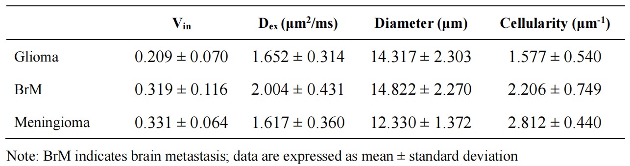
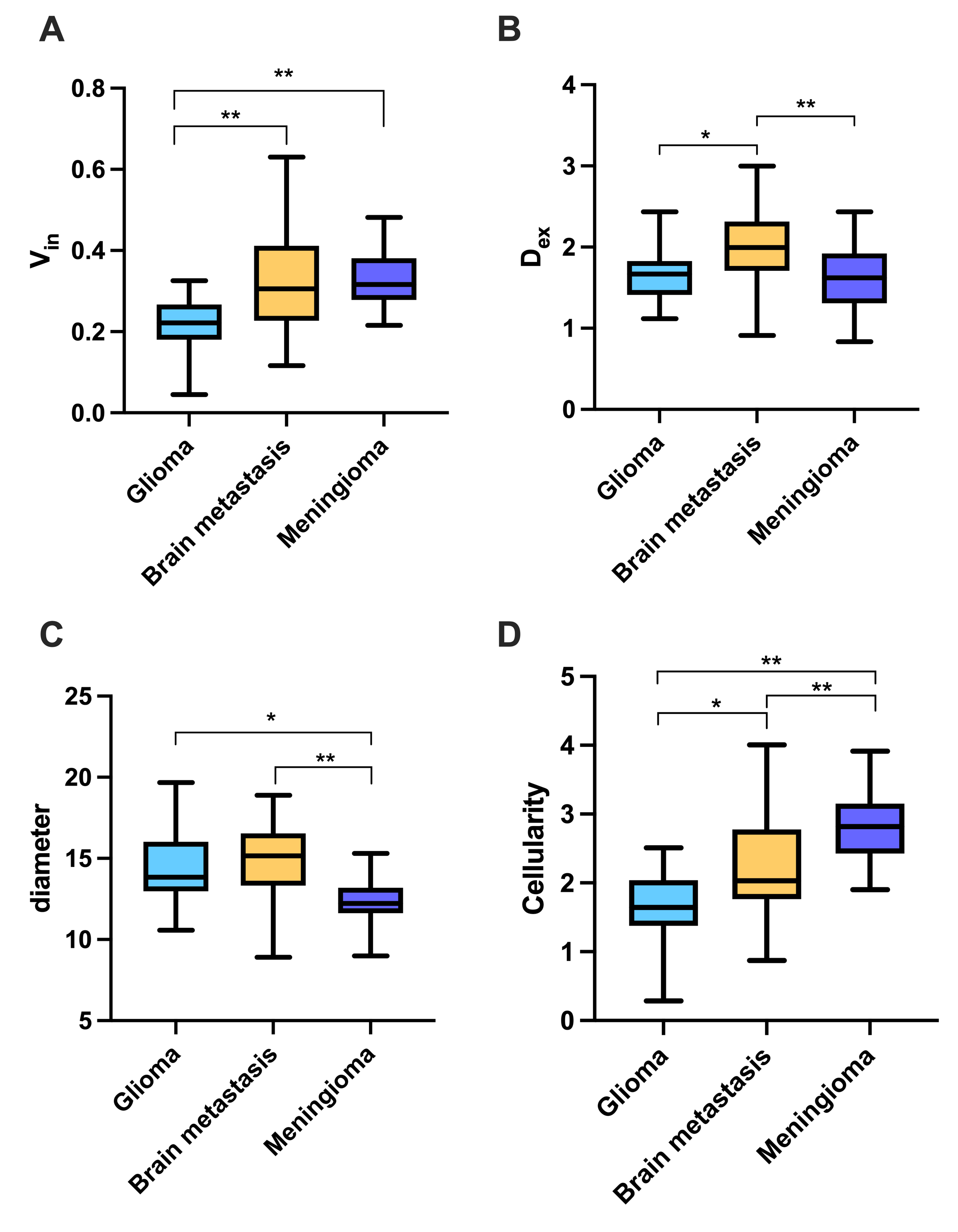
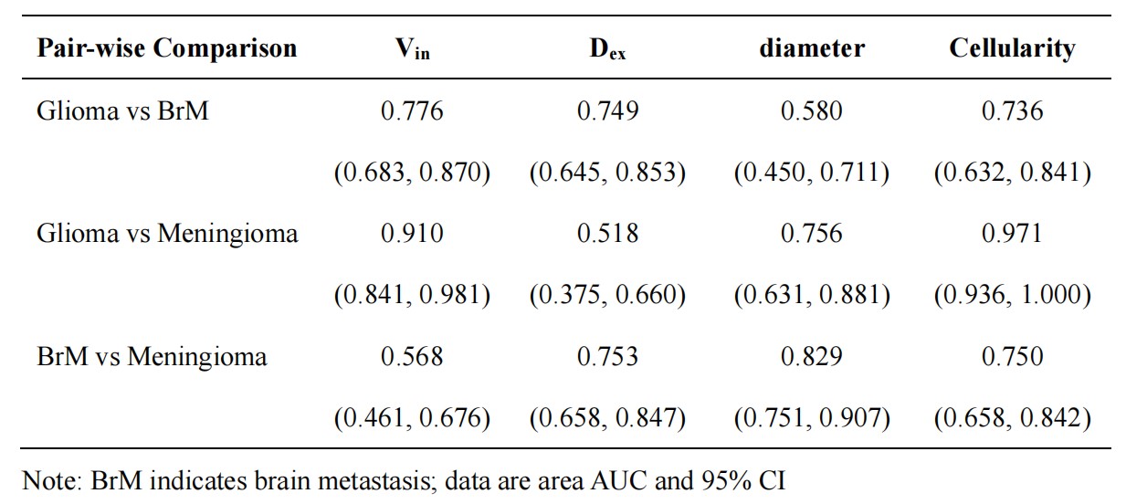
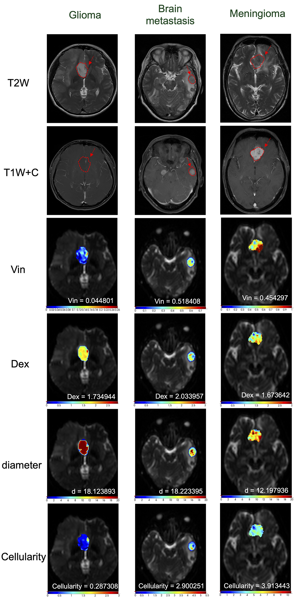
The IMPULSED parameters for gliomas, brain metastases, and meningiomas are summarized in Table 1. Brain metastases and meningiomas both had considerably higher Vin than gliomas (both P<0.001, Figure 1A), but there was no significant difference in Vin between brain metastases and meningiomas (P > 0.05). Dex was considerably greater in brain metastases than in gliomas and meningiomas (P<0.01, P<0.001, respectively, Figure 1B), while there was no significant difference in gliomas and meningiomas for Dex (P > 0.05). Gliomas and brain metastases were found to be larger in diameter than meningiomas (P<0.01, P<0.001, respectively, Figure 1C), whereas there was no significant difference in diameter between gliomas and brain metastases (P > 0.05). Meningiomas showed higher cellularity than brain metastases (P<0.001), and brain metastases were found to have higher cellularity than gliomas (P<0.001) (Figure 1D). Representative IMPULSED parametric maps of each tumor type are shown in Figure 2. The pair-wise discrimination performance of IMPULSED-based parameters is summarized in the Table 2. According to receiver operating characteristic analysis, Vin performed best in distinguishing gliomas from brain metastases (AUC, 0.776; 95% CI, 0.683, 0.870). Cellularity performed best in distinguishing gliomas from meningiomas (AUC, 0.971; 95% CI, 0.936, 1.000). Diameter distinguished brain metastases from meningiomas best (AUC, 0.829; 95% CI, 0.751, 0.907).Discussion and conclusion
Glioma, brain metastasis, and meningioma treatment differ significantly; therefore, a definitive diagnosis at the time of initial imaging could have an impact on patient treatment. Time-dependent diffusion MRI that utilizes oscillating gradient decoding is highly suitable for the visualization of cellular microstructures4. This approach exploits the time-dependent diffusion characteristics of isotropic diffusion to investigate cellular properties5. This pilot demonstrates that glioma, brain metastasis, and meningioma have distinctive IMPULSED-based parameters. Time-dependent diffusion MRI is a promising method for characterizing brain tumor microstructures.Acknowledgements
NoneReferences
- Schaff LR, Mellinghoff IK. Glioblastoma and Other Primary Brain Malignancies in Adults: A Review. JAMA. 2023;329(7):574-587.
- Xu J, Jiang X, Devan SP, et al. MRI-cytometry: Mapping nonparametric cell size distributions using diffusion MRI. Magn Reson Med. 2021;85(2):748-761.
- Xu J, Jiang X, Li H, et al. Magnetic resonance imaging of mean cell size in human breast tumors. Magn Reson Med. 2020;83(6):2002-2014.
- Wu D, Jiang K, Li H, et al. Time-Dependent Diffusion MRI for Quantitative Microstructural Mapping of Prostate Cancer. Radiology. 2022;303(3):578-587.
- Jiang X, Li H, Devan SP, Gore JC, Xu J. MR cell size imaging with temporal diffusion spectroscopy. Magn Reson Imaging. 2021;77:109-123.
Figures