2139
Harmonization of multi-site diffusion MRI data of Human Connectome Project Lifespan and Disease Studies from 2545 subjects1Harvard Medical School and Brigham and Women's Hospital, Boston, MA, United States, 2Brigham and Women's Hospital, Boston, MA, United States
Synopsis
Keywords: Diffusion Analysis & Visualization, Diffusion/other diffusion imaging techniques, Harmonization
Motivation: The Human Connectome Project (HCP) is a multi-site neuroimaging initiative that studies brain connections across the lifespan and in diseases. Scanner variability, especially in diffusion MRI (dMRI) data, can introduce bias and prevent reliable pooling of data.
Goal(s): We present our harmonization efforts on the dMRI data from 11 HCP datasets using a well-validated harmonization algorithm based on rotation-invariant spherical harmonics.
Approach: Using several diffusion measures, we demonstrate that harmonization removes significant statistical differences between datasets.
Results: Harmonized HCP dMRI data will be shared in the NIMH Data Archive and facilitate large-scale analysis and potentially enhance our understanding of neurological and psychiatric disorders.
Impact: Harmonizing diffusion MRI data from 11 Human Connectome Project scanners enables more reliable cross-site brain connectivity analyses. Leveraging large-scale harmonized diffusion MRI data can enhance statistical power, paving the way for advanced neurological and psychiatric insights within the HCP study.
Introduction
The Human Connectome Project (HCP) is a multi-site neuroimaging initiative that aims to study the connections of the human brain1–3. The HCP initially focused on the healthy adult brain4 but has expanded to study different life stages and diseases5. The HCP lifespan project explores how brain connectivity changes during typical development and aging, while the HCP disease projects explore how brain connectivity changes in various neurological and psychiatric disorders2,6. By comparing the connectomes of individuals with specific disorders to those of healthy individuals, researchers hope to pinpoint changes associated with each disease. However, combining neuroimaging data from multiple sites requires careful handling of scanner-related measurement bias before further analysis. Despite consistent imaging protocols across sites in the HCP, intrinsic hardware variabilities and software versions can introduce scanner-related bias7,8. This bias is particularly significant in diffusion MRI (dMRI), reducing statistical power and reliability of multi-site dMRI data analysis. Harmonization is an image processing technique that standardizes dMRI datasets from different sources, enabling pooling of data for joint analysis7,9. This is especially important for neuroimaging studies of psychiatric disorders, where effect sizes associated with psychiatric disorders are often small10. This study summarizes our harmonization efforts for the HCP lifespan and disease projects.Methods
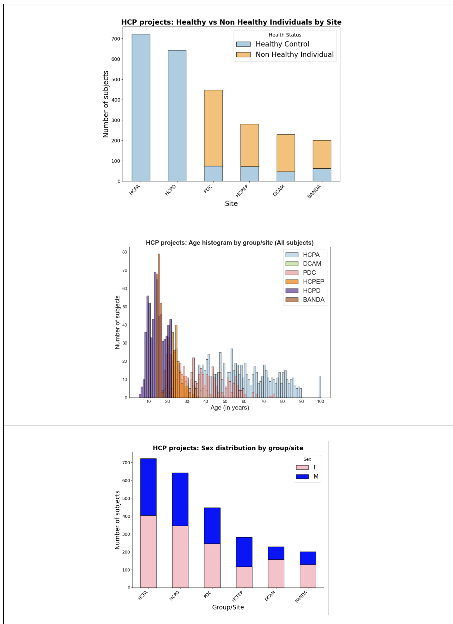
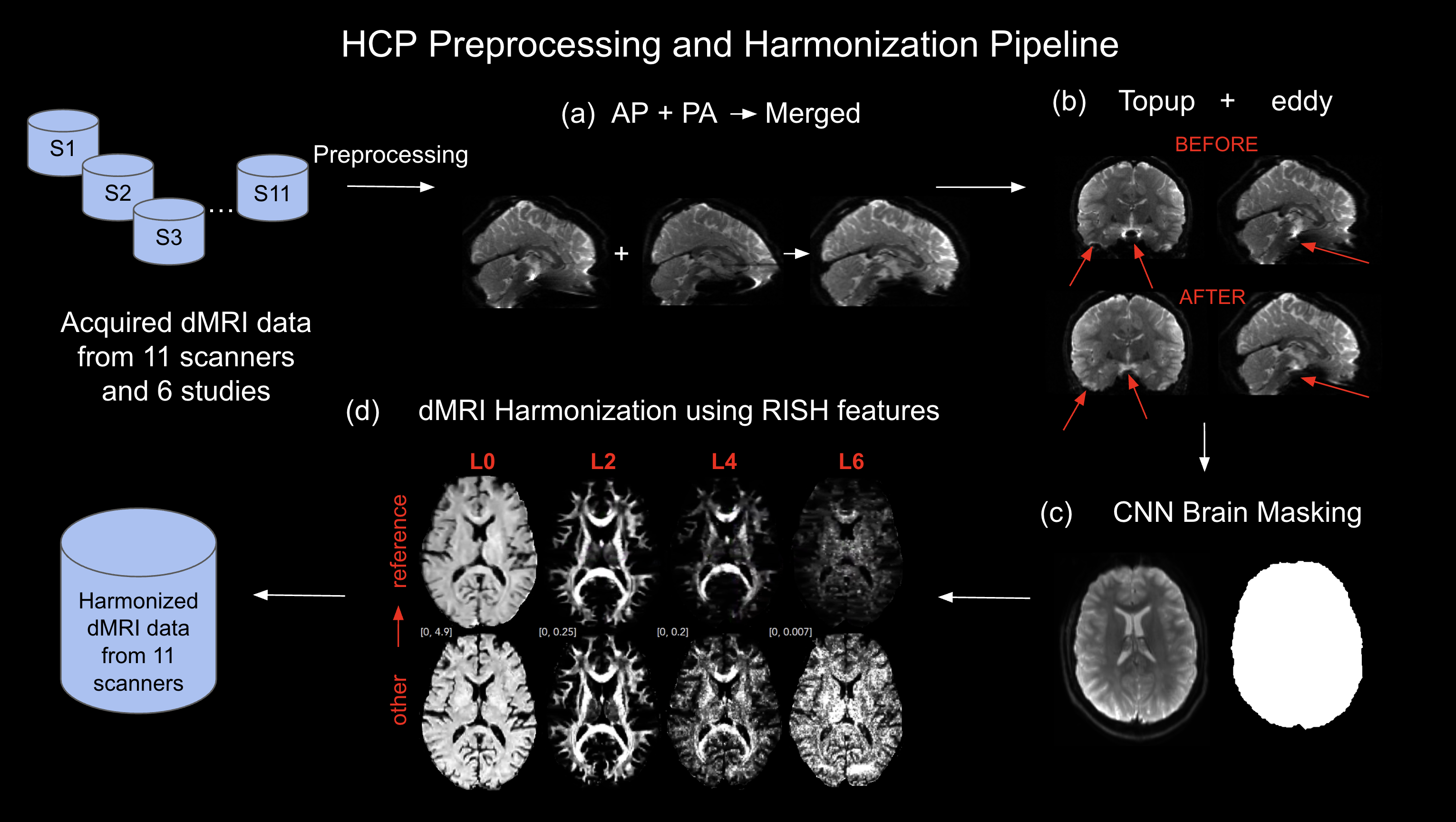
a) Dataset. We sourced unprocessed dMRI data from the NIMH Data Archive (NDA), which included HCP lifespan (HCPD and HCPA) and disease datasets: PDC, HCPEP, DCAM, and BANDA. Figure 1 provides a participant overview by study. Diffusion MRI data was acquired on 11 scanners: 3T Siemens Prisma or Prisma fit scanners with similar acquisition parameters (two anterior-posterior (AP) and two posterior-anterior (PA) phase-encode directions with 99 and 98 gradient directions and b=1500, 3000 s/mm^2).b) DMRI data pre-harmonization procedure. To standardize the preprocessing applied to each dataset and minimize preprocessing-related variability across HCP datasets, we used the same preprocessing pipeline (See Figure 2). First, for each subject’s dMRI data, we used the AP and PA phase-encode directions within FSL’s eddy and topup tool to remove susceptibility-induced distortions and eddy and motion artifacts. Then, we applied our deep learning-based brain masking tool, which robustly extracts the brain from dMRI data in a few seconds on a GPU. This made the dMRI data ready for harmonization9,11.
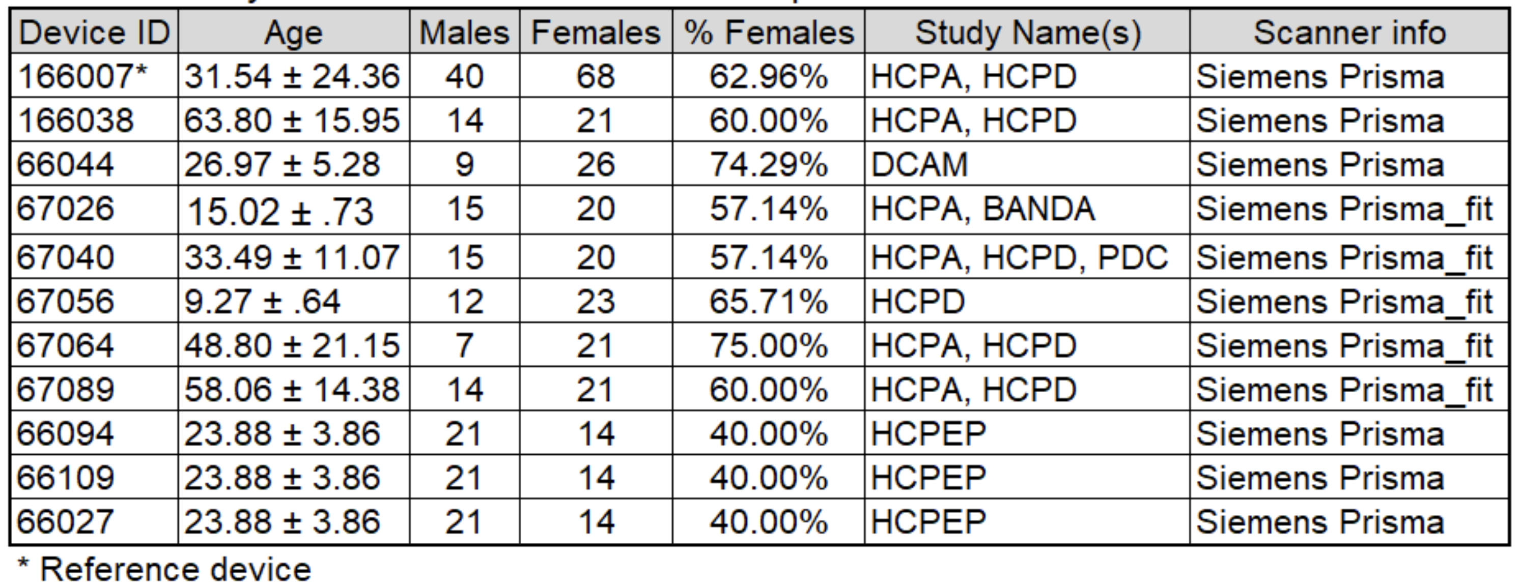
c) DMRI data harmonization. Next, we applied our retrospective harmonization algorithm, which aligns dMRI data from different scanners directly on the dMRI data by leveraging unique tissue properties using Rotation-Invariant-Spherical-Harmonics (RISH) features7,12. We validated the performance of this algorithm on multiple datasets, including a large schizophrenia database, the harmonization community challenge, and the Adolescent Brain Cognitive Development study in the past7–10. We selected 30 healthy subjects from each scanner, matched by age, sex, and IQ, to one reference dataset to which all other scanner data were harmonized (Table 1). We chose the reference device based on its wide range of characteristics (e.g., age, sex) and sample size, which allowed us to obtain representative samples across different scanners. We used the RISH features of these subjects to create templates representing scanner differences. After determining these mappings, we applied these templates to the full dMRI dataset for harmonization. More details of these steps can be found in7–10.
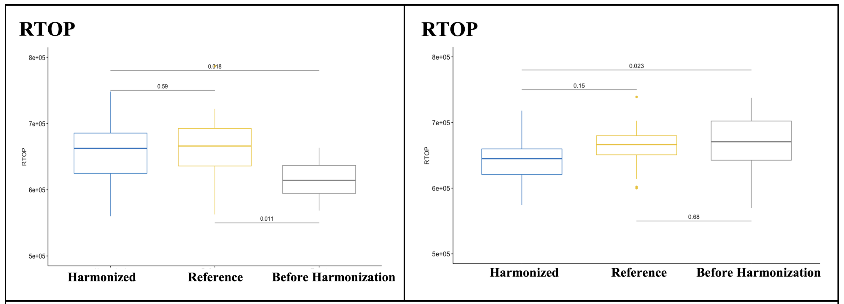
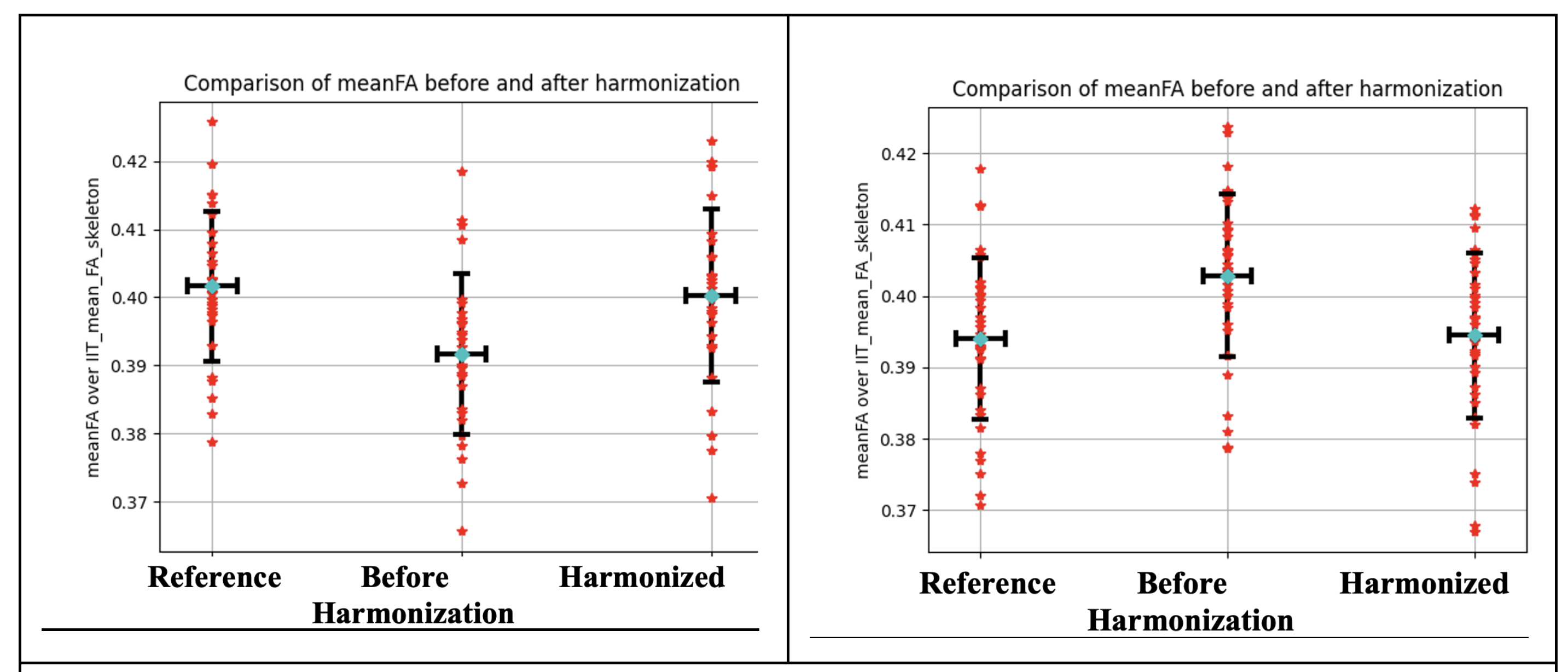
d) Demonstration of harmonization performance. Various dMRI measures, such as Return-To-Origin-Probability (RTOP) and Fractional-Anisotropy (FA), were calculated pre- and post-harmonization and compared with the reference dataset as a baseline. We assessed the harmonization performance in 30 matched subjects. Averages measures were calculated over the whole brain white matter skeleton and 42 white matter regions of interest13,14. To evaluate the performance of the harmonization, we compared the original and harmonized datasets to the reference dataset using unpaired t-tests. To further verify the harmonization, we conducted unpaired t-tests using 30 newly matched subjects, which were not part of creating templates.
Results
In template creation, we selected the scanner with deviceid=166007 as the reference scanner since it included the largest number of healthy controls and a wide range of age distribution (N=30). Figures 3 and 4 demonstrate the performance of the harmonization for two datasets (dataset 1 and dataset 2). Any existing statistical differences across datasets were removed during harmonization (p>0.53).Discussion and Conclusion.
We present our harmonization efforts on the dMRI data of the HCP study collected from 11 scanners. The harmonized data will be shared in the NDA, enabling large-scale data analysis as if the data came from the same scanner. This should significantly increase statistical power, allowing the researchers to better characterize the connectomes of individuals with specific disorders relative to those of healthy controls and potentially identify neuroanatomical changes associated with each disease.Acknowledgements
We gratefully acknowledge funding from the following National Institutes of Health (NIH) grant: R01 MH119222 (PIs: Dr. Yogesh Rathi, Dr. Lauren J. O’Donnell). We also acknowledge funding provided by the Brigham and Women’s Hospital Program for Interdisciplinary Neurosciences through a gift from Lawrence and Tina Rand and the Brain and Behavior Research Foundation NARSAD Young Investigator Award (PI: Dr. Suheyla Cetin-Karayumak).References
1. Uğurbil, K. et al. Pushing spatial and temporal resolution for functional and diffusion MRI in the Human Connectome Project. Neuroimage 80, 80–104 (2013).
2. Bookheimer, S. Y. et al. The Lifespan Human Connectome Project in Aging: An overview. Neuroimage 185, 335–348 (2019).
3. Glasser, M. F. et al. The minimal preprocessing pipelines for the Human Connectome Project. Neuroimage 80, 105–124 (2013).
4. Van Essen, D. C. et al. The WU-Minn Human Connectome Project: an overview. Neuroimage 80, 62–79 (2013).
5. Harms, M. P. et al. Extending the Human Connectome Project across ages: Imaging protocols for the Lifespan Development and Aging projects. Neuroimage 183, 972–984 (2018).
6. Somerville, L. H. et al. The Lifespan Human Connectome Project in Development: A large-scale study of brain connectivity development in 5-21 year olds. Neuroimage 183, 456–468 (2018).
7. Cetin Karayumak, S. et al. Retrospective harmonization of multi-site diffusion MRI data acquired with different acquisition parameters. Neuroimage 184, 180–200 (2019).
8. Ning, L. et al. Cross-scanner and cross-protocol multi-shell diffusion MRI data harmonization: Algorithms and results. Neuroimage 221, 117128 (2020).
9. Cetin-Karayumak, S. et al. Harmonized diffusion MRI data and white matter measures from the Adolescent Brain Cognitive Development Study. bioRxiv (2023) doi:10.1101/2023.04.04.535587.
10. Cetin-Karayumak, S. et al. White matter abnormalities across the lifespan of schizophrenia: a harmonized multi-site diffusion MRI study. Mol. Psychiatry 25, 3208–3219 (2020).
11. Palanivelu S*, Cetin Karayumak S*, Billah T, Bouix S, Rathi Y. CNN based diffusion MRI brain segmentation tool. (2020) doi:10.5281/zenodo.3665739.
12. Cetin Karayumak S*, Billah T*, Bouix S, Rathi Y. Multi-site Diffusion MRI Harmonization. (2019) doi:10.5281/zenodo.2584275.
13. Varentsova, A., Zhang, S. & Arfanakis, K. Development of a high angular resolution diffusion imaging human brain template. Neuroimage 91, 177–186 (2014).14. Qi, X. & Arfanakis, K. Regionconnect: Rapidly extracting standardized brain connectivity information in voxel-wise neuroimaging studies. Neuroimage 225, 117462 (2021).
Figures