2138
Observation of morphochemical changes and water molecule dynamics of Trypoxylus dichotomus in the pupal stage using 9.4-T MRI1Course of Science and Technology, Graduate School of Science and Technology, Tokai University, Hiratsuka, Japan, 2Course of Electrical and Engineering, Graduate School of Engineering, Tokai University, Hiratsuka, Japan, 3Department of Human and Information Science, School of Information Science and Technology, Tokai University, Hiratsuka, Japan, 4Technical Joint Management Office, Tokai University, Hiratsuka, Japan, 5Department of Natural Sciences, Faculty of Science and Engineering, Tokyo City University, Setagaya, Japan, 6Division of Natural Sciences, Graduate School of Integrative Science and Engineering, Tokyo City University, Setagaya, Japan, 7Saijo Ecology Institute, Higashi-hiroshima, Japan
Synopsis
Keywords: Diffusion Analysis & Visualization, Diffusion Tensor Imaging, Spectroscopy, Morphology
Motivation: Clarification of tissue destruction and reconstruction process of holometabolous insects might lead to innovative technology for regenerative medicine.
Goal(s): Observation of Morpholochemical changes and water molecule dynamics in the pupal body.
Approach: We applied T2W, DTI and PRESS to the pupal body of Trypoxylus dichotomus.
Results: From the pupation, the digestive tract swelled and became a liquid reservoir. It has high ADC and FA in the Head-Foot direction. After the pupation, the structure was narrower and elongated. The results suggest that destructed tissues were stored temporarily in the form of reservoir, and through the liquid channel around the reservoir to reconstruction tissues.
Impact: In the pupal body, the tissue absorbed into the digestive tract while some of the nervous system and muscles are retained formed a liquid reservoir, used to form adult tissues such as flight muscles through certain liquid channel.
Introduction
Tissue destruction and reconstruction in the pupal body of holometabolous insects is considered to be a kind of cell reprogramming process. Clarification of this process might lead to innovative technology for regenerative medicine. What is already known, the larval tissues are rapidly replaced by adult tissues only once [1], and decision conversion sometimes occur in this process [2]. The internal contents of the pupa is extremely soft and invasive resection is not appropriate for observing the temporal change of the tissue. X-CT is also not suitable for continuous observation [3] because of the inherent ionizing property. Magnetic resonance imaging (MRI)seems to be optimal by virtue of its non-ionizing feature as well as high soft-tissue contrast. Thus, in this study, we observed the tissue changes within the pupa of the Trypoxylus dichotomus from the viewpoints of morphochemistry and water molecule dynamics using 9.4T vertical MR system.Materials and Methods
Forty subjects of T. dichotomus grown at two different districts in Japan. Each subject was placed in a homemade cradle and observed continuously from prepupa to just before emergence using 9.4-T MRI: T2-Weighted image - Fast Spin Echo, T2W-FSE (TR, 4292.9; TE, 64 ms; ST, 1 mm; slice, 24; FOV, 40×50 mm; matrix, 320×400; spatial resolution, 125×125μm;), Diffusion Tensor Imaging - Echo Planer Imaging, DTI-EPI (TR, 4000 ms; TE, 21.15 ms; ST, 1 mm; slice, 16; FOV, 42×30 mm; Matrix, 168×120; spatial resolution, 250×250μm; δ, 2.5 ms; Δ, 8.5 ms; b-value, 640s/mm2; MPG application axis, 30), PRESS (TR, 4000ms; TE, 21.15 ms; voxel size, 3×3×3mm3; data points, 2048; Bandwidth, 10 ppm; Water suppression Bandwidth, 2 ppm). In order to suppress artifacts due to body movements during imaging, some individuals were given diethyl ether inhalation anesthesia for 10 to 15 minutes and fixed using a plastic straw and silicone plate.Results
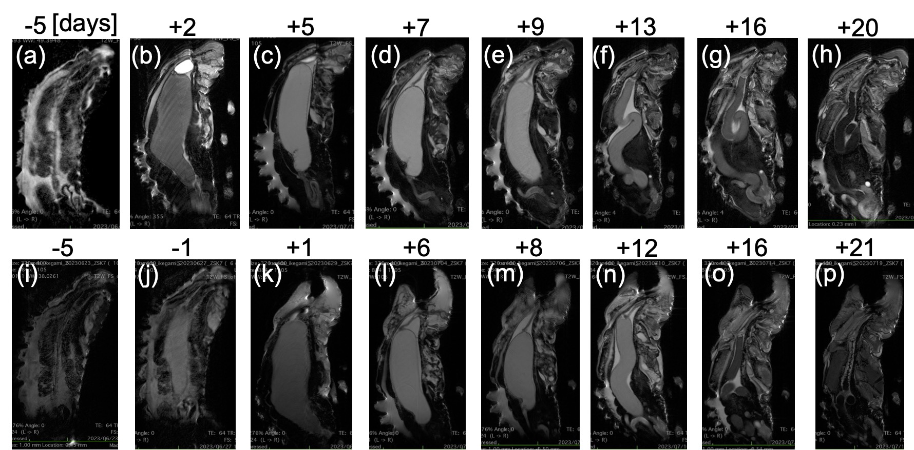
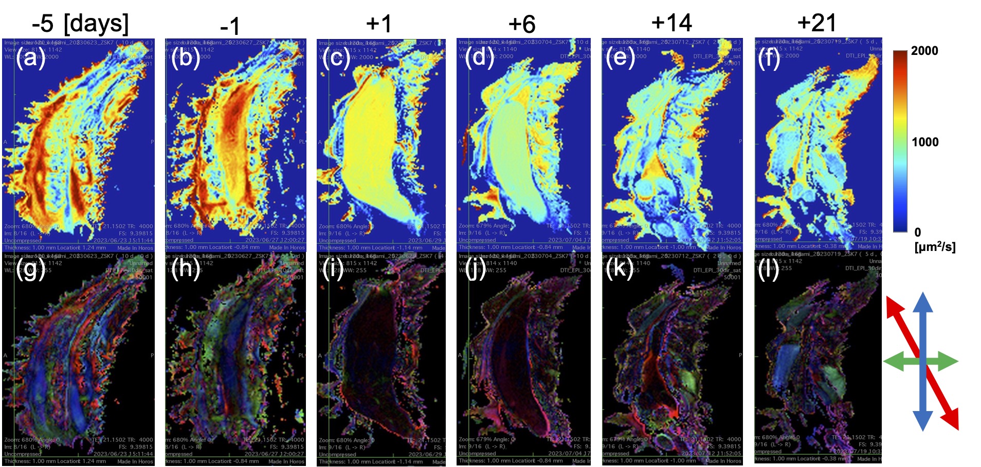
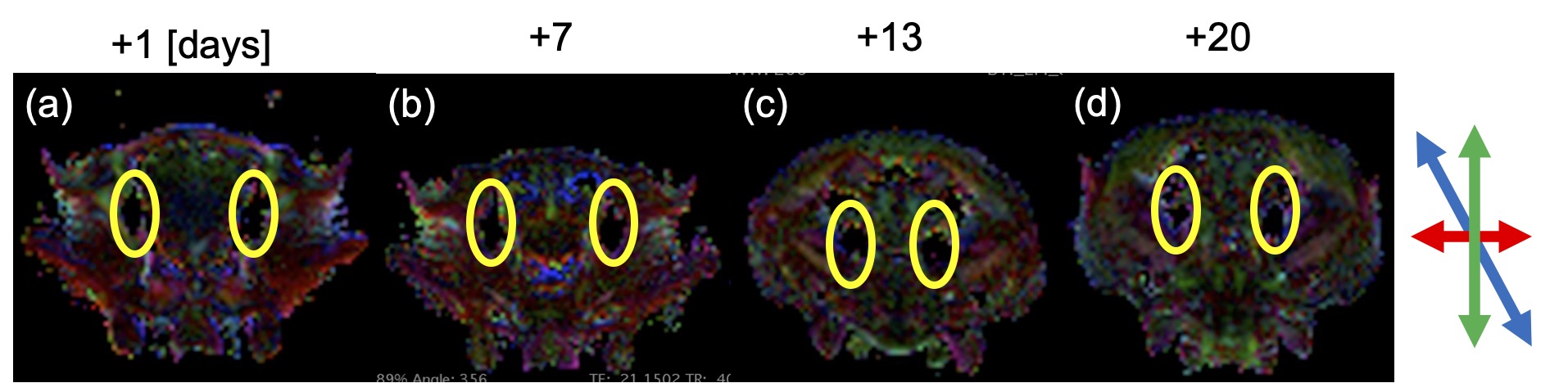
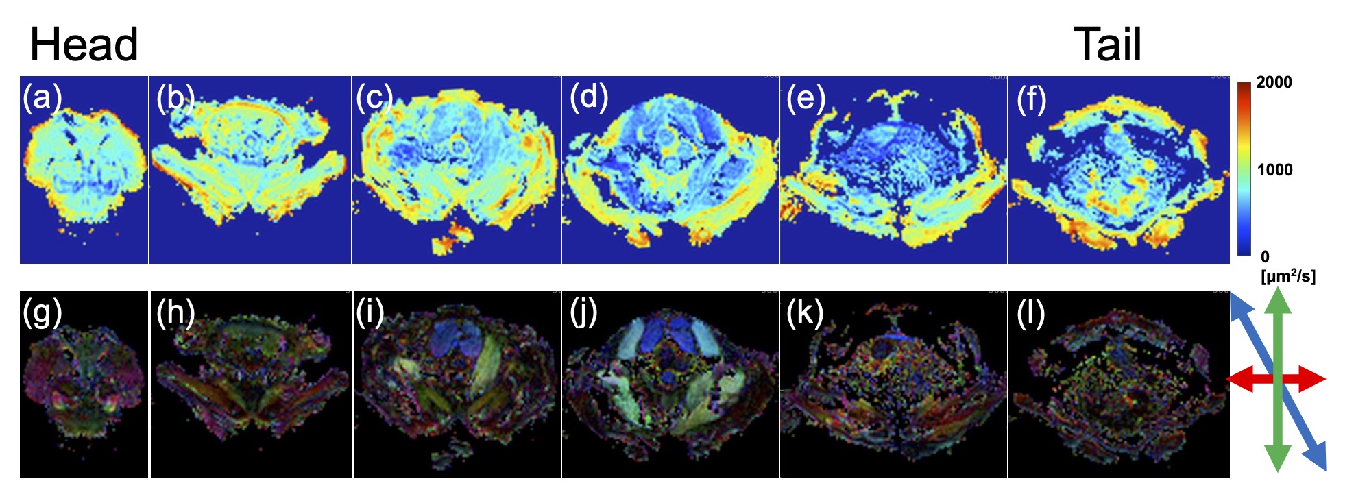
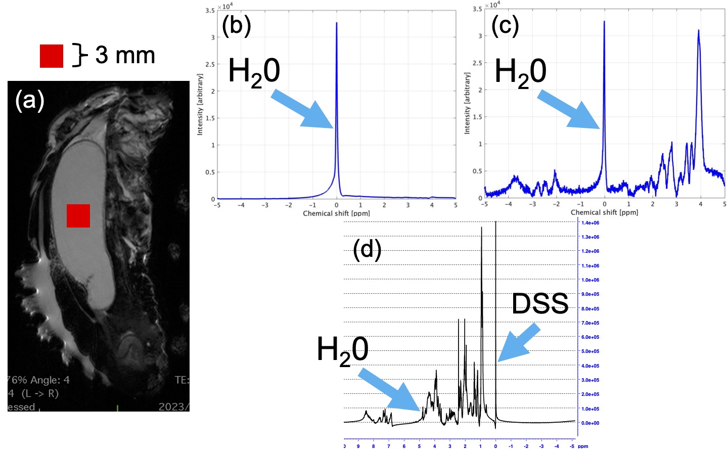
Figure1(a-h) and 1(i-p) show T2W of the female and the male which pupation day is ±0. The most remarkable feature of the prepupal to initial pupal stage was that there developed a huge liquid reservoir dominating the trunk part in every individual. The liquid reservoir became narrower and elongated day by day, and the adult tissues such as flight muscles were formed. Figure2(a-f) and 2(g-l) show the sagittal slices are the ADC map and FA map of the male. Just before pupation, the ADC was approximately 2000 square μm per seconds, and the anisotropy was in the Head-Foot direction. After the pupation, no specific anisotropy in the reservoir, although the ADC becomes lower. Figure3(a-d) show the axial slices in the head part of the female at 1 and 7 and 13 and 20 days after pupation. The yellow oval seems to represent part of the central nervous system. The nervous system did not appear to change significantly from pupation to emergence. Figure4(a-f) and 4(g-l) show the axial slices at 20 days after pupation. The flight muscles were observed in detail. The dorsal longitudinal muscle, DLM which is blue, and the dorsal ventral muscle, DVM which is indicated by the inside green, whereas the direct muscle is the outer green. Figure5(a) shows the sagittal slice of T2W at 6 days after pupation, 5(b) and (c) show the proton spectrum based on PRESS which is 5(c) was water signal suppressed. In addition, 5(d) shows the proton spectrum based on NMR which extracted the liquid from reservoir. The component inside the liquid reservoir is almost only water(b). However, when the water signal was suppressed then it was smaller peaks(c). In other trial, we extracted the liquid, and the NMR spectrum showed more detailed multiple peaks. As to the reason why there are smaller peaks, we still don’t know.Discussion
In the pupation period of the beetle, it seems that the destructed larval tissues were absorbed into the digestive tract and stored temporarily in the form of liquid reservoir. Proton chemical shift observation demonstrated that the liquid inside the reservoir were dominated by water with some smaller amounts of contents including aromatic and lipid protons. The contents of the reservoir were then used to form adult tissues possibly through certain liquid channel around the reservoir. The beetle pupae exhibit body movements when touched, hence it is thought that some central nervous system and muscle larval tissues are retained while larval tissues disintegrate. When we looking at the nervous system, we couldn't find any papers on beetles, but we don't think there is much difference in how the nervous system runs [4].Conclusion
In the pupal body of holometabolous insects, tissue changes occur while some of the nervous system and muscles remain.Acknowledgements
No acknowledgement found.References
[1] Hoshika S, Sun X, Kuranaga E, Umetsu D. Reduction of endocytic activity accelerates cell elimination during tissue remodeling of the Drosophila epidermal epithelium. Development. 2020;147(7).
[2] Hadorn E. Transdetermination in cells. Sci Am. 1968;219(5):110-4.
[3] Lowe T, Garwood RJ, Simonsen TJ, Bradley RS, Withers PJ. Metamorphosis revealed: time-lapse three-dimensional imaging inside a living chrysalis. J R Soc Interface. 2013;10(84):20130304.
[4] Rittschof C Clare, Schirmeier S. Insect models of central nervous system energy metabolism and its links to behavior. Glia. 2018;66(6):1160-1175.
Figures