2137
Approaches for correcting motion in diffusion-weighted imaging acquired using diffusion gradient cycling1MR Research, GE Healthcare, Beijing, China, 2Brainnetome Center, Institute of Automation, Chinese Academy of Sciences, Beijing, China, 3University of the Chinese Academy of Sciences, Beijing, China, 4Laboratory of Brain Atlas and Brain-inspired Intelligence, Institute of Automation, Chinese Academy of Sciences, Beijing, China
Synopsis
Keywords: Diffusion Analysis & Visualization, Motion Correction, Diffusion Gradient Cycling
Motivation: Diffusion gradient cycling (DGC) enables more efficient diffusion-weighted imaging (DWI) scanning, but it is not compatible with typical preprocssing pipelines.
Goal(s): Our goal is to propose two approaches for correcting motion in DWI data acquired using DGC: slice-to-volume registration and volume-to-volume registration with the assistance of slice reordering.
Approach: Intentional motion was introduced during the DWI acquisition, and the proposed approaches were implemented and applied to remove artifacts caused by this motion.
Results: Both approaches effectively eliminated motion-induced artifacts, and the intentional motion was estimated correctly.
Impact: Motion-induced artifacts can be eliminated, and correct motion estimation can be achieved in DWI acquired with DGC. Our proposed approaches are publicly available and can be easily integrated into preprocessing pipelines.
Introduction
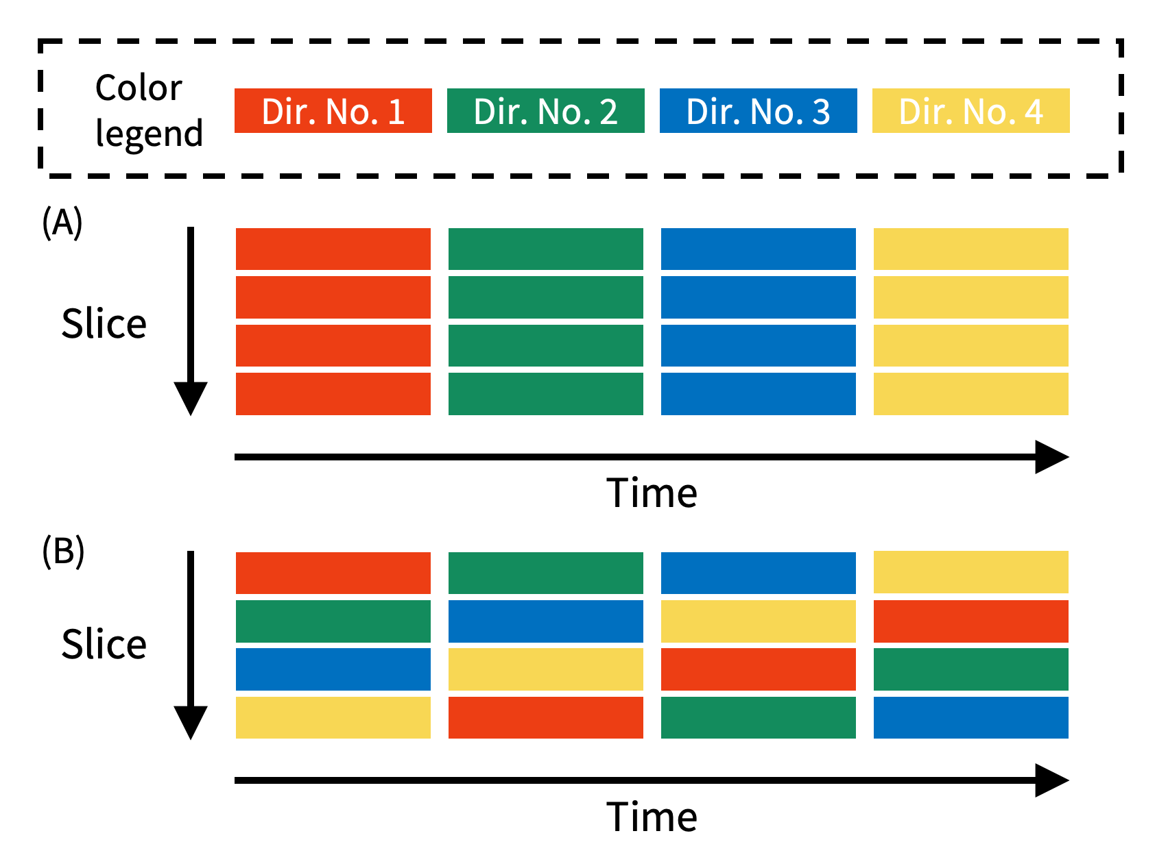
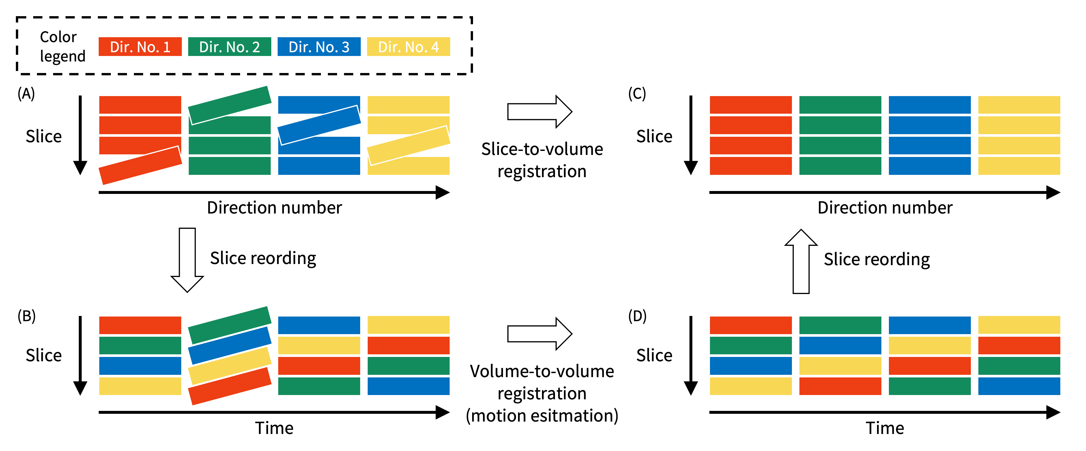
The need to explore the microstructure of neural tissue and connectional anatomy across various length scales drives the advancement of high-performance gradient technology1–3. The required strong gradients pose significant demands regarding thermal heating and power supply, resulting in longer acquisition times to allow for cooling periods.To address this issue, diffusion gradient cycling (DGC) has been implemented to control the playout order of the slice stack and different diffusion weighting directions so that the required add-on time for gradient cooling is reduced when diffusion directions are interleaved across the series (Figure 1). This hence enables more efficient scanning. However, DGC distributes the slices affected with motion. This slice misalignment results in the corruption in all volumes (Figure 2A). Existing motion correction tools based on volume-to-volume registration are incompatible with DGC4.
In this study, two approaches were proposed and evaluated for correcting motion in diffusion-weighted imaging (DWI) acquired using DGC.
Methods
Two approaches for motion correction are illustrated in Figure 2. Slice-to-volume-based registration methods have been implemented to correct artifacts caused by within-volume motion-induced artifacts4,5, which is also expected to correct the slice misalignment introduced by DGC. An alternative approach involves reordering all slices before and after volume-to-volume registration to prevent slice misalignment during the registration precedure.One participant underwent scanning on a 3T GE SIGNA UHP scanner (GE Healthcare, Waukesha, Wisconsin, US) for two runs using a single-shot spin-echo echo planar imaging sequence with the following parameters: TE/TR=47.7ms/10s, acquisition matrix=120×120, 60 slices, 2mm isotropic voxel size, 1 image with b-value=0s/mm2 and 30 noncolinear directions with b-value=1000s/mm2. Automated DGC was disabled in the first run but enabled in the second run. During each run, the subject was instructed to rapidly move to different positions.
Before motion correction, image denoising (MRtrix’s dwidenoise) and removing Gibbs ringing artefacts (MRtrix’s mrdegibbs) were applied. Volume-to-volume-based correction and movement estimation were performed using FSL's eddy6. Slice-to-volume registration was performed using both FSL’s eddy and the SHARD reconstruction software7,8. The slice reordering was implemented in python and can be found at GitHub (https://github.com/xu-boyan/gradient_cycling_reorder). Finally, DTI parameter maps were calculated to evaluate the effectiveness of preprocessing.
Results
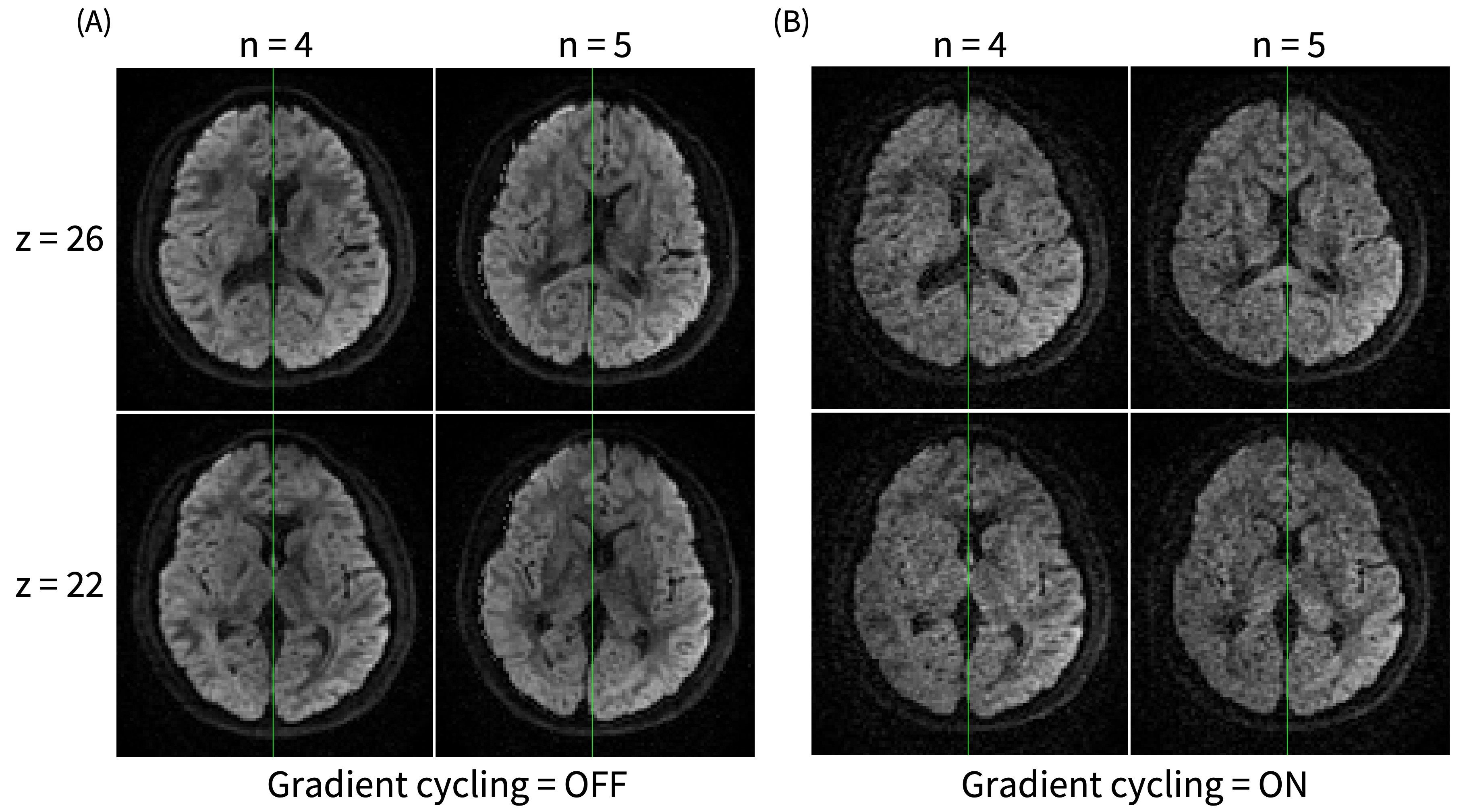
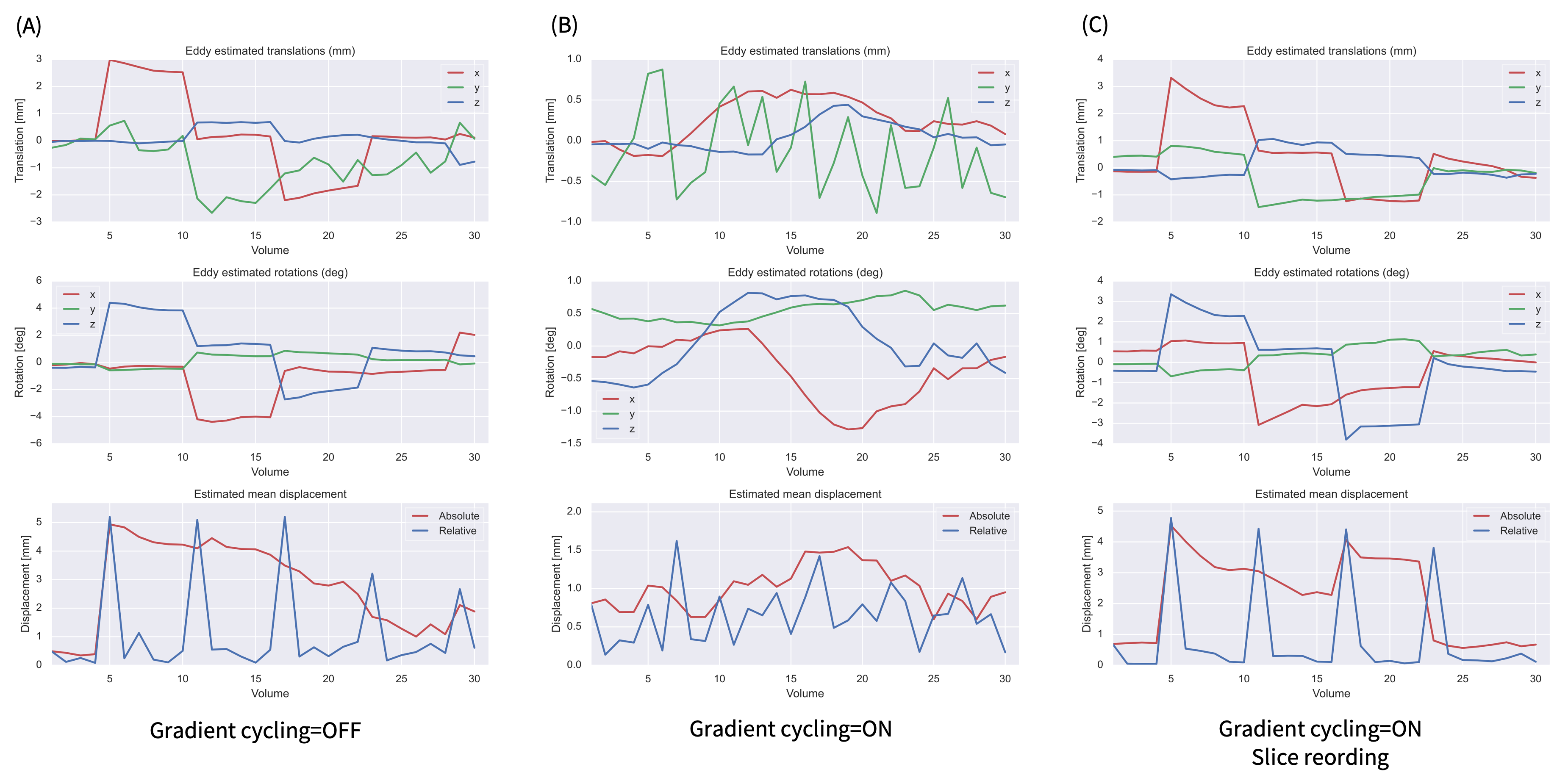
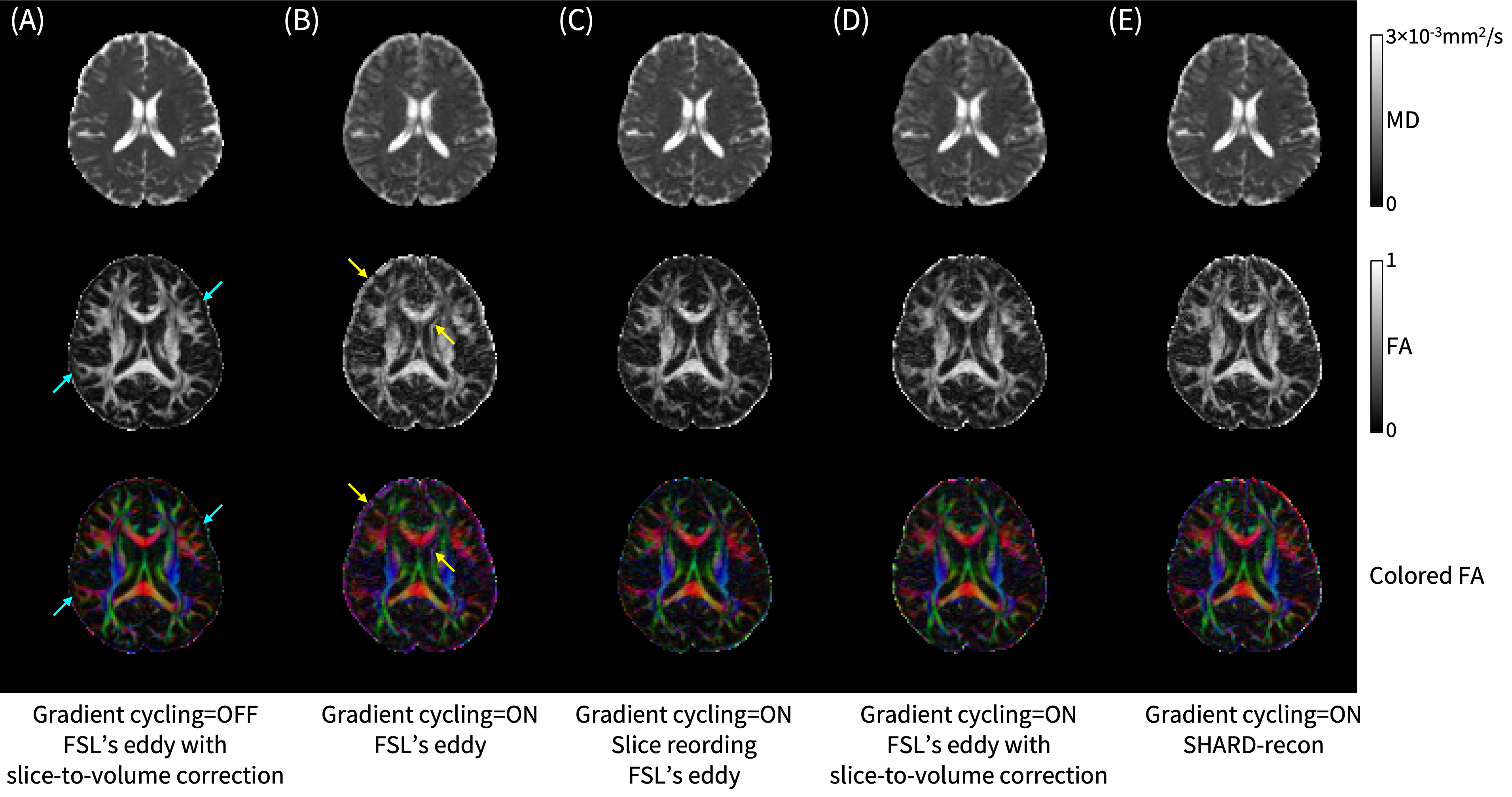
In the case of DGC, motion-induced positional differences were distributed across slices within each volume, resulting in the misalignment between slices within the same volume (Figure 3). The estimation of intentional motion was erroneous due to this slice misalignment. However, with our proposed slice reordering method, correct estimation of intentional motion was achieved (Figure 4). After applying the regular preprocessing pipeline to data acquired with DGC, motion-induced artifacts were evident in the calculated parameter maps. These artifacts can be eliminated with our suggested motion correction approaches (Figure 5).Discussion
Two approaches for correcting motion in DWI acquired using DGC have been proposed and evaluated in this study. Both effectively eliminated motion-induced artifacts compared to the typical volume-to-volume-based method. Slice-to-volume-based registration methods have been implemented in various software packages, and our source code for slice reordering is publicly available. Therefore, these approaches can be easily integrated into preprocessing pipelines. Estimating subject motion correctly is beneficial for quality control in large imaging datasets.It should be noted that further calculation may also be affected since the b-matrix cannot be properly rotated when correcting for subject motion in data acquired with DGC9. Slice-wise calculation is therefore necessary to achieve a high degree of precision.
Conclusion
Motion-induced artifacts can be eliminated, and correct motion estimation can be achieved in DWI acquired with DGC. Our proposed approaches are publicly available and can be easily integrated into preprocessing pipelines.Acknowledgements
No acknowledgement found.References
1. Foo TKF, Tan ET, Vermilyea ME, et al. Highly Efficient Head-Only Magnetic Field Insert Gradient Coil for Achieving Simultaneous High Gradient Amplitude and Slew Rate at 3.0T (MAGNUS) for Brain Microstructure Imaging. Magn. Reson. Med. 2020;83:2356–2369.2. Huang SY, Witzel T, Keil B, et al. Connectome 2.0: Developing the next-Generation Ultra-High Gradient Strength Human MRI Scanner for Bridging Studies of the Micro-, Meso- and Macro-Connectome. NeuroImage 2021;243:118530.
3. Fan Q, Eichner C, Afzali M, et al. Mapping the Human Connectome Using Diffusion MRI at 300 mT/m Gradient Strength: Methodological Advances and Scientific Impact. NeuroImage 2022;254:118958.
4. Tax CMW, Bastiani M, Veraart J, Garyfallidis E, Okan Irfanoglu M. What’s New and What’s next in Diffusion MRI Preprocessing. NeuroImage 2022;249:118830.
5. Ferrante E, Paragios N. Slice-to-Volume Medical Image Registration: A Survey. Med. Image Anal. 2017;39:101–123.
6. Andersson JLR, Sotiropoulos SN. An Integrated Approach to Correction for Off-Resonance Effects and Subject Movement in Diffusion MR Imaging. NeuroImage 2016;125:1063–1078.
7. Andersson JLR, Graham MS, Drobnjak I, Zhang H, Filippini N, Bastiani M. Towards a Comprehensive Framework for Movement and Distortion Correction of Diffusion MR Images: Within Volume Movement. NeuroImage 2017;152:450–466.
8. Christiaens D, Cordero-Grande L, Pietsch M, et al. Scattered Slice SHARD Reconstruction for Motion Correction in Multi-Shell Diffusion MRI. NeuroImage 2021;225:117437.
9. Leemans A, Jones DK. The B-Matrix Must Be Rotated When Correcting for Subject Motion in DTI Data. Magn. Reson. Med. 2009;61:1336–1349.
Figures