2135
The use of multiparametric brain tumor segmentation for investigating 1H MRSI-detected, fasting-induced ketone body accumulation in glioma1Institute of Neuroradiology, Goethe University Frankfurt, University Hospital, Frankfurt am Main, Germany, 2University Cancer Center Frankfurt (UCT), Frankfurt am Main, Germany, 3Frankfurt Cancer Institute (FCI), Frankfurt am Main, Germany, 4German Cancer Research Center (DKFZ) Heidelberg, Germany and German Cancer Consortium (DKTK), Partner Site Frankfurt/Mainz, Frankfurt am Main, Germany, 5Dr. Senckenberg Institute of Neurooncology, Goethe University Frankfurt, University Hospital, Frankfurt am Main, Germany, 6Department of Neurosurgery, Goethe University Frankfurt, University Hospital, Frankfurt am Main, Germany
Synopsis
Keywords: Cancer, Tumor, Nutritional Intervention, MR spectroscopy, Tumor segmentation
Motivation: The evaluation of MR spectroscopy imaging findings with multiparametric brain tumor segmentation might facilitate the understanding of altered tumor metabolism induced by intervention on an individual patient/tumor level.
Goal(s): In this study, we used this approach to elucidate the glioma metabolism under nutritional intervention.
Approach: The concentrations of ketone bodies in brain tumor after 72-hour-fasting were correlated with the volume of glioma sub-regions for 13 brain tumor patients.
Results: The outcome indicates that the accumulation of ketone bodies in solid tumors and necrotic areas after fasting might be a result of neovascularization and the blood-brain barrier compromise.
Impact: Here, we report on the validation of a dedicated, multi-voxel MRSI protocol with fully automated multiparametric segmentation of glioma sub-regions for monitoring fasting-induced changes.
Introduction
Emerging evidence suggests that fasting could play a key role in cancer treatment1,2. However, its metabolic effects on gliomas with a high degree of intratumoral heterogeneity remain equivocal3-5. A better understanding of diet-induced changes in tumor tissue on an individual patient/tumor level is therefore crucial to successfully incorporate dietary interventions into current treatment options. Multi-voxel MR-spectroscopic imaging is a promising tool for non-invasive monitoring of such changes6,7. During periods of fasting, ketone bodies (KBs) are generated from β-oxidation of fatty acids. Mapping these metabolites along with other MRS-detectable metabolites in glioma patients might allow us to identify patient subgroups eligible for fasting as a therapeutic strategy.As tumors represent a heterogenous mixture of tissues on a macroscopic and microscopic level, the correlation of MRSI findings with macroscopic tumor subregions (i.e., edema, necrotic region/non-enhancing tumor core, and contrast-enhancing tumor) obtained from multiparametric brain tumor segmentation8 might be a first step towards an improved interpretation of our metabolic maps. Here, we merge these two modalities to study the glioma metabolism under fasting.
Methods
The investigation is part of a prospective, single-group study investigating effects of one 72-hour-fasting cycle on glioma tumor tissue (ClinicalTrials.gov Identifier: NCT04461938). Patients ≥18 years of age with MRI-suspected glioma WHO grade II-IV (at the time 2016 WHO classification) and a recommendation for biopsy/resection were eligible. MRSI measurements were performed prior to fasting (Session 1) and after 72 hours of fasting (Session 2) on a clinical whole-body 3T MR Scanner (MAGNETOM Prisma, Siemens Healthineers, Erlangen, Germany) in order to non-invasively assess metabolic flexibility of tumor cells. The protocol included 3D-T1W and 2D-T2W reference images and 2D 1H sLASER CSI at an echo time of 144 ms. In addition, B1 maps and 2D 1H FID CSI sequences without water suppression were recorded for absolute quantification of metabolite concentrations. Surgically collected tumor tissue after Session 2 included a standard neuropathological workup and additional metabolic analyses.LCModel was used for the 1H spectral analysis. Metabolite signals for the basis set were simulated for the sLASER pulse sequence at TE=144 ms with prior knowledge of chemical shift and J-coupling, using the jMRUI plug-in NMR-ScopeB (Version 6.0). Metabolites were obtained in voxels selected from tumor region and the contralateral hemisphere considering partial volume effect.
The BraTS Toolkit was used for fully-automated segmentation of glioma sub-regions on co-registered routine brain tumor imaging data obtained during the trial screening process which includes T1w, T1w contrast-enhanced, T2w, and FLAIR images. The tumor volume was segmented into three classes: edema, necrotic region/non-enhancing tumor core, and contrast-enhancing tumor.
Results
22 patients (16 males, 6 females) were examined with our MRSI protocol at baseline and after 72 hours of fasting. 9 patients were excluded from the analysis: 1 patient diagnosed with a secondary brain tumor, 2 patients who did not complete the required 72 hours of fasting, and 6 patients with insufficient spectral quality due to temporal lobe location of tumor (n=13).β-hydroxybutyrate (β-OHB) and acetoacetate (AcAc) were detected with less than 80% of Cramér Rao Lower Bounds in the volume of the contrast-enhanced tumor of all thirteen patients. The third KB, acetone (Ace), however, was observed only in 7/13 patients’ tumor volume.
The calculated β-OHB concentration for each voxel was used to generate β-OHB maps. These maps are demonstrated in Figure 1a for three patients with high blood β-OHB levels (> 4 mmol/L) after 72 hours of fasting. β-OHB and AcAc were not only detected in the volume of the contrast-enhanced tumor but also in the necrotic/non-contrast-enhanced tumor core (Figure 1b). The inverse spectral pattern of β-OHB, as well as lactate (Lac), allowed us to distinguish both from elevated lipid peaks (associated with necrosis and membrane breakdown in glioma) arising in the 0.9 – 1.3 ppm range as shown in Figure 1c.
The averaged concentrations of β-OHB and AcAc in solid tumors were found to be significantly correlated with the volume of contrast-enhanced tumor (ρ = 0.76, p = 0.003 and ρ = 0.56, p < 0.05, respectively) and the necrosis/non-contrast-enhanced tumor core (ρ = 0.81, p = 0.0007 and ρ = 0.76, p = 0.003, respectively). Exemplary tumor segmentations and results of the correlation analyses are shown in Figure 2.
Discussion
The correlation between the size of a segmentation subgroup and KB concentrations led us to consider that the high concentrations, especially of β-OHB, may be a result of neovascularization and the blood-brain barrier compromise. The integration of multiparametric brain tumor segmentation with MRSI might facilitate understanding of altered tumor metabolism under intervention despite intratumoraö heterogeneity.Acknowledgements
KJW and SA were funded by the Mildred Scheel Career Center Frankfurt (Deutsche Krebshilfe). KJW was funded by the Else Kröner-Fresenius-Stiftung (EKFS).References
1. Nencioni, A., Caffa, I., Cortellino, S. & Longo, V. D. Fasting and cancer: molecular mechanisms and clinical application. Nat Rev Cancer 18, 707–719 (2018).
2. Pietrocola, F. et al. Caloric Restriction Mimetics Enhance Anticancer Immunosurveillance. Cancer Cell 30, 147–160 (2016).
3. Maurer, G. D. et al. Differential utilization of ketone bodies by neurons and glioma cell lines: a rationale for ketogenic diet as experimental glioma therapy. BMC Cancer 11, 315 (2011).
4. De Feyter, H. M. et al. A ketogenic diet increases transport and oxidation of ketone bodies in RG2 and 9L gliomas without affecting tumor growth. Neuro Oncol 18, 1079–1087 (2016).
5. Sperry, J. et al. Glioblastoma Utilizes Fatty Acids and Ketone Bodies for Growth Allowing Progression during Ketogenic Diet Therapy. iScience 23, 101453 (2020).
6. Wenger, K. J. et al. Maintenance of Energy Homeostasis during Calorically Restricted Ketogenic Diet and Fasting-MR-Spectroscopic Insights from the ERGO2 Trial. Cancers 12, 3549 (2020).
7. Berrington, A. et al. Cerebral Ketones Detected by 3T MR Spectroscopy in Patients with High-Grade Glioma on an Atkins-Based Diet. AJNR 1908–1915 (2019).
8. Kofler, F. et al. BraTS Toolkit: Translating BraTS Brain Tumor Segmentation Algorithms Into Clinical and Scientific Practice. Frontiers in Neuroscience 14, (2020).
Figures
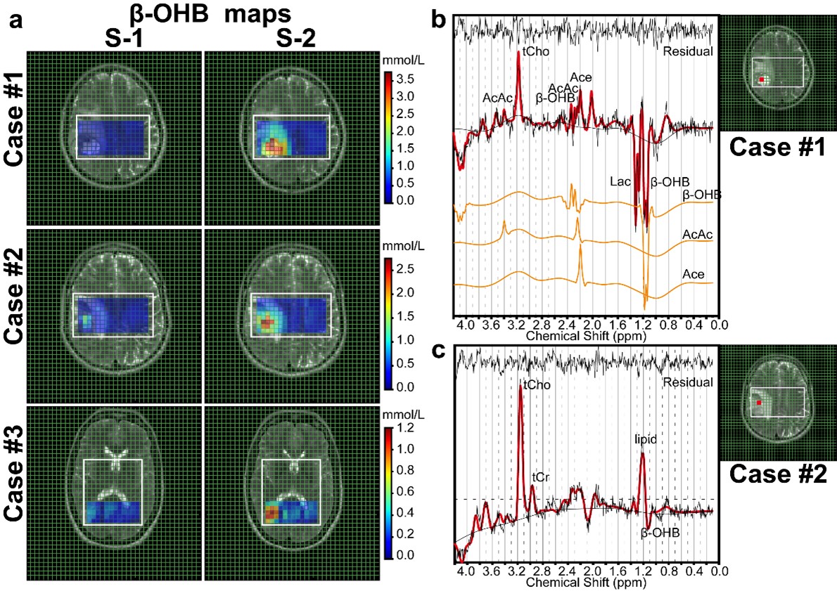
Figure 1. (a) β-OHB maps registered on T2WI of three patients with high blood β-OHB levels (> 4 mmol/L) obtained from 1H sLASER CSI with 144 ms measurement at baseline (left, S-1) and after 72 hours of fasting (right, S-2). (b, c) 1H CSI spectra of two patients obtained after 72 hours of fasting from the necrotic core of a glioblastoma. tCho indicates total Cho; tCr, total Cr.
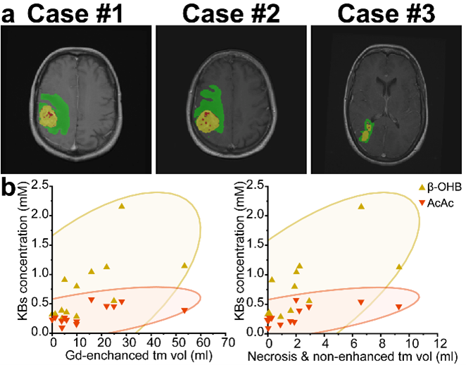
Figure 2. (a) Tumor segmentation results on three patients with high blood β-OHB levels (> 4 mmol/L) and KBs’ accumulation in tumor. The results are overlaid on post-contrast T1W images. Each color indicates a tumor class: red color represents necrosis/non-contrast-enhanced tumor, green edema and yellow contrast-enhanced tumor. (b) Spearman’s correlation analysis between KBs’ accumulation in tumor after 72-hour-fasting and existing tumor volume. The ellipses represent a 95% confidence interval.