2134
Thresholding to remove fluid from FreeSurfer segmentations of quantitative brain maps – a simple solution to a painful problem1Bioengineering, Imperial College London, London, United Kingdom, 2Radiotherapy and Imaging, Institute of Cancer Research, London, United Kingdom, 3Department of Imaging, Imperial College Healthcare NHS Trust, London, United Kingdom, 4London Institute of Medical Sciences, Medical Research Council, London, United Kingdom, 5Surgery and Cancer, Imperial College London, London, United Kingdom, 6Precision Imaging Beacon, School of Medicine, University of Nottingham, Nottingham, United Kingdom, 7School of Physics and Astronomy, University of Nottingham, Nottingham, United Kingdom, 8Dept. of Brian Rehabilitation and Repair, UCL Institute of Neurology, London, United Kingdom, 9Research and Collaborations UK, Siemens Healthcare Ltd, Camberley, United Kingdom, 10Computer and Electrical Engineering, Boise State University, Boise, ID, United States, 11Centre for Medical Imaging and Computing, UCL, London, United Kingdom, 12University College London Hospitals NHS Foundation Trust, London, United Kingdom
Synopsis
Keywords: Segmentation, MR Fingerprinting
Motivation: Segmentation of quantitative maps is required to compute anatomical region mean relaxation times. FreeSurfer is designed to automatically segment conventional weighted images. Voxels of CSF contaminate tissue segmentations in some healthy volunteers, skewing the mean T1 or T2.
Goal(s): To remove contaminant fluid from tissue regions of interest in T1 and T2 maps.
Approach: Mean T1 or T2 of CSF in ventricles is used as a maximum threshold within brain tissue.
Results: Thresholding prior to 2D erosion of masks removes contaminant CSF and prevents erroneous variation between 10 healthy volunteers.
Impact: A simple threshold-based correction of FreeSurfer segmentation applied to quantitative T1 and T2 maps. The same threshold can be applied to all subjects in one step, eliminating the need for laborious manual adjustment.
Introduction
FreeSurfer has been developed to segment conventional weighted images of the brain. Segmentations can be imperfect, including voxels of CSF and other fluid within tissue ROIs. Very few fluid voxels can skew means of quantitative relaxometry data but do not affect geometric measurements of volumes and thickness that FreeSurfer has been optimised for. We present a simple threshold-based correction of FreeSurfer segmentation that eliminates the need for laborious manual adjustment.Methods
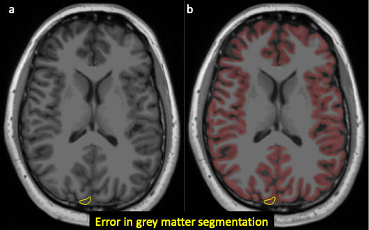
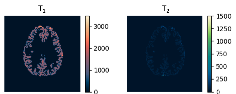
10 volunteers were recruited following informed consent and ethics board approval. The scanning protocol included a 1x1x1mm3 T1 MPRAGE structural volumetric sequence and a 1x1x5mm3 Siemens FISP MRF[1] sequence.The FreeSurfer recon-all command[2] was performed on the high resolution MPRAGE. In 4/10 volunteers, extra cranial non-grey matter like cerebrospinal fluid (CSF) was erroneously included within the ROI of grey matter, particularly in immediate proximity to the sagittal sinus. An example is shown in Figure 1. The FreeSurfer output segmentations were registered to the quantitative maps using a rigid-body registration in FSL[3] and were used to extract data from the quantitative maps. Grey matter segmentations of the quantitative maps are shown in Figure 2.
To correct these segmentations, maximum thresholds are applied based on mean values of CSF in ventricles, also segmented by FreeSurfer. For T1, the CSF mean across 10 volunteers and therefore the maximum threshold for tissue was 2700ms. For T2, it was 255ms.
Results and Discussion
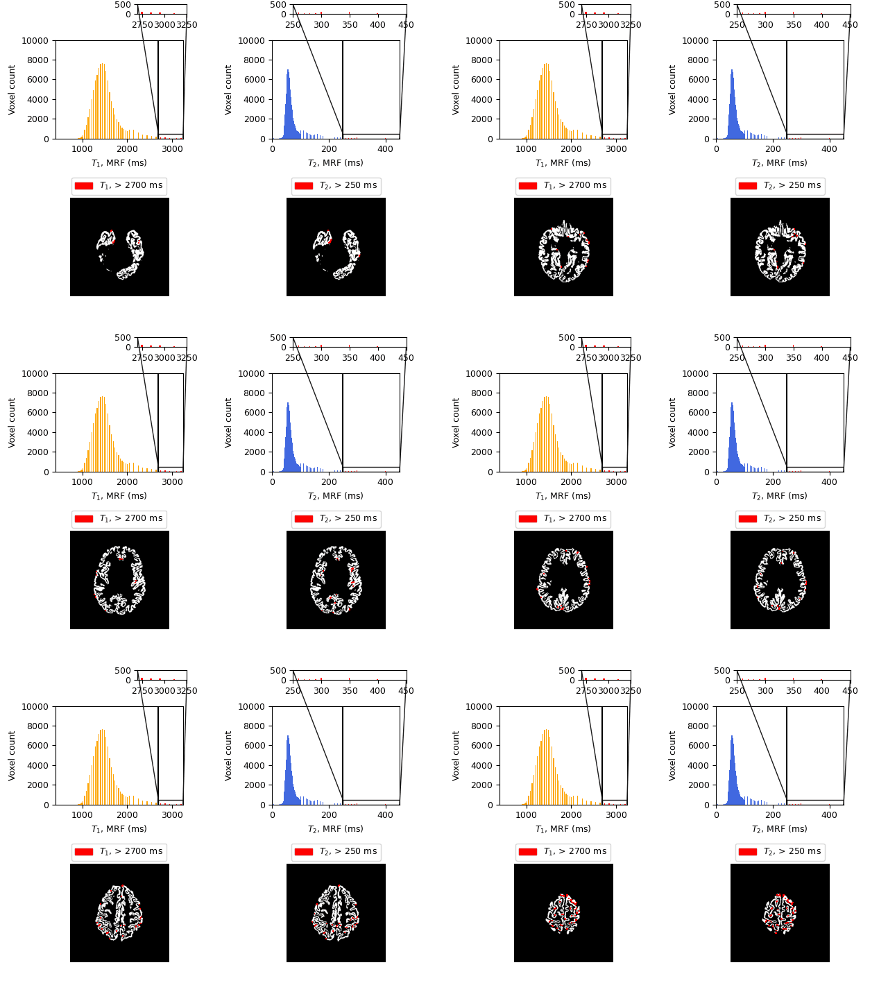
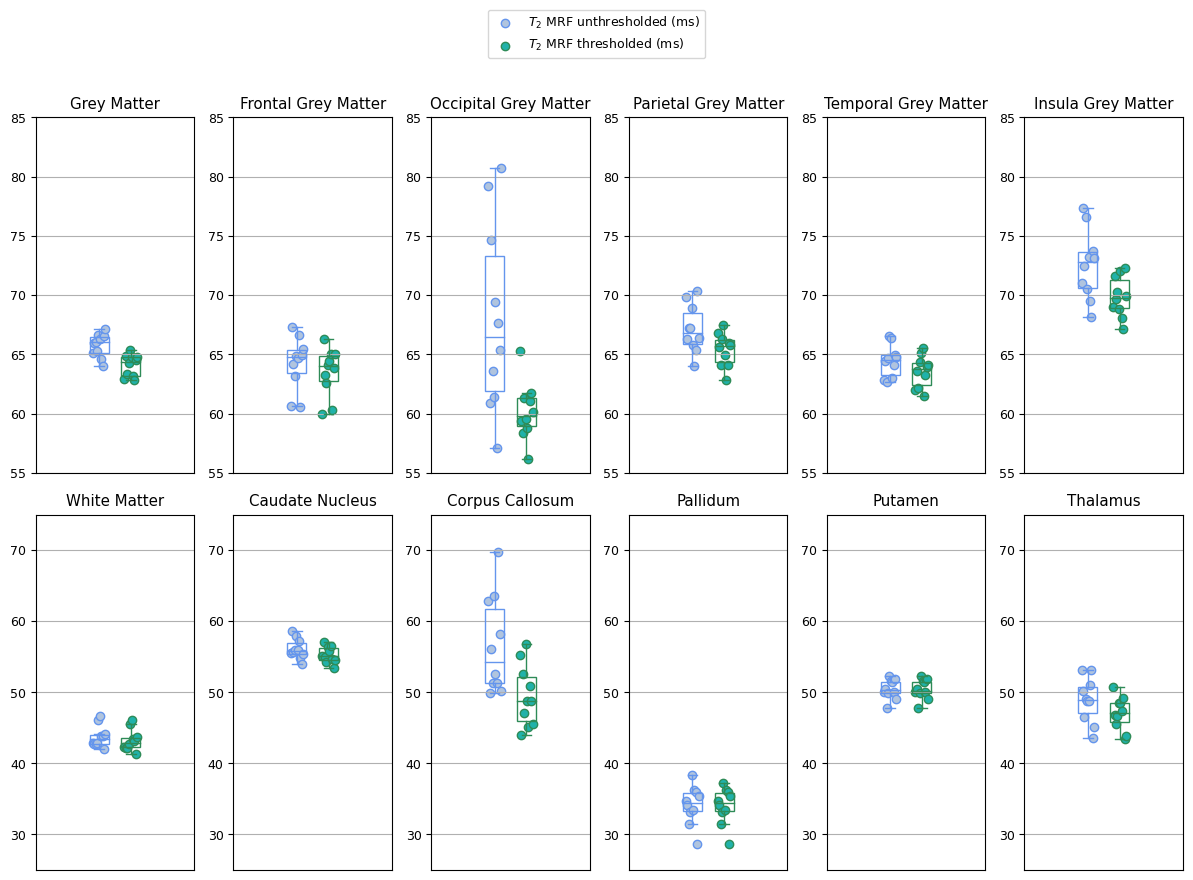
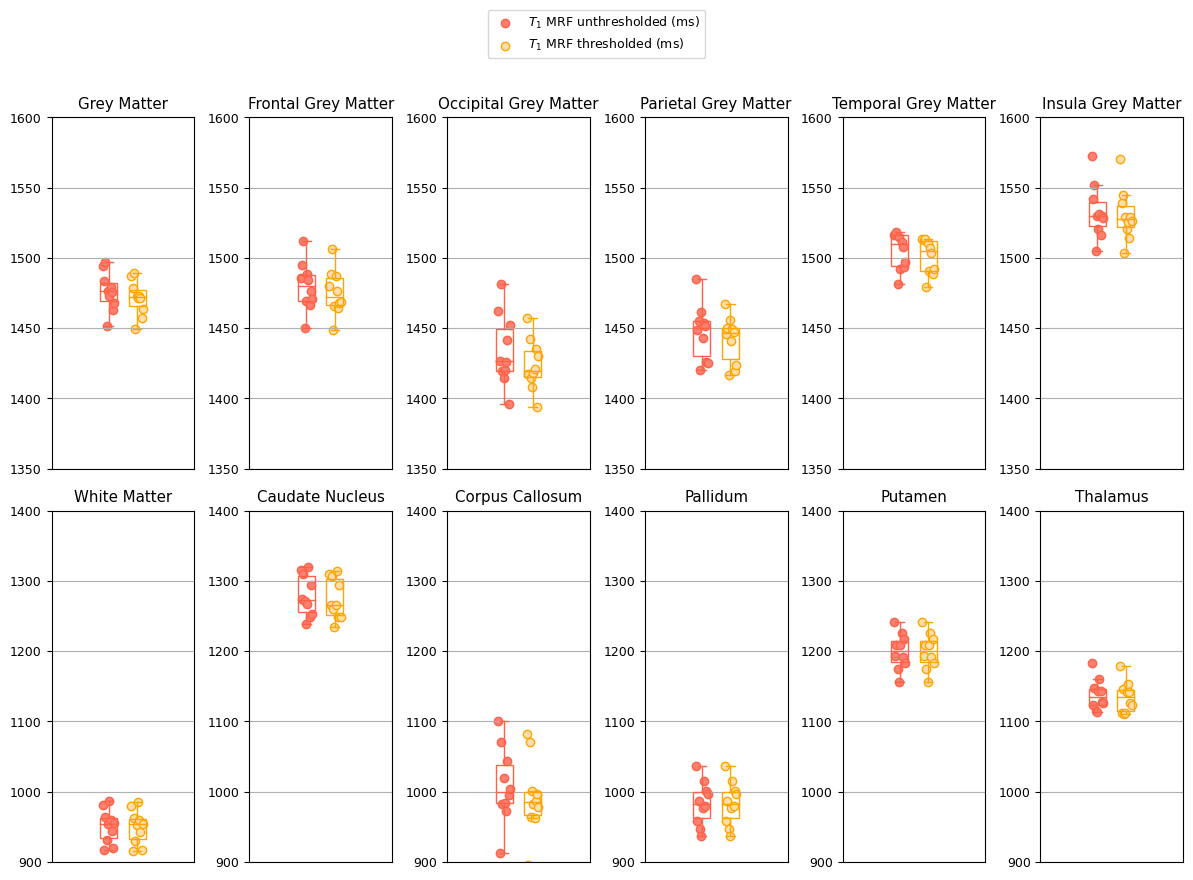
T1 and T2 histograms of grey matter voxels for one healthy volunteer are shown in Figure 3, along with the location of the removed voxels. All voxels in tissue segmentations exceeding mean relaxation times in CSF were removed; these are physically unrealistic for tissue and more consistent with CSF or cortical vessels. Maximum thresholds were computed for both T1 and T2 maps. Significant inclusion of CSF in grey matter occurred in 4/10 healthy volunteers. Biologically unrealistic T1s up to 3500ms and T2s up to 1500ms in the grey matter, shown in Figure 3, are not due to partial voluming of CSF within tissue voxels but due to segmentation errors.Box plots of T2 means in each ROI for each volunteer prior to thresholding are shown in Figure 4a, with thresholding to remove CSF voxels are shown in Figure 4b. Mean values are computed after eroding once (removing voxels in the ROI that are not surrounded by other voxels in the ROI) for every image slice. T1 data are shown in Figure 5.
Comparing Figures 4 and 5, ROIs with variance between volunteers that was impacted most by thresholding were the same for T1 and T2. The corpus callosum is a small white matter region neighbouring CSF in the ventricles, and the occipital grey matter partially surrounds the sagittal sinus, where segmentation errors have been demonstrated in Figure 3. Here, we apply thresholds to T1 and T2 separately to demonstrate applicability to any quantitative mapping method, but since MRF T1 and T2 maps are co-registered, masks of voxels exceeding thresholds of T1 or T2 could be combined for accuracy.
In smaller ROIs such as occipital grey matter which neighbour CSF in the brain, thresholding has a clear impact: less variation in ROI means between volunteers. The inter-volunteer variance of mean T2 in occipital grey matter is reduced by 92% from 57ms to 5.37ms after thresholding. Thresholding by the mean of CSF removes up to 1.8% of the voxels in the occipital grey matter T2 segmentation, however removing these voxels leads to a 22% decrease in mean T2.
The exact placement of the threshold is somewhat arbitrary but requires a meaningful basis. Removing all voxels with a T1 or T2 exceeding the minimum value of the CSF in the ventricles removes up to 40% data. The mean of the CSF is a conservative threshold; we can be certain any discarded voxels are fluid not tissue. Some fluid may still be present, but enough is removed to prevent erroneous variation between subjects that results from CSF and other fluid contamination in ROIs of some volunteers.
Conclusion
Removing voxels from tissue ROIs that exceed the mean relaxation times of CSF in quantitative T1 and T2 maps leads to reduced variation in affected ROIs across 10 volunteers. Thresholds are easily computed and can be applied in a batch process. Removing only up to 1.8% voxels in ROIs with segmentation errors leads to a more than 15% change in T2 average in occipital grey matter for 4/10 volunteers. Where ROIs do not neighbour CSF, there are no voxels removed and therefore no change in mean; all data was processed in one step.Acknowledgements
The authors would like to thank the volunteers who participated in the study and acknowledge funding from the Imperial NHS Imaging Department and the CRUK Convergence Science Centre. The authors would also like to thank the Bangerter Lally group, Imperial NHS ImRes Group, the Imperial MRI Physics Collective, and Mathias Nittka (Siemens Healthineers, Germany).References
1. Jiang Y, et al. MRI Fingerprinting Using Fast Imaging with Steady State Precision (FISP) with Spiral Readout. (2015). Magn Reson Med. 74, 1621-1631.
2. Fischl, B. FreeSurfer. (2012). NeuroImage, 62(2), 774–781.
3. Jenkinson, M., Bannister, P., Brady, M., & Smith, S. (2002). Improved Optimization for the Robust and Accurate Linear Registration and Motion Correction of Brain Images. NeuroImage, 17(2), 825–841.
Figures