2132
Deep learning segmentation of I-125 brachytherapy seeds in prostate cancer patients based on synthetically generated multi-echo training data1Institute of Radiology, University Hospital Erlangen, Erlangen, Germany, 2Department of Radiation Oncology, University Hospital Erlangen, Erlangen, Germany, 3Comprehensive Cancer Center Erlangen-EMN, Erlangen, Germany
Synopsis
Keywords: Electromagnetic Tissue Properties, Machine Learning/Artificial Intelligence, Brachytherapy, Segmentation, Susceptibility
Motivation: Deep learning (DL) networks trained with synthetically generated data enable the visualization of I-125 brachytherapy seeds in prostate cancer patients in quantitative susceptibility mapping (QSM), possibly eliminating the need for a CT-scan in future.
Goal(s): The Goal was to automatically detect and segment I-125 seeds in-vivo by using a DL network directly (without QSM) on gradient-echo-sequence (GRE) data.
Approach: A U-Net was trained with synthetically generated multi-echo GRE magnitude and phase input data and corresponding target seed segmentations.
Results: The seed segmentations were of high visual quality and showed good agreement (85% detection rate) with corresponding CT-scans in five prostate cancer patients.
Impact: This work proposes a fast and completely automatic MRI-only based workflow for segmenting in-vivo brachytherapy seeds in prostate cancer patients. This approach has the potential to eliminate the need for a CT-scan, thereby reducing the use of ionizing radiation.
Introduction
A common approach in treating prostate cancer is the application of low-dose-radiation brachytherapy using I-125 seeds, which contain Ti and Ag1,2. Usually a CT-scan is performed to monitor local implantation. While the CT offers seed visualization, it provides only poor soft tissue contrast and requires ionizing radiation exposure.Recently, I-125 seeds were visualized with MRI by using a deep learning (DL) quantitative susceptibility mapping (QSM) reconstruction method based on synthetically generated training data.3 Subsequent seed segmentation can be performed either manually, or automatically via a second DL network.2
The aim of this work, parts of which have been presented previously4, was to further investigate the possibility of detecting and localizing I-125 seeds by using a DL network directly (without QSM) on gradient-echo (GRE) phase and magnitude input data.
Methods
To train a neural network for I-125 seed segmentation, 250 synthetic phase maps and magnitude images with 3TE each, were generated together with their corresponding seed segmentation targets. The phase maps and magnitudes images consisted of a large ellipsoid simulating the pelvis, which was filled with smaller ellipsoids and rectangles of different sizes. Each shape was assigned a normal distributed magnetic susceptibility value and a Rician distributed magnitude value.3,5 Additionally, cylindrical seeds were simulated and randomly positioned. Values of uniformly distributed susceptibility ($$$\chi\in[1,6]$$$ ppm) and magnitude were designated to each seed. For each data set, random background fields were simulated.3The corresponding phase $$$\phi$$$ was calculated via convolution with $$$d_z$$$ the z-component of a point-dipole response:6
$$\phi=\gamma B_0(\chi\ast d_z)TE\quad,$$
with $$$B_0$$$=1.5T and the gyromagnetic ratio $$$\gamma$$$. Then Laplacian-based phase unwrapping3,6 was applied to all generated maps, before the TE related seed phase maps, shape phase maps and background field were added on top of each other. Between 0%-50% of these shapes were selected to mimic fat tissue by adding an offset. Phase maps and magnitude images were calculated at TE=2.8/ 5.82/ 8.84 ms. Lastly, Gaussian and Rician noise was added to phase and magnitude respectively.5 Figure 1 shows representative input training data. Corresponding seed segmentation targets were generated.
A U-Net3,7 was trained on patches of the synthetic input and target maps for 250.000 training pairs (Figure 2). A Tversky loss function8 was chosen with the weight $$$\beta$$$=0.25.
To evaluate the network, in-vivo prostate scans of five brachytherapy patients were acquired on a 1.5T MRI scanner (MAGNETOM Sola, Siemens Healthineers, Erlangen). A GRE imaging sequence with TE=2.8/ 5.82/ 8.84/ 11.86/ 14.88/ 17.89 ms, TR=21 ms, bandwidth=780 Hz/px, voxel size=1×1×2 mm³, and TA=5:07 min was used.2,3 Furthermore, a CT-scan from the same day was used as seed segmentation ground truth (GT).2
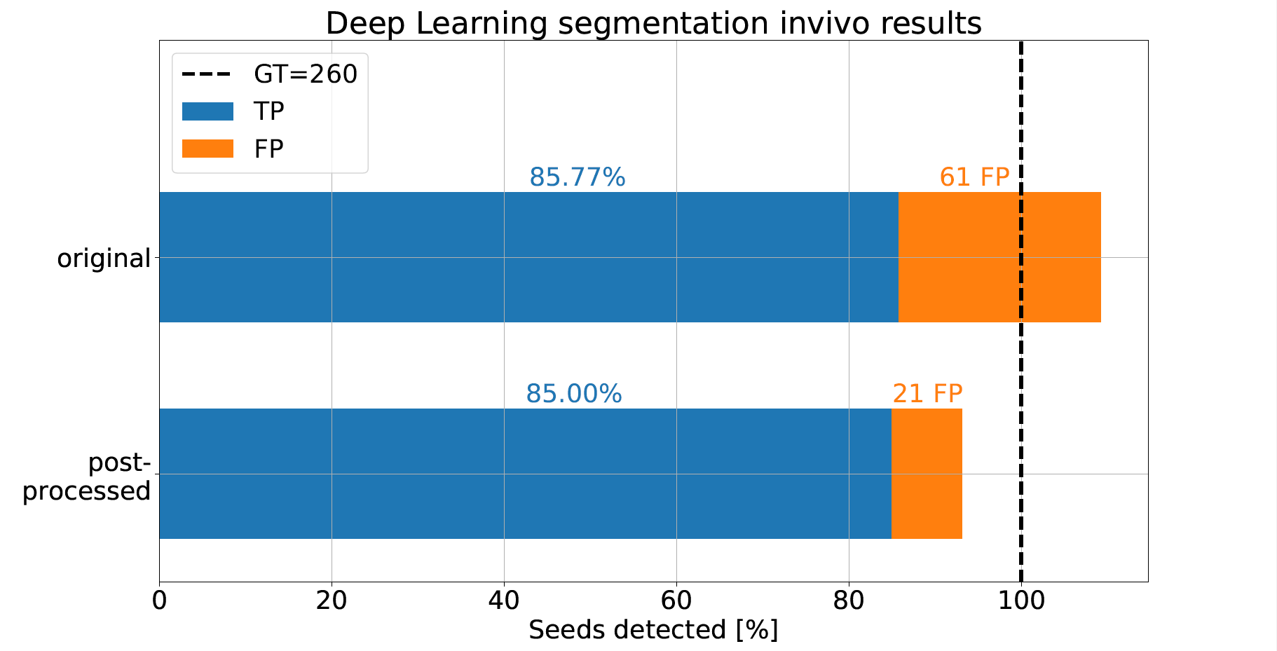
After applying the network to the in-vivo data of the first three TEs, a filter eliminated all predictions with cluster size below 52 connected voxels. The amount of true positives (TP, seeds detected correctly) consisted of all seed predictions intersecting with the CT based GT, yielding the number of false positives (FP) and false negatives (FN) accordingly.
Results
Figure 3 shows the 3D segmentation results of all five patients. The predicted TP seeds were in good agreement with the GT regarding size and position, apart from minor offsets/mismatches.The network correctly identified 85% of the seeds (Figure 4), where most patients had a TP rate of 84%-96% and 1-4 FPs. Patient 2 posed an exception, with a detection rate of 49% and 14 FPs. The FPs of all patients amounted to 21.
Discussion
The proposed approach allows a fast and completely automatic seed detection in-vivo, potentially accelerating dosimetric analysis in prostate cancer treatment. An advantage of using synthetically generated training data is that the network training becomes independent of in-vivo patient data, allowing the flexible development of a DL seed segmentation tool. The network can be adjusted to different sequence parameters and field strengths and does not rely on gathering vast amounts of in-vivo training data beforehand.There are still challenges that need to be tackled, as a TP rate of 100% would be desirable. The phase map quality could be increased by using novel denoising algorithms or higher field strengths. The CT seeds appear shifted to the associated dipole field position in the phase maps (Figure 3). This is presumably a result of misregistration between CT- and MRI-scan, caused by slightly different organ alignment, e.g., by a filled bladder.
Conclusion
Training a DL segmentation network on synthetically generated GRE phase and magnitude data allows for automatic detection of in-vivo brachytherapy seeds in prostate cancer patients with high visual quality, possibly eliminating the need for a CT-scan.Acknowledgements
We thank the Imaging Science Institute (Erlangen, Germany) for providing us with measurement time.References
1. Eckert&Ziegler BEBIG GmbH. Fact sheet IsoSeed I25.S17plus. https://medical.ezag.com /de/products/isoseed-i25-s17plus/
2. Grigo J, Karius A, Bert C, Putz F. Towards a deep learning based magnetic resonance imaging only workflow for postimplant dosimetry in I-125 seed brachytherapy for prostate cancer. Proc Intl Soc Brachytherapy
3. Hanspach J, Laun F. Deep learning-based quantitative susceptibility mapping (QSM) in the presence of fat using synthetically generated multi-echo phase training data. Magn. Reson. Med. 2022; 88:1548-1560
4. Mücke L, Grigo J, Karius A, Bert C, Uder M, Laun F, Hanspach J. Training a deep learning based segmentation network for I-125 brachytherapy seed detection in prostate cancer patients using synthetically generated multi-echo MRI training data. Proc Intl DS-ISMRM 2023
5. Gudbjartsson H, Patz S. The rician distribution of noisy MRI data. Magn. Reson. Med. 1995; 34.6:910-914
6. Deistung A, Schweser F, Reichenbach J. Overview of quantitative susceptibility mapping. NMR Biomed. 2017, 30:e3569.
7. Ronneberger O, Fischer P, Brox T. U-Net: Convolutional Networks for Biomedical Image Segmentation. MICCAI 2015; 234-241
8. Jadon S. A survey of loss functions for semantic segmentation. IEEE (CIBCB) 2020
Figures
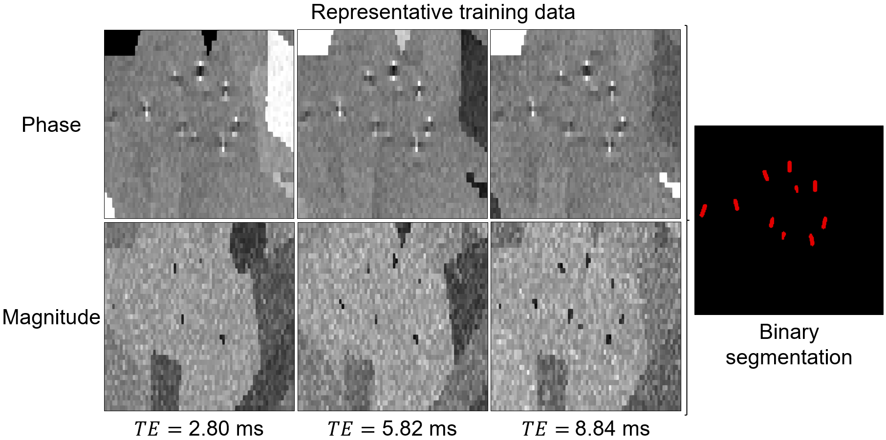
Figure 1: Representative example of one synthetically generated training data set consisting of phase maps (upper row) and magnitude images (lower row) at three TEs. These, together with the corresponding high resolution segmentation target (right), were used as training data for a U-Net. The phase maps include random fat offsets and seed related dipole fields, while in the magnitude images the seeds are marked by hypointensities.
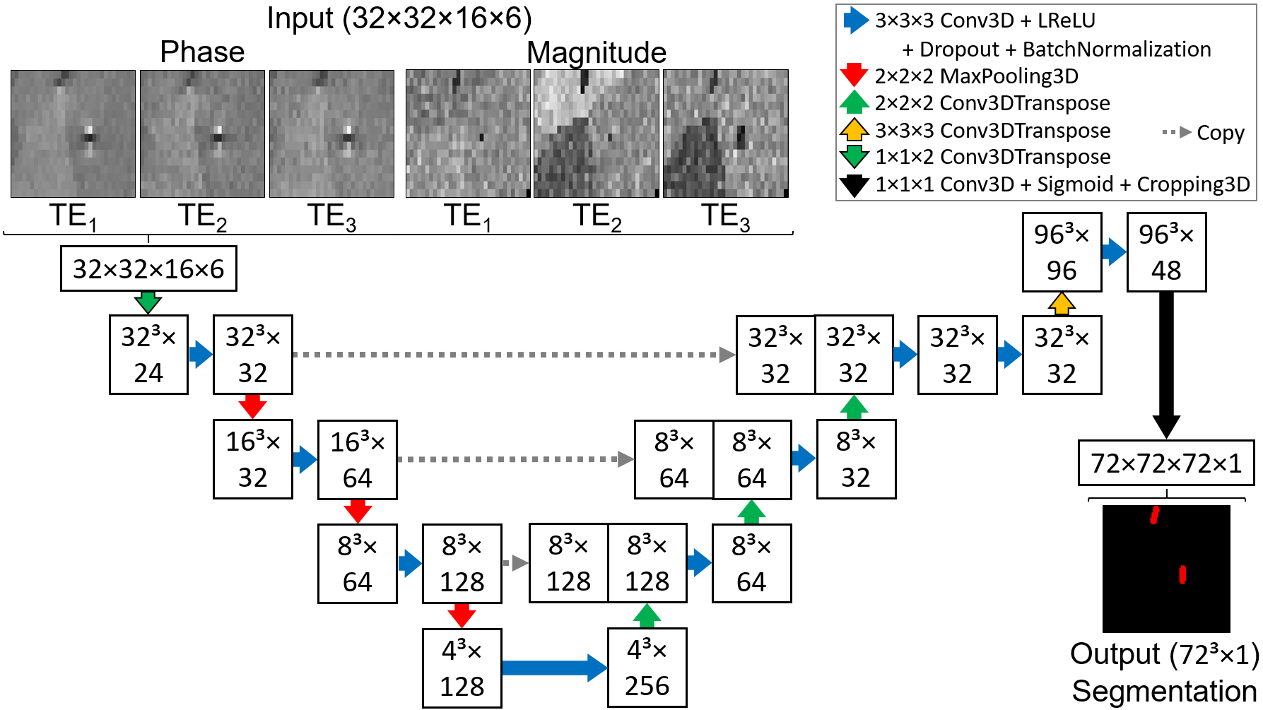
Figure 2: Illustration of the U-Net architecture, which was enhanced by a super resolution extension to increase the resolution of the seed segmentation. Unwrapped phase maps and magnitude patches (32×32×16×6) at three TE served as input data, while the seed segmentation target was cropped3 (72×72×72).
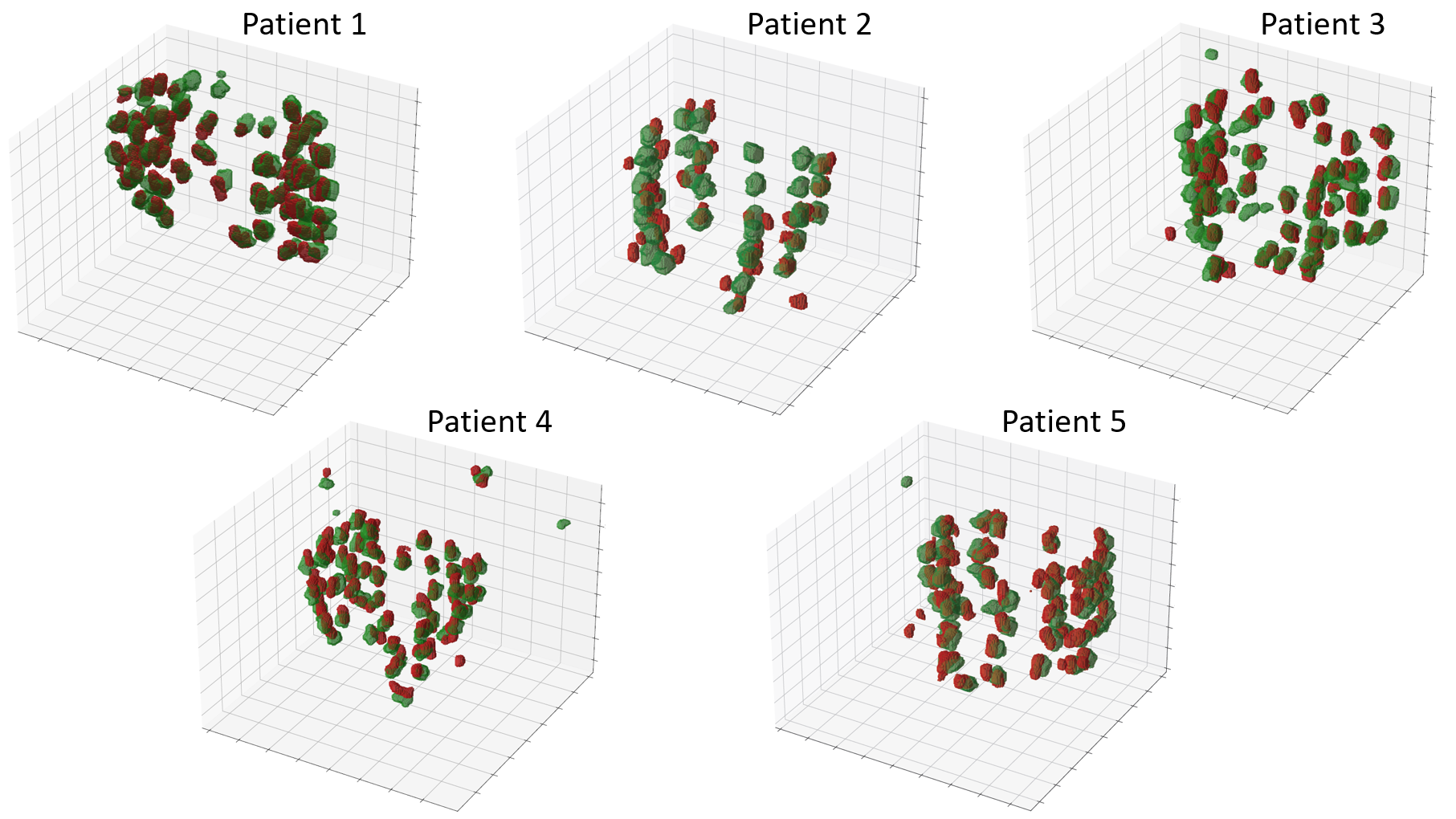
Figure 3: DL segmentation results in 3D of the five patients, comparing the network prediction (red) with the CT-scan based GT (green). For each patient there is a slight systematic offset in position between prediction and GT. Apart from this minor mismatch the predictions are in good visual agreement with the GT.