2124
MRF-Synth: An Image Generation Framework for Learning Contrast-Invariant Brain Segmentation1Biomedical Engineering, Case Western Reserve University, Cleveland, OH, United States, 2Radiology, Case Western Reserve University, Cleveland, OH, United States, 3Radiology, University of North Carolina at Chapel Hill, Chapel Hill, NC, United States
Synopsis
Keywords: Segmentation, Brain, Magnetic Resonance Fingerprinting, Contrast-Invariant, Age-Agnostic
Motivation: Intensity-based brain segmentation methods face challenges with generalizability, as they are susceptible to site, age, and contrast variations.
Goal(s): Develop an efficient, unified framework to train segmentation networks that are insensitive to contrast variations.
Approach: MRF sequences encode image time series that include both common and uncommon contrasts. We develop MRF-Synth, a framework to generate contrasts from MRF quantitative maps for training and evaluating contrast-invariant networks.
Results: We show that a segmentation U-Net trained with MRF-Synth yields highly consistent results across contrasts, vendors, and ages (DICE > 0.86 in adults).
Impact: MRF-Synth represents an efficient, generalizable framework for developing and evaluating contrast-invariant segmentation networks. We demonstrate the utility of MRF-Synth in training a U-Net to segment healthy MR brain images into 18 anatomical regions regardless of contrast, scanner, vendor, or age.
Introduction
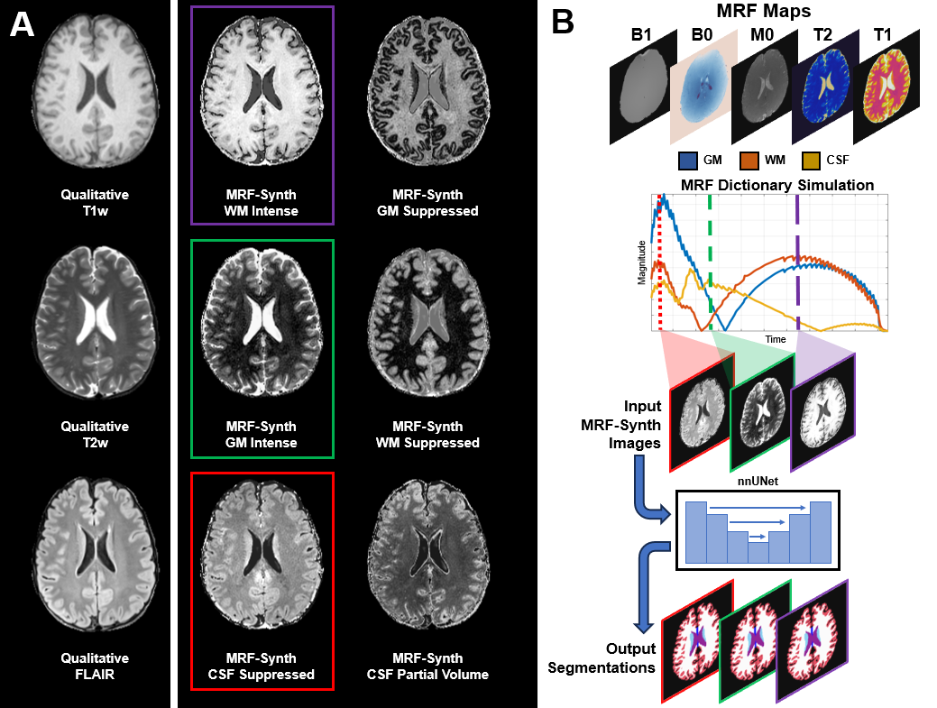
MR images commonly exhibit contrasts that vary between tissues (Fig. 1A), sites, and scanners1,2. Downstream tasks, like segmentation, can struggle to generalize to images with variable contrasts3,4. Existing approaches for contrast generation have important limitations. Contrast synthesis from quantitative maps5,6 is currently limited to simulating specific sequences. Contrast synthesis based on domain randomization7 disregards how MR physics and tissue parameters affect contrasts.MR Fingerprinting8 (MRF) allows simultaneous quantification of tissue parameter maps such as T1, T2, and M0, as well as B0 and B1, from which an arbitrary number of contrasts can be synthesized (Fig. 1A). We developed a framework, called MRF-Synth, to generate multiple contrasts for a single anatomy based on MRF parameter matching and dictionary simulation for training nnUNet9 to segment MR brain images with widely different contrasts.
Methods
MRF-Synth images represent simulations of MRF time series by modeling voxel-wise signals using the Bloch equations and parameter maps. These images can be saved during MRF reconstruction pattern matching or reproduced from quantitative maps with a dictionary (Fig. 1B). Using MRF-Synth images, we trained a network for anatomical segmentation. We deployed an off-the-shelf U-Net (nnUNetv29) to demonstrate that performance is driven by MRF-derived contrasts, not novel network architecture.Training data comprised 32 single-site healthy volunteer 3D MRF scans sharing a 1,440 time point dictionary10. 145 time points per subject were randomly sampled for a total of 4,640 variable-contrast brain volumes. Images were skull stripped using FreeSurfer11 (v7.4.1) and converted to size 256x256x256 with 1mm isotropic resolution. A 3d_fullres nnUNetv2 was trained with an 80/20 training/validation data split. Training used a summed Cross Entropy and DICE loss with FreeSurfer (v7.4.1) reference labels of 18 brain regions derived from MRF-synthesized T1w images. Training was performed for 1,000 epochs on the CWRU HPC using an Nvidia V100 GPU (32GB memory), taking a total of 68.9 hours.
Testing data were 8 subjects with clinical and MRF images10, as well as cross-vendor clinical images from 4 additional subjects12 and 2 infant subjects13. We investigated the performance of a segmentation network trained with MRF-Synth in four experiments:
- Evaluation of the segmentation of 145 MRF-Synth images with varying image contrasts with respect to FreeSurfer and voxel-wise label consensus across MRF-Synth segmentations.
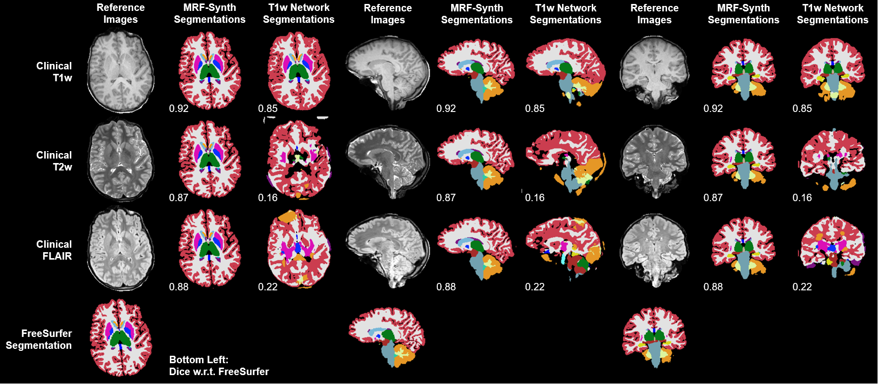
- Comparison of segmentation results of multi-contrast clinical images based on U-Nets trained only with T1w images and trained using the whole spectrum of contrasts.
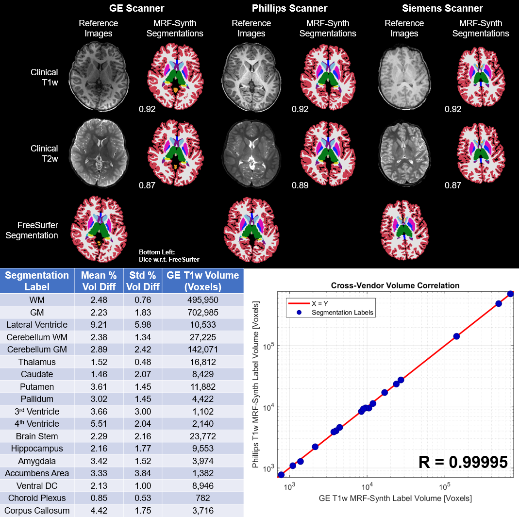
- Evaluation of segmentation using clinical scans from three vendors. Single-subject label volume consistency was evaluated as the percent volume differences between a GE T1w MRF-Synth segmentation and three other variable contrast/vendor results.
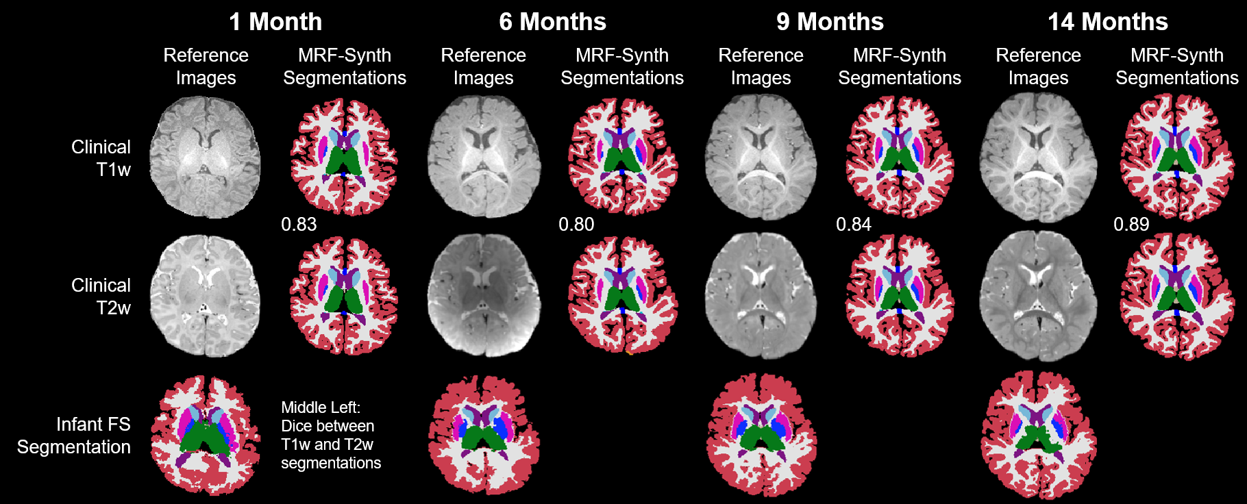
- Evaluation of generalizability to infant scans unseen in training with respect to infant FreeSurfer14.
$$$DICE=\sum_{k=1}^{K}\left(\frac{N_{X,k}}{N_{X}}\right)\left(\frac{2\left|X_{k}\bigcap_{}^{}Y_{k}\right|}{\left|X_{k}\right|+\left|Y_{k}\right|}\right)$$$
where K is the number of labels in images X and Y, NX is the number of voxels in X, NX,k is the number of voxels assigned label k in X, and Xk and Yk are binary equivalents of X and Y for k.
Results
MRF-Synth Multi-Contrast (Fig. 2)MRF-Synth segmentations of 145 MRF-Synth test images were compared to FreeSurfer reference (DICE ≥ 0.92). Segmentation variability was quantified by comparing individual labels with respect to a voxel-wise label consensus computed across all images. Learning with MRF-Synth yielded consistently high accuracy despite variations in contrast (DICE ≥ 0.95).
Clinical Multi-Contrast (Fig. 3)
Evaluations based on clinical images with three contrasts indicated MRF-Synth produced segmentations comparable to FreeSurfer (DICE ≥ 0.87). Unlike contrast-invariant learning with MRF-Synth, an nnUNet trained on T1w-like images performs poorly on T2w and FLAIR images (DICE ≥ 0.16).
Clinical Cross-Vendor (Fig. 4)
MRF-Synth segmentation generalized across three different vendors (DICE ≥ 0.87), despite substantial contrast and intensity variations. Segmentation agreement of one subject between vendors indicated appreciable segmentation repeatability with MRF-Synth (T1w DICE 0.89, T2w DICE 0.88, Label Volume Difference 3.14 ± 2.66 %, Cross-Vendor Label Volume Pearson Correlation Coefficient 0.9995).
Clinical Cross-Age (Fig. 5)
Generalizability was further assessed by applying the segmentation network trained with adult MRF-Synth data to longitudinal infant images. MRF-Synth segmentations are consistent across ages (DICE ≥ 0.80) despite the inverted WM/GM contrasts and low contrast-to-noise ratio in neonate images. Single-timepoint T1w vs T2w segmentation DICE comparisons range from 0.80-0.89, even in a near-isointense 6-month-old subject.
Conclusion
We present MRF-Synth, a contrast-invariant network training framework leveraging the spectrum of image contrasts inherent to MRF. Despite training with single-site adult MRF data, our segmentation network was generalizable across vendors, ages, and contrasts, and was resilient to spatial intensity biases. Future iterations using manual refinements of the FreeSurfer labels should further improve segmentation results.Acknowledgements
This work was supported by NIH grants R01 CA269604, R01 CA282516, R01 NS109439, and UKRI MR/W031566.
This work was also supported in part by National Institutes of Health (NIH) under grants R01EB008374, R01MH125479, and R01CA266702.
This work made use of the High Performance Computing Resource in the Core Facility for Advanced Research Computing at Case Western Reserve University.
References
- Biberacher V, Schmidt P, Keshavan A, et al. Intra- and interscanner variability of magnetic resonance imaging based volumetry in multiple sclerosis. Neuroimage 2016; 142:188-197.
- Shinohara RT, Oh J, Nair G, et al. Volumetric Analysis from a Harmonized Multisite Brain MRI Study of a Single Subject with Multiple Sclerosis. American Journal of Neuroradiology 2017; 38(8):1501-1509.
- Jovicich J, Marizzoni M, Sala-Llonch R, et al. Brain morphometry reproducibility in multi-center 3T MRI studies: A comparison of cross-sectional and longitudinal segmentations. NeuroImage 2013; 83:472-484.
- Clark K, Woods R, Rottenberg D, et al. Impact of acquisition protocols and processing streams on tissue segmentation of T1 weighted MR images. NeuroImage 2006; 29:185-202.
- Borges P, Shaw R, Varsavsky T, et al. Acquisition-invariant brain MRI segmentation with informative uncertainties. Arxiv 2021; 2111.04094.
- Jog A, Hoopes A, Greve D, et al. PSACNN: Pulse sequence adaptive fast whole brain segmentation. NeuroImage 2019; 199:553-569.
- Billot B, Greve D, Puonti O, et al. SynthSeg: Segmentation of brain MRI scans of any contrast and resolution without retraining. Medical Image Analysis 2023; 86:102789.
- Ma D, Gulani V, Seiberlich, N, et al. Magnetic Resonance Fingerprinting. Nature 2013; 495(7440):187-192.
- Isensee F, Jaeger P, Kohl S, et al. nnU-Net: a self-configuring method for deep learning-based biomedical image segmentation. Nature Methods 2021; 18(2):203-211.
- Ma D, Jones S, Deshmane A, et al. Development of High-Resolution 3D MR Fingerprinting for Detection and Characterization of Epileptic Lesions. JMRI 2019; 49:1333-1346.
- Fischl, B. FreeSurfer. NeuroImage 2012; 62 (2) 774-781.
- Jernigan T, Brown S, Dowling G. The Adolescent Brain Cognitive Development Study. Journal of Research on Adolescence 2018; 28 (1) 154-156.
- Howell B, Styner M, Gao W, et al. The UNC/UMN Baby Connectome Project (BCP): An overview of the study design and protocol development. NeuroImage 2019; 185:891-905.
- Zöllei L, Iglesias J, Ou Y, et al. Infant FreeSurfer: An automated segmentation and surface extraction pipeline for T1-weighted neuroimaging data of infants 0-2 years. NeuroImage 2020; 218:116946.
Figures