2123
High quality brain segmentation from synthetic MPRAGE images at 7T MRI1Department of Physics, NTNU, Trondheim, Norway, 2DZNE, Bonn, Germany, 3GIGA-CRC In Vivo Imaging, University of Liège, Liège, Belgium, 4Department of Physics and Astronomy, University of Bonn, Bonn, Germany
Synopsis
Keywords: Segmentation, Brain, High-Field MRI, Data Analsyis, Anaysis/Processing, Segementation, Multi-Contrast, Neuro-imaging, synthetic MPRAGE
Motivation: Brain segmentation and multiparameter mapping (MPM) are important for neurodegenerative disease characterization. Acquiring sub-millimeter images increases scan time and patient discomfort. At 7T, B1+ inhomogeneities challenge brain segmentation.
Goal(s): The quality of brain segmentations produced from FastSurferVINN was evaluated and compared between a 7T MPRAGE protocol and two synthetic MPRAGE approaches.
Approach: MPRAGE and MPM images were acquired on 16 subjects across three 7T sites using pTx pulses. MPRAGElike and synMPRAGE images were generated from MPM. All images were segmented with FastSurferVINN.
Results: FastSurferVINN seems to be a robust technique to segment sub-millimeter 7T images. MPRAGElike generated superior segmentations compared to synMPRAGE.
Impact: Neuroscientists with Multi-Parameter Mapping sequences in their imaging protocol can approximate an MPRAGElike image (preferably over synthetic synMPRAGE). Acquiring an MPRAGE sequence solely for brain segmentation can be avoided resulting in a considerable amount of scan time saved.
Introduction
Most brain segmentation algorithms are not optimized to handle the B1+ inhomogeneities and sub-millimeter resolution typically obtained at 7T1. Moreover, most of them require an anatomical T1-weighted image like MPRAGE2. Often brain segmentation is the only purpose of such T1w images. Thus, high-resolution neuro-imaging protocols for neurodegenerative disease characterization that contain both multiparameter mapping and MPRAGE can become quite long.Recently, Deep-Learning-based segmentation techniques have been proposed like FastSurferVINN3 which produces sub-millimeter brain segmentations of MPRAGE images. Even if optimized for 3T, FastSurferVINN then becomes an interesting option for 7T images.
Additionally, recent developments of Universal Pulses4 (UP) at Ultra-High-Field MRI (UHF-MRI) allows the straightforward usage of parallel transmit (pTx) pulses5, resulting in a significant reduction of B1+ inhomogeneities.
The objective of this study was twofold. First, to assess the performance of FastSurferVINN in an untested setting with (0.6mm)3 7T images acquired with UPs. Second, to compare the quality of segmentations calculated from two ‘synthetic MPRAGE’6 techniques based on Multi-Parametric Mapping7 (MPM) against the segmentation quality from MPRAGE images.
Methods
16 healthy volunteers were scanned on two 7T Terra and one 7TPlus Magnetom systems (Siemens Healthineers, Erlangen, Germany) at three sites equipped with 8Tx-32Rx head coils (Nova Medical, Wilmington, Delaware) in pTx mode. The study was approved by the local review boards of each site and volunteers signed a written informed consent form before scanning.MPM, MPRAGE and B1+ map were acquired for each subject. The MPM protocol consisted of a multi-echo skipped-CAIPI 3D-EPI8 at (0.6mm)3 resolution with MTw, PDw and T1w contrasts (4 TEs each). MPM and MPRAGE used UPs created from a database of B0-B1 maps acquired at all sites. A B1+ map was acquired with the Actual Flip Angle Imaging9 (AFI) technique. Acquisition parameters are shown in Table 1.
Marchenko-Pastur PCA denoising10-13 was applied utilizing high redundancy in the twelve raw MPM images. Then, the MPM images and B1+ map were given to the hMRI toolbox14 (v0.3.0) embedded in SPM1215 to calculate B1+ corrected R1, PD, MTsat, and R2* quantitative maps (qMaps) in addition to coregistering MPM and MPRAGE images together.
Two techniques were utilized to generate ‘synthetic MPRAGE’ images. First, a technique deriving an MPRAGElike16 image from the 1st TE MPM images with the following equation was used:
$$MPRAGE_{like} = \frac{T_{1w,TE1} - 100}{0.5 \times (PD_{w,TE1} + MT_{w,TE1}) + 100}$$
where 100 is a regularization factor to avoid noise in non-tissue areas17.
The second approach synthesizes synMPRAGE6 images from the MPM-derived R1 map using parameters matched with the reference MPRAGE.
All images were segmented with FastSurferVINN into 35 brain subregions and quantitatively evaluated with and without preceding N4 bias-field correction18-19. The complete pipeline is shown on Figure 2.
Results and Discussion
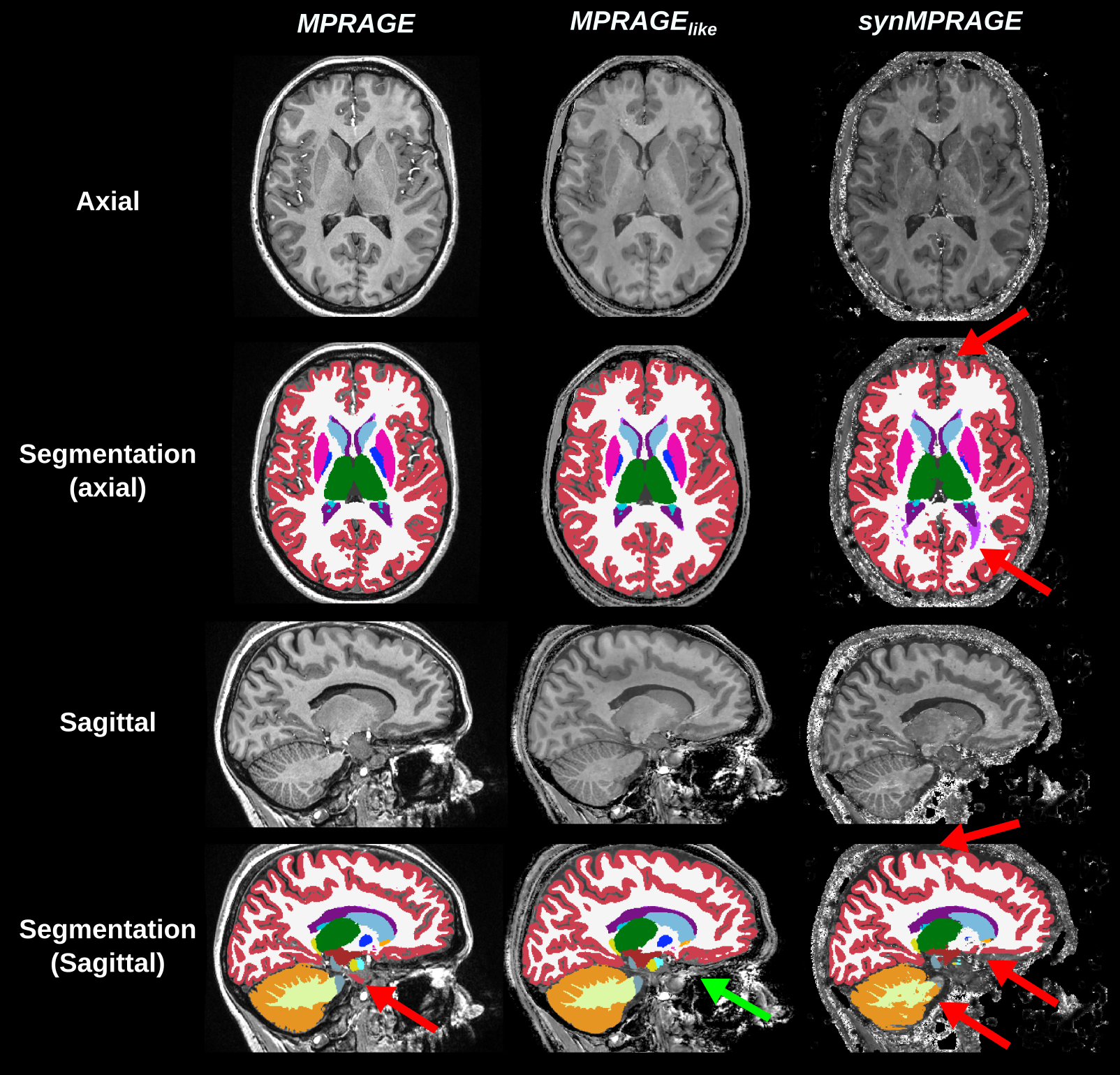
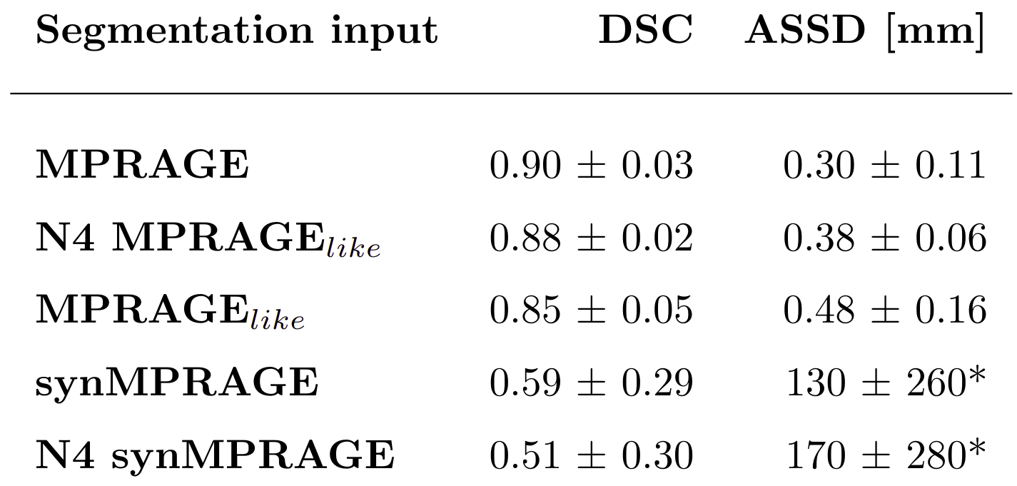
FastSurferVINN properly segmented both the uncorrected and N4-corrected MPRAGE images as shown in Figure 3. Fine structures like cortical sulci benefited from the (0.6mm)3 resolution whereas thin cerebellum white matter (WM) branches were not fully segmented. The cortical region next to sinuses appeared to be challenging to segment even for the N4-corrected MPRAGE where no improvement was observed. Nevertheless, N4-correction helped in regions with low WM-GM contrast.Furthermore, FastSurferVINN successfully segmented the MPRAGElike and synMPRAGE as seen with Figure 4. MPRAGElike showed great similarities with MPRAGE visually and quantitatively according to the mean Dice Similarity Coefficient (DSC) and Average Symmetric Surface Distance (ASSD) values reported in Table 2. The ASSD values reported for the uncorrected MPRAGE and both versions of MPRAGElike were smaller than one voxel. SynMPRAGE gave noisier segmentations with errors in WM and cortex. The DSC and ASSD were substantially inferior compared to the other techniques besides also having large standard deviations. Particularly large ASSD values reported for both synMPRAGEs are due to missing segmentations by FastSurferVINN. Ultimately, the inferior metric values for synMPRAGEs were due to higher variability in image quality. This is presumably due to a higher sensitivity to motion artifacts compared to MPRAGElike.
The bias correction detrimentally affected the synMPRAGE segmentations compared to MPRAGE and MPRAGElike. This adverse effect could come from the fact that R1 maps (hence synMPRAGE) do not exhibit typical B1 variations across the image.
It is worth noting that the N4-corrected MPRAGE used as the reference corresponds to a ‘silver standard’ since no manual segmentations nor expert visual assessment were performed.
Conclusion
FastSurferVINN allows segmenting sub-millimeter 7T T1w images across the whole brain in a previously untested setting (UPs, (0.6mm)3 & synthetic MPRAGEs). Moreover, MPRAGElike offers a more robust and simpler approximation than synMPRAGE. Thus, neuroscientists using MPM protocols can consider calculating MPRAGElike images instead of acquiring an MPRAGE solely for brain segmentation.Acknowledgements
No acknowledgement found.References
Svanera, M., Benini, S., Bontempi, D., & Muckli, L. (2021). CEREBRUM‐7T: Fast and Fully Volumetric Brain Segmentation of 7 Tesla MR Volumes. Human brain mapping, 42(17), 5563-5580.
Mugler III, J. P., & Brookeman, J. R. (1990). Three‐dimensional magnetization‐prepared rapid gradient‐echo imaging (3D MP RAGE). Magnetic resonance in medicine, 15(1), 152-157.
Henschel, L., Kügler, D., & Reuter, M. (2022). FastSurferVINN: Building resolution-independence into deep learning segmentation methods—A solution for HighRes brain MRI. NeuroImage, 251, 118933.
Gras, V., Vignaud, A., Amadon, A., Le Bihan, D., & Boulant, N. (2017). Universal pulses: a new concept for calibration‐free parallel transmission. Magnetic resonance in medicine, 77(2), 635-643.
Padormo, F., Beqiri, A., Hajnal, J. V., & Malik, S. J. (2016). Parallel transmission for ultrahigh‐field imaging. NMR in Biomedicine, 29(9), 1145-1161.
Nöth, U., Hattingen, E., Bähr, O., Tichy, J., & Deichmann, R. (2015). Improved visibility of brain tumors in synthetic MP‐RAGE anatomies with pure T1 weighting. NMR in Biomedicine, 28(7), 818-830.
Weiskopf, N., & Helms, G. (2008). Multi-parameter mapping of the human brain at 1mm resolution in less than 20 minutes. Proceedings of 16th ISMRM, Toronto, Canada, 16, 2241.
Stirnberg, R., & Stöcker, T. (2021). Segmented K-space blipped-controlled aliasing in parallel imaging for high spatiotemporal resolution EPI. Magnetic resonance in medicine, 85(3), 1540–1551.
Yarnykh, V. L. (2007). Actual flip‐angle imaging in the pulsed steady state: a method for rapid three‐dimensional mapping of the transmitted radiofrequency field. Magnetic Resonance in Medicine: An Official Journal of the International Society for Magnetic Resonance in Medicine, 57(1), 192-200.
Veraart, J., Novikov, D. S., Christiaens, D., Ades-Aron, B., Sijbers, J., & Fieremans, E. (2016). Denoising of diffusion MRI using random matrix theory. Neuroimage, 142, 394-406.
Veraart, J., Fieremans, E., & Novikov, D. S. (2016). Diffusion MRI noise mapping using random matrix theory. Magnetic resonance in medicine, 76(5), 1582-1593.
Cordero-Grande, L., Christiaens, D., Hutter, J., Price, A. N., & Hajnal, J. V. (2019). Complex diffusion-weighted image estimation via matrix recovery under general noise models. Neuroimage, 200, 391-404.
Tournier, J. D., Smith, R., Raffelt, D., Tabbara, R., Dhollander, T., Pietsch, M., ... & Connelly, A. (2019). MRtrix3: A fast, flexible and open software framework for medical image processing and visualisation. Neuroimage, 202, 116137.
Tabelow, K., Balteau, E., Ashburner, J., Callaghan, M. F., Draganski, B., Helms, G., ... & Mohammadi, S. (2019). hMRI–A toolbox for quantitative MRI in neuroscience and clinical research. Neuroimage, 194, 191-210.
Ashburner, J., Barnes, G., Chen, C. C., Daunizeau, J., Flandin, G., Friston, K., ... & Penny, W. (2014). SPM12 manual. Wellcome Trust Centre for Neuroimaging, London, UK, 2464(4).
Stirnberg R., Völzke Y., Löwen D., Pracht E., … & Stöcker T. (2023). High-resolution whole-brain multi-parameter mapping at 7 Tesla with interleaved fly-back 3D-EPI and universal pTX pulses. Proceedings of the International Society of Magnetic Resonance in Medicine, 2023, Toronto. 0272.
O'Brien, K. R., Kober, T., Hagmann, P., Maeder, P., Marques, J., Lazeyras, F., ... & Roche, A. (2014). Robust T1-weighted structural brain imaging and morphometry at 7T using MP2RAGE. PloS one, 9(6), e99676.
N4: Tustison, N. J., Avants, B. B., Cook, P. A., Zheng, Y., Egan, A., Yushkevich, P. A., & Gee, J. C. (2010). N4ITK: improved N3 bias correction. IEEE transactions on medical imaging, 29(6), 1310–1320. https://doi.org/10.1109/TMI.2010.2046908
Figures
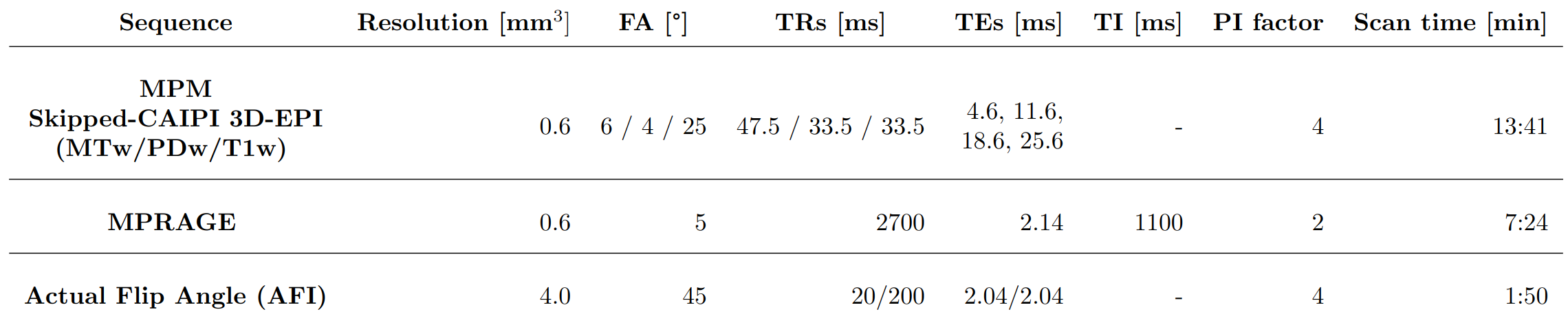
Table 1: Acquisition parameters of the sequences acquired in this study.
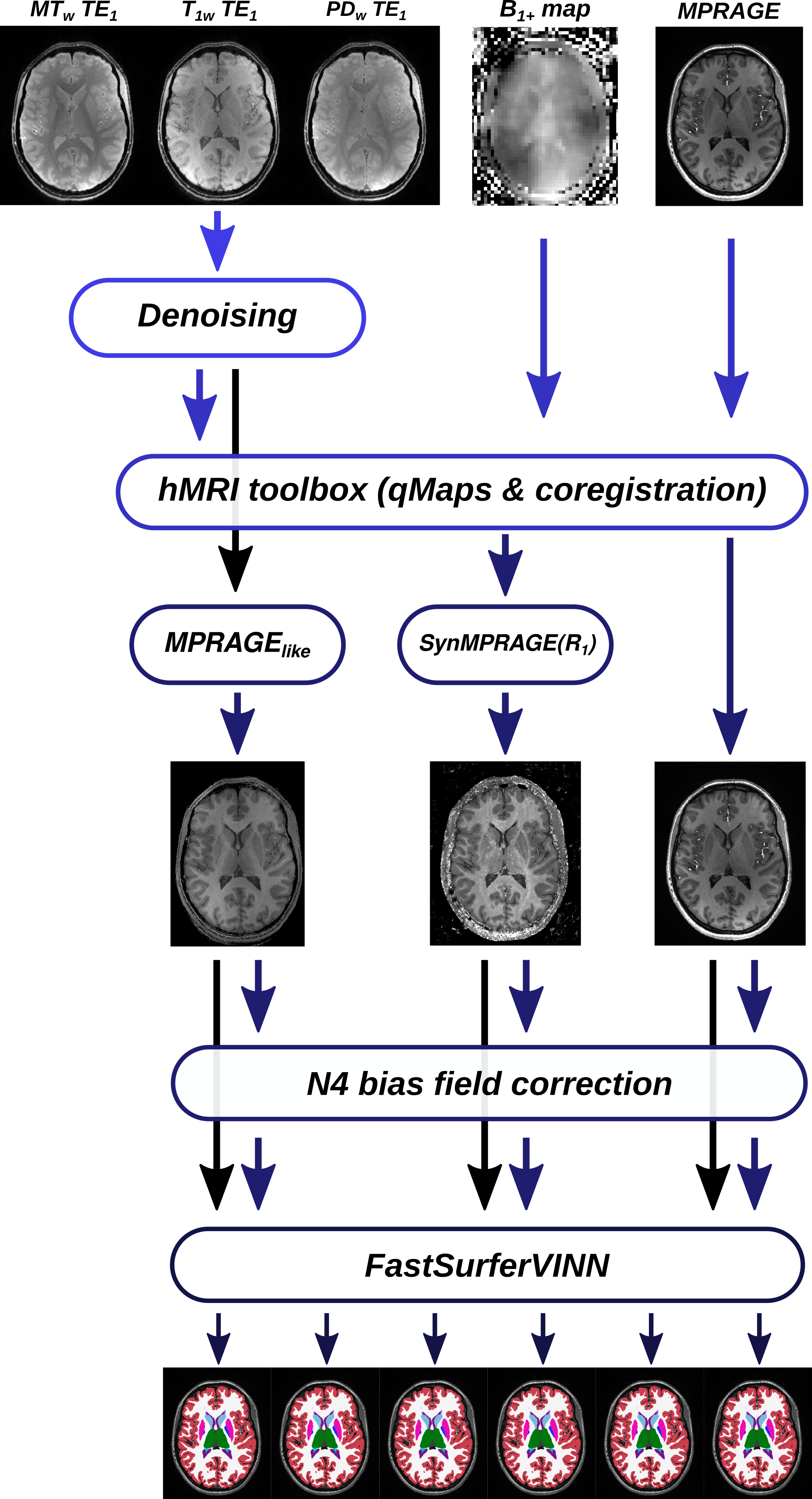
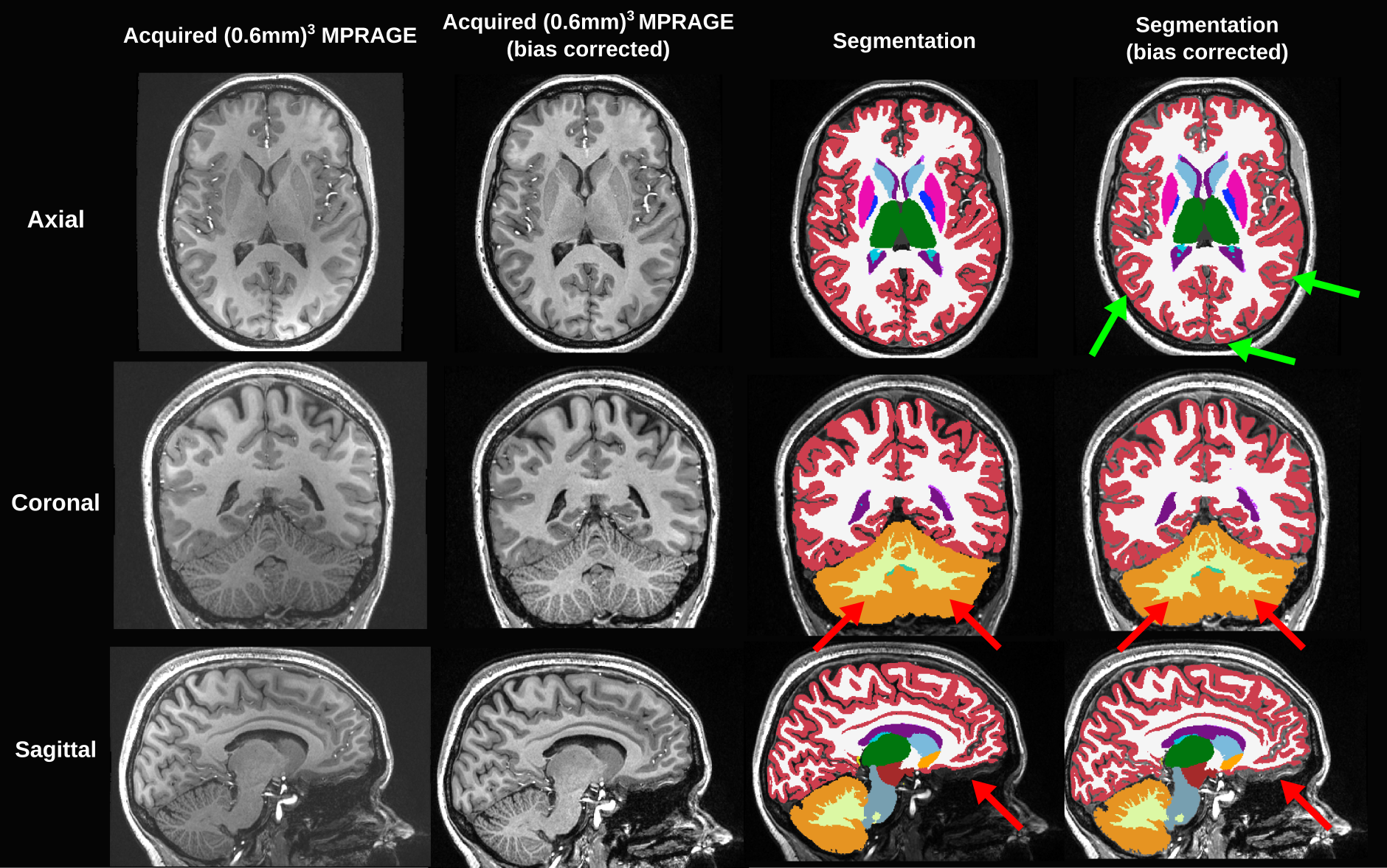
Figure 3: Segmentation results for one subject produced from FastSurferVINN for the (0.6mm)3 acquired MPRAGE images and its bias corrected version. The input images and corresponding segmentations are shown in the three anatomical planes.