2120
Quantitative Analysis of DCE-MRI Data using DL model based on Signal Intensity vs Concentration Curves1Centre for Biomedical Engineering, Indian Institute of Technology Delhi, New Delhi, India, 2Department of Radiology, Fotis Memorial Research Institute, Gurugram, India, 3Yardi School of Artificial Intelligence, Indian Institute of Technology Delhi, New Delhi, India, 4Department of Biomedical Engineering, All India Institute of Medical Sciences, New Delhi, India
Synopsis
Keywords: Analysis/Processing, DSC & DCE Perfusion, DEEP LEARNING, BRAIN TUMOR, PERFUSION PARAMETERS
Motivation: Quantitative analysis of dynamic-contrast-enhanced MRI(DCE-MRI) is valuable approach for mapping tumor physiology; however, traditional non-linear-least squares(NLLS) methods are slow and provide noisy maps. Deep-learning(DL) approach offers solutions, yet reported models rely on signal-intensity-time-curves(SIC) which are MRI-acquisition protocol dependent.
Goal(s): To develop DL network(CNNCON) that uses concentration-time-curves(CTCs) to estimate perfusion-parameters(GTKM) and compare with SIC-based DL network(CNNSIGNAL).
Approach: Two CNN networks were developed using CTC and SIC data(simulations and in-vivo). Performance of models was evaluated on simulated data with different protocols and experimental data.
Results: The CNNCONC outperforms NLLS & CNNSIGNAL in terms of speed, accuracy and smoothness of maps.
Impact: The proposed DL framework improves DCE-MRI analysis by providing more accurate and robust results in less time. It eliminates protocol dependence and holds the potential for routine clinical use in the diagnosis and treatment planning of brain tumor patients.
Introduction
Tracer-kinetic(TK) analysis(using GTKM, LTKM, etc) of DCE-MRI data provide valuable parameters related to physiology of tumor, which have shown great potential in diagnosis of tumors including gliomas1. Traditionally, these models use non-linear-least squares(NLLS) fitting approach for computing TK parameter maps. However, NLLS fitting takes long processing time and generates noisy maps.Recently, deep-learning(DL) frameworks have been explored to overcome these challenges2–6. However, reported DL models rely on signal-intensity-time-curves(SIC) which remain susceptible to acquisition protocol variations. Moreover, concentration-time-curves(CTC) provide a more direct representation of the actual contrast agent concentration in tissues and can be easily computed from SIC without taking much time.
Here we hypothesise that DL models based on CTC are less susceptible to imaging protocol and have better generalizability compared to SIC-based models.
Methods
First, we implemented CNN-based framework(CNNSIGNAL)6, which takes SICs, arterial-input-function(AIF) and T10 as inputs. The proposed model (CNNCON) takes input as a fusion of AIF and CTCs only to estimate TK parameters(Ktrans, Ve & Vp). Figure 1 shows overview of the methodology. DCE-MRI data consisted of 32-time frames.Both frameworks used Toft’s GTKM model parameter values estimated using NLLS fitting as ground truth. The training was done with batch size of 32 for 200 epochs; learning rate 2x10⁻⁴ with decay of 10-4 for both the networks with same activation function and optimizer. GTKM physics informed loss function was used for both networks.
Synthetic Data
We synthesized 3,600,000 CTCs with synthetic noise(SNR = 5-80). Synthetic CTCs were generated using GTKM from randomly sampled TK parameters in gray matter, white matter and tumor tissues (Ktrans =[1x10⁻⁵- 2x10⁻¹ min-1], Vp=[0.0005-1] and Ve=[0.02-0.6]). These CTCs were then converted to SICs using GTKM and population-based AIF7; where TR=6.3ms, Flip Angle(FA)θ=20°, r1 = 3.5ms, So=1000 & T10=1500ms. Each CTC and SIC size was 1X32 with time resolution of 0.025 min-1.
Patient Data
51 pre-operative glioma patients were used in this study. MR scans were performed on a 3.0T MRI scanner(Ingenia, Philips Healthcare, and The Netherlands). DCE-MRI data was acquired for 20 slices and 32 time points with FOV=230×230, TR/TE = 3.0/6.27s, FA = 20° in addition to conventional MRI sequences and pre-contrast T10 maps. SICs were converted to CTCs using pre-contrast T10.
For training and testing purposes, 80:20 split was performed randomly on both synthetic and patient data. Additionally, variations of SICs were synthesized with different TR and FA (-25%, +25%, 50% & 100%) from baseline CTCs to mimic different acquisition protocol (Figure 2). Both the networks were first trained and tested on synthetic data followed by transfer learning for patient data. Mean Absolute Error (MAE), structural-similarity-index-measure(SSIM) and root-mean-square(RMSE) were calculated for the three predicted parameters to assess the performance of both networks.
Results
The training and testing for synthetic data shows that proposed model predicted parameters with lower MAEs for Ktrans, Vp and Ve(Figure 3). Similar performance translated onto patient dataset as well (Table 1) with better SSIM and RMSE scores for CNNCON. The average processing time reduced from 45min (NLLS) to less than 5 min (for both CNN models) on regular desktop computer. Figure 4 shows that proposed network maps are even less noisy than other two approaches with higher precision and accuracy.Discussion
The proposed network reduces the computational time substantially when compared with NLLS method. It additionally reduces variability associated with acquisition protocols by choosing CTCs over SICs(Figure 2) for capturing reliable physiological information from the tissues. Experimentation on synthetic data demonstrates how error propagates by variations in TR & FA when SICs are used as the input(Table 1).The study suggest that CTCs should be preferred choice for DL frameworks to estimate faster and less noisy TK maps. However, it has been assessed on the simulation data and needs to be investigated on same patient’s data acquired with variations in acquisition protocols. In future, proposed CNNCON framework will be used to evaluate performance on other brain tumors from multiple centres and DCE-MRI data of different time frames or temporal resolutions.
Conclusion
The proposed DL network (CNNCON) performs quantitative analysis of DCE-MRI data rapidly and with more accuracy compared to traditional NLLS fitting. It is insensitive to imaging protocols compared to reported DL model(CNNSIGNAL). And hence offer better generalizability.Acknowledgements
Authors acknowledge the funding support of SERB, DST (project number: CRG/2019/005032).
References
1. Wu, J. et al. Glioma grade discrimination with dynamic contrast-enhanced MRI: An accurate analysis based on MRI guided stereotactic biopsy. Magn Reson Imaging 99, 91–97 (2023).
2. Ottens, T. et al. Deep learning DCE-MRI parameter estimation: Application in pancreatic cancer. Med Image Anal 80, (2022).
3. Ulas, C. et al. Direct estimation of pharmacokinetic parameters from DCE-MRI using deep CNN with forward physical model loss. in Lecture Notes in Computer Science (including subseries Lecture Notes in Artificial Intelligence and Lecture Notes in Bioinformatics) vol. 11070 LNCS 39–47 (Springer Verlag, 2018).
4. Zeng, Q. & Zhou, W. An Attention Based Deep Learning Model for Direct Estimation of Pharmacokinetic Maps from DCE-MRI Images. in Proceedings - 2021 IEEE International Conference on Bioinformatics and Biomedicine, BIBM 2021 2368–2375 (Institute of Electrical and Electronics Engineers Inc., 2021). doi:10.1109/BIBM52615.2021.9669582.
5. Zou, J., Balter, J. M. & Cao, Y. Estimation of pharmacokinetic parameters from DCE-MRI by extracting long and short time-dependent features using an LSTM network. Med Phys 47, 3447–3457 (2020).
6. Fang, K. et al. Convolutional neural network for accelerating the computation of the extended Tofts model in dynamic contrast-enhanced magnetic resonance imaging. Journal of Magnetic Resonance Imaging 53, 1898–1910 (2021).
7. Parker, G. J. M. et al. Experimentally-derived functional form for a population-averaged high-temporal-resolution arterial input function for dynamic contrast-enhanced MRI. Magn Reson Med 56, 993–1000 (2006).
Figures
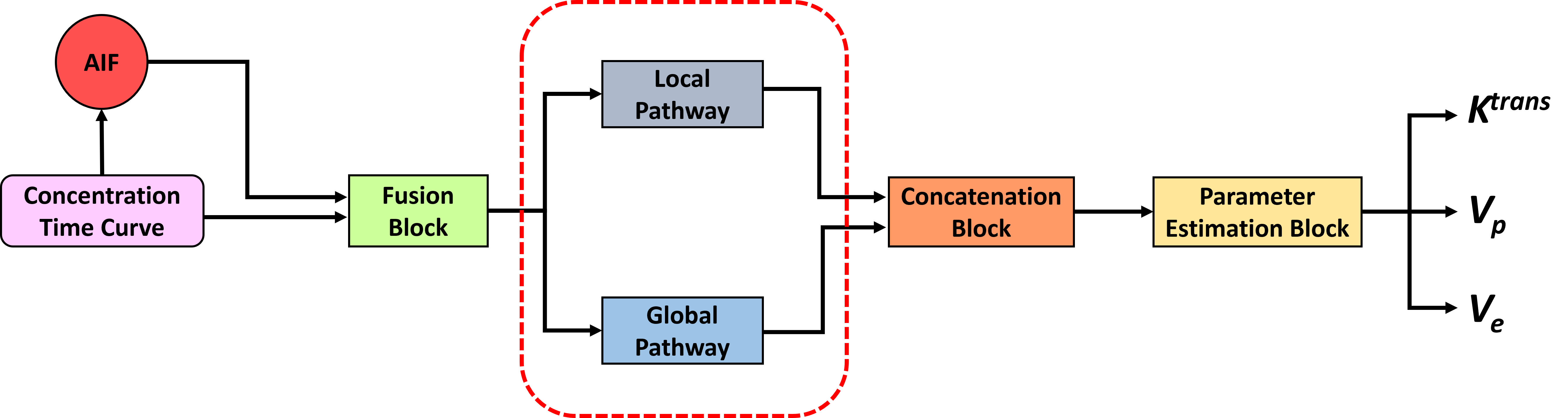
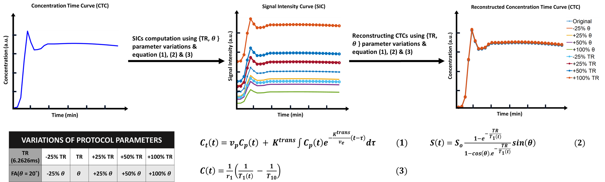
Figure 2 : Comparison between SICs and CTCs(baseline and reconstructed) computed using different variations of protocol parameters (TR & θ)
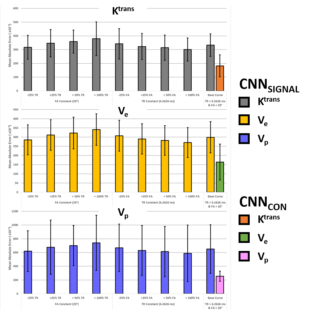
Figure 3 : Mean Absolute Error of CNNSIGNAL and CNNCON (all protocol parameter variations will have same MAEs)for Ktrans, Vp & Ve
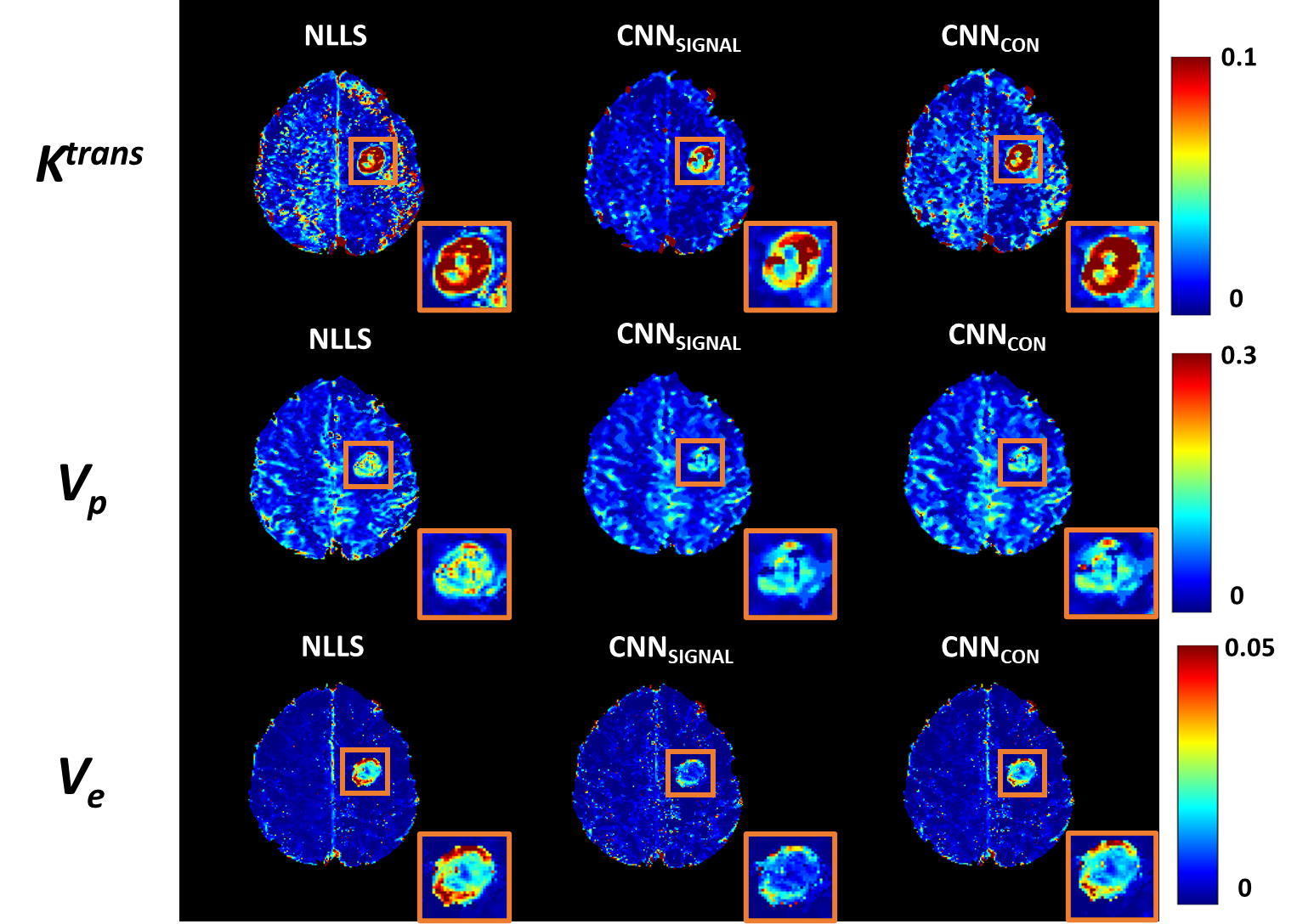
Figure 4 : TK Parametric maps : each row shows Ktrans, Vp & Ve of NLLS(ground truth), CNNSIGNAL and CNNCONCENTRATION respectively of representative case of High Grade Glioma(HGG)
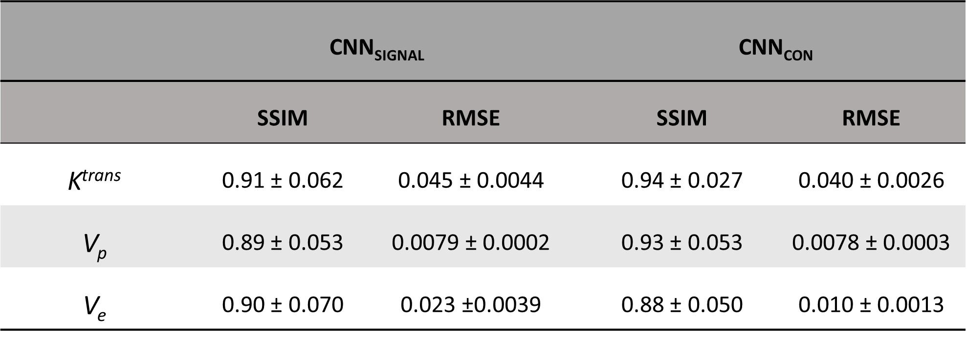
Table 1: SSIM & RMSE values of predicted TK parametric maps (Ktrans, Vp & Ve respectively) on the test patient data for both the networks CNNSIGNAL and CNNCON