2113
Deep learning-based quantification of myocardial oxygen extraction fraction and blood volume in health: reproducibility, sex, and homogeneity1Washington University in St. Louis, St. Louis, MO, United States
Synopsis
Keywords: Analysis/Processing, Machine Learning/Artificial Intelligence
Motivation: A previously developed MRI method for quantitative myocardial oxygen extraction mapping (mOEF) showed promising results, but image quality suffered from distortion and inhomogeneity artifacts.
Goal(s): The objective of this study is to evaluate a new CMR method for in vivo measurement of mOEF utilizing on a deep-learning quantification approach in healthy controls.
Approach: A new pulse sequence and a novel deep learning-based analysis method were created and evaluated on a group of healthy subjects.
Results: This investigation yielded dramatically improved image quality, which allowed reliable evaluation of reproducibility and distribution of mOEF within the heart.
Impact: Our study, involving 20 healthy volunteers, showcased outstanding reproducibility in the measurements, suggesting its potential for translation into imaging studies for patients with myocardial metabolic dysfunction.
Introduction
Impaired myocardial oxygen metabolism dysfunction usually precedes ventricular mechanical dysfunction 1. Quantification of myocardial oxygen extraction fraction (mOEF) can allow direct measurement of myocardial oxygen metabolism. The objective of this study is to evaluate a new CMR method for in vivo measurement of mOEF utilizing on a deep-learning quantification approach in healthy controls.Methods
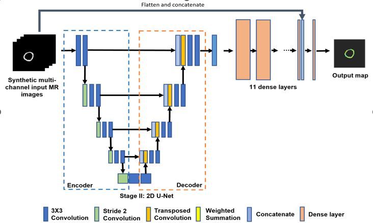
A new CMR sequence for mOEF data acquisition was implemented using an asymmetric-spin-echo prepared sequence with multiple single-short balanced steady state readout (ASEprep). Eighteen single-shot images were collected with different asymmetric-spin-echo shifts to provide oxygen-sensitive weightings. For the quantification of mOEF and myocardial blood volume (MBV) 3, synthetic mOEF and MBV training sets were simulated based on theoretical model developed previously 2, with a variety of imperfect conditions (noise and inhomogeneity) being added. A total of 1800 simulated data sets were created, in which 80% was used for training and 20% for testing. These data were fed to an UNet-based fully connected neural network (UFCN) that was comprised of an encoder, and decoder, and a set of dense layers (Figure 1). The final output was mOEF and MBV maps. The human study was approved by local human study committee. Twenty young healthy volunteers (age, 22.9 ± 3.3 yrs, 10 female) were recruited and they underwent the same MRI scans twice at different days for assessing reproducibility. Three slice scans at short-axis views were obtained during each session (basal, mid, apex). Each mOEF map was acquired with a breath-holding time of 18 RR intervals, using the ASEprep sequence with a spatial resolution was 1.7 x 1.7 mm2. The coefficient of variance (CoV) was calculated for assessing reproducibility of mOEF or MBV, on the basis of slice and subject. Analysis of Variance (ANOVA) was employed to compare mOEF and MBV between male and female, as well as among different locations of the heart.Results
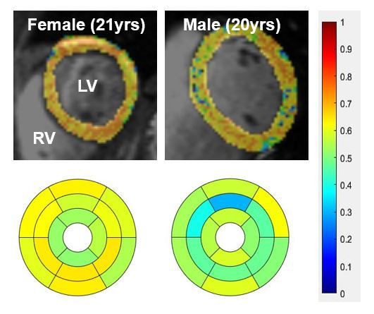
The mOEF maps quantified by utilizing the new physics based UFCN model showed relatively uniform distribution of mOEF and MBV signals across the whole heart. The CoV for mOEF was 6.3% [4.1,7.9] % and 4.0% [1.6, 5.4] % on the basis of slice and subject, respectively. The corresponding CoV for MBV was 11.1% [7.8, 13.7] % and 5.9% [2.9, 7.8] %. There was no significant difference in MBV between male and female. However, mOEF in female was significantly higher than that in male (F: 0.60 ± 0.07 vs. M: 0.56 ± 0.03, p < 0.05). Within the heart, neither mOEF nor MBV shows any significant difference among three slices. Figure 2 shows basal slices of mOEF maps and bullseye display of one male and one female.Discussion & Conclusion
The quantification of mOEF and MBV are reproducible by utilizing the new physics based artificial intelligence network. The image quality of mOEF and MBF appears to allow clinical tests. While both mOEF and MBV are uniformly distributed in whole hearts of healthy subjects, female subjects show higher mOEF values, which needs to be considered in the future patient study.Acknowledgements
The research is supported in part by National Institutes of Health grant HL165238 and UL1TR002345, as well as American Heart Association grant 23SCISA1145192.References
1. Karwi QG, Uddin GM, Ho KL, Lopaschuk GD. Loss of Metabolic Flexibility in the Failing Heart. Front Cardiovasc Med. 2018 Jun 6. PMCID: PMC5997788.
2. An H, Lin W. Impact of intravascular signal on quantitative measures of cerebral oxygen extraction and blood volume under normo- and hypercapnic conditions using an asymmetric spin echo approach. Magn Reson Med. 2003;50:708-716.
3. McCommis KS, Zhang H, Goldstein TA, Misselwitz B, Abendschein DR, Gropler RJ, Zheng J. Myocardial blood volume is associated with myocardial oxygen consumption: an experimental study with cardiac magnetic resonance in a canine model. JACC Cardiovasc Imaging. 2009;2:1313-1320.
Figures