2104
MRI-Based Deep Learning for Automatic Segmentation of Punctate White Matter Injury in Neonates1Department of Radiology, The First Affiliated Hospital of Xi'an Jiaotong University, Xi’an, China, 2Department of Research and Development, Shanghai United Imaging Intelligence Co., Ltd., Shanghai 200232, China
Synopsis
Keywords: Analysis/Processing, Segmentation, Punctate white matter injury
Motivation: Punctate white matter injury (PWMI) in neonates is characterized by small lesions and significant sample variability, posing a challenge for quantification.
Goal(s): We introduce a novel approach that combines the 3D nnU-Net framework for semantic segmentation of PWMI using neonatal brain MR images.
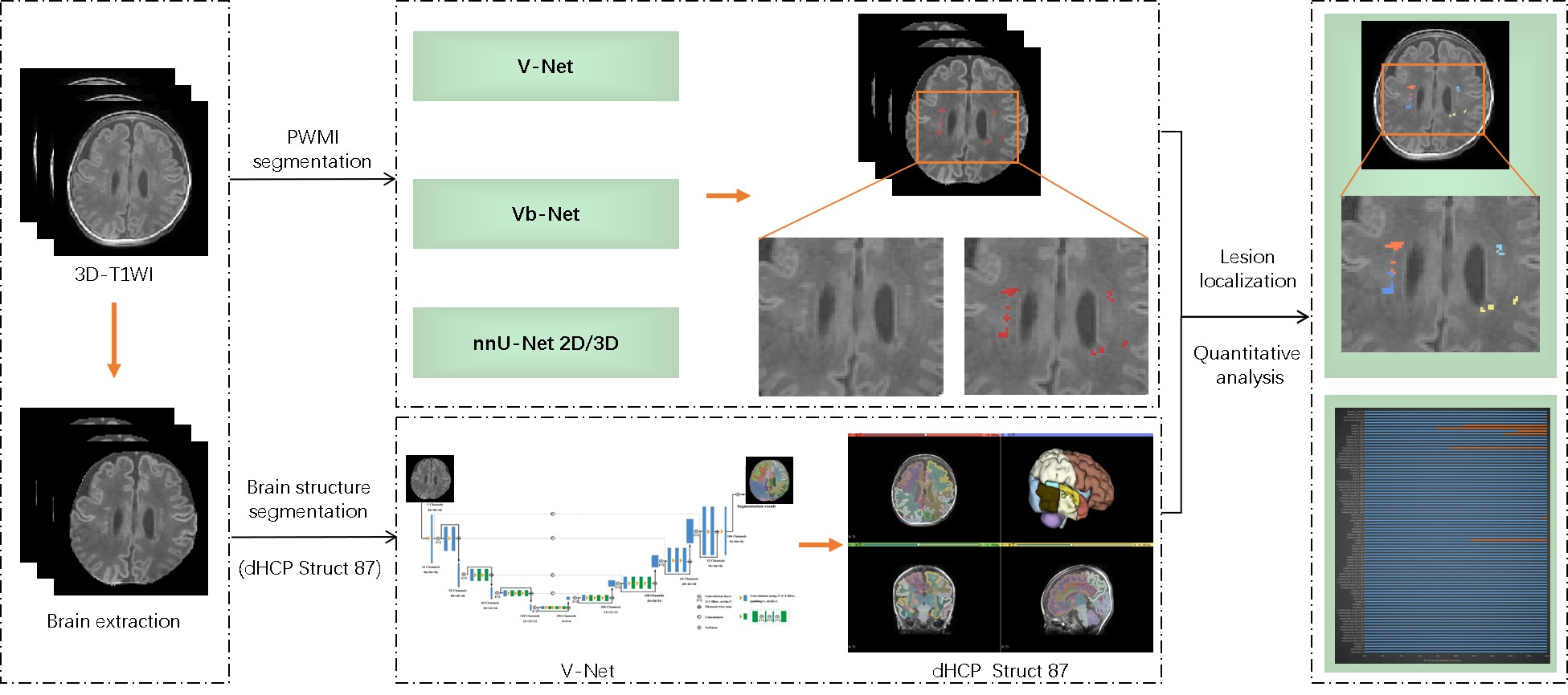
Approach: The PWML automatic segmentation models, based on 3D-T1WI, was developed utilizing V-Net, VB-Net, 2D nnU-Net and 3D nnU-Net. Automatic localization of lesions and quantitative analysis of the brain regions were further realized by segmentation of dHCP template brain regions.
Results: The automatic segmentation model demonstrated robust performance, achieving a median Dice Similarity Coefficientn of 0.865 on the test set.
Impact: This innovation offers an automatic and accurate segmentation of PWMI regions, potentially providing clinicians with a powerful tool for the automatic localization and classification model construction, quantitative analysis and grading prognostic study of PWMI in neonates.
Introduction
Punctate white matter injury (PWMI) in neonates presents with small lesions and substantial sample variability, posing a challenge for quantification. The nnU-Net is an advanced segmentation technique recognized for its excellent performance in biomedical image segmentation tasks. This adaptability is primarily attributed to its capacity to dynamically adjust to varying input image sizes and manual label numbers, rather than utilizing a fixed network structure. This study introduces a deep learning algorithm that integrates the 3D nnU-Net1 for the segmentation of PWMI regions in neonatal MR images. This approach facilitates automatic lesion localization and quantitative analysis via dHCP template for brain region segmentation.Methods
This retrospective study included 115 neonatal brain 3D-TIWI MR images with PWML from the First Affiliated Hospital of Xi'an Jiaotong University, comprising training set (n= 93, PMA 37.93±2.95 weeks, 47 females), validation set (n=22, PMA 37.58±2.27 weeks, 10 females). Furthermore, we collected additional PWMI data from the dataset as independent test set (n=59, PMA 37.31±2.11 weeks, 29 females). 3D-FSPGR T1WI were performed on a 3T scanner (Signa HDxt, General Electric Medical System, Milwaukee, WI, USA) with 8-channel head coil: TR/TE=10/4.6ms, slice thickness=1mm, acquisition time=5 minute 10 seconds. All images were processed using an image analysis tool named uAI Research Portal (Shanghai United Imaging Intelligence Co. Ltd)2. Briefly, the N4 algorithm was applied to 3D-T1WI images for bias field correction to handle the inhomogeneity of the magnetic field. Brain extraction was also performed. Two skilled observers independently manually delineated PWMI regions guided by a neuroradiologist with 15 years of experience. The PWML automatic segmentation models, based on 3D-T1WI, was developed utilizing V-Net, VB-Net, 2D nnU-Net and 3D nnU-Net. Automatic localization of lesions and quantitative analysis of the brain regions were further realized by segmentation of dHCP template brain regions (Figure 1). Model performance was assessed through Dice similarity coefficient (DSC), Hausdorff distance (HD), and average surface distance (ASD).Results
The DSC, HD and ASD of V-Net, VB-Net, 2D nnU-Net and 3D nnU-Net were shown in Table 1. The automatic segmentation model of 3D nnU-Net demonstrated robust performance, with a median DSC of 0.865 on the validation set. And the model's median DSC independent test set was 0.650.Discussion
We utilized the powerful and unparalleled 3D nnU-Net network to be the backbone and rebuilt a new training pipeline adopting our data so as to PWMI automatic segmentation approach. Under this framework, detailed features at different scales are captured using a contracting path, and these features are then combined using an expansive path. We employed a series of 3D convolutional layers appended with non-linear ReLU activation layers to extract multi-scale features from MR images. Furthermore, these features from the deeper layers contain a more abstract semantic information, which is effective for lesion location recognition. The features from shallower layers, meanwhile, preserve better spatial information, making them suitable for precise boundary recognition. We used the strategy of data augmentation with 5-fold cross-validation to avoid overfitting when predicting. To further improve the performance and generalization of our model, we randomly split the dataset, replaced batch normalization with group normalization, and changed larger convolution kernels. The automatic segmentation model of 3D nnU-Net enabled automatic lesion localization and quantitative analysis via dHCP template and demonstrated robust performance.Conclusion
The MRI-based deep learning model for automatic segmentation of PWMI regions offers a significant path to augment clinical diagnosis and evaluation capabilities. There is substantial potential for the developed model to be integrated into routine practice for better management and therapeutic decision-making for PWMI. Considering the enormous implications and impact on neonatal healthcare, validations of the proposed model in multi-center studies with a larger sample volume could further add to its robustness and real-world applicability.Table 1 The PWMI automatic segmentation result.
| Method | Validation set (n=22) | Independent test set (n=59) | ||||
| DSC | HD | ASD | DSC | HD | ASD | |
| V-Net | 0.058 | 0.150 | 55.582 | 61.141 | 14.543 | 31.813 | 0.362 | 0.332 | 28.671 | 29.843 | 8.030 | 7.034 |
| Vb-Net | 0.430 | 0.366 | 47.034 | 41.464 | 9.300 | 9.650 | 0.423 | 0.379 | 7.810 | 15.682 | 0.540 | 2.593 |
| 2D nnU-Net | 0.743 | 0.704 | 8.943 | 14.659 | 0.311 | 1.646 | 0.568 | 0.559 | 19.755 | 23.132 | 1.947 | 3.825 |
| 3D nnU-Net | 0.865 | 0.856 | 4.780 | 11.298 | 0.193 | 0.924 | 0.650 | 0.637 | 14.177 | 20.373 | 1.064 | 3.026 |
DSC indicates Dice similarity coefficient; HD, Hausdorff distance; ASD, average surface distance.
Acknowledgements
This work was supported by the National Natural Science Foundation of China (82272618, 81971581) and the Key Research and Development Program of Shaanxi(2021SF-092). Please address correspondence to Jian Yang, e-mail: yj1118@mail.xjtu.edu.cn.References
[1] Isensee F, Jaeger PF, Kohl SAA, Petersen J, Maier-Hein KH. nnU-Net: a self-configuring method for deep learning-based biomedical image segmentation [J]. Nat Methods. 2021;18(2):203-211.
[2]Wu J, Xia Y, Wang X, Wei Y, Liu A, Innanje A, Zheng M, Chen L, Shi J, Wang L, Zhan Y, Zhou XS, Xue Z, Shi F and Shen D (2023) uRP: An integrated research platform for one-stop analysis of medical images. Front. Radiol. 3:1153784. doi: 10.3389/fradi.2023.1153784.
Figures