2103
Fully-Automated Segmentation algorithm of Rectal Cancer and mesorectum on Multiparametric MR1Department of MRI Diagnosis, The Second Affiliated Hospital of Harbin Medical University, Harbin, China, 2The Second Affiliated Hospital of Harbin Medical University, Harbin, China, 3MR Research China, GE HealthCare, Beijing, China
Synopsis
Keywords: Analysis/Processing, Cancer
Motivation: Developing a fast solution to segment rectal tumors and mesorectal tissue instead of the current manual labeling.
Goal(s): The goal was to develop an automated segmentation model using nnU-Net for fully-automated segmentation of rectal cancer and mesorectum on MR images.
Approach: The dataset was divided into training and testing sets, and pre-processing steps were conducted to minimize computational burden. The nnU-Net deep learning network was employed to train the model.
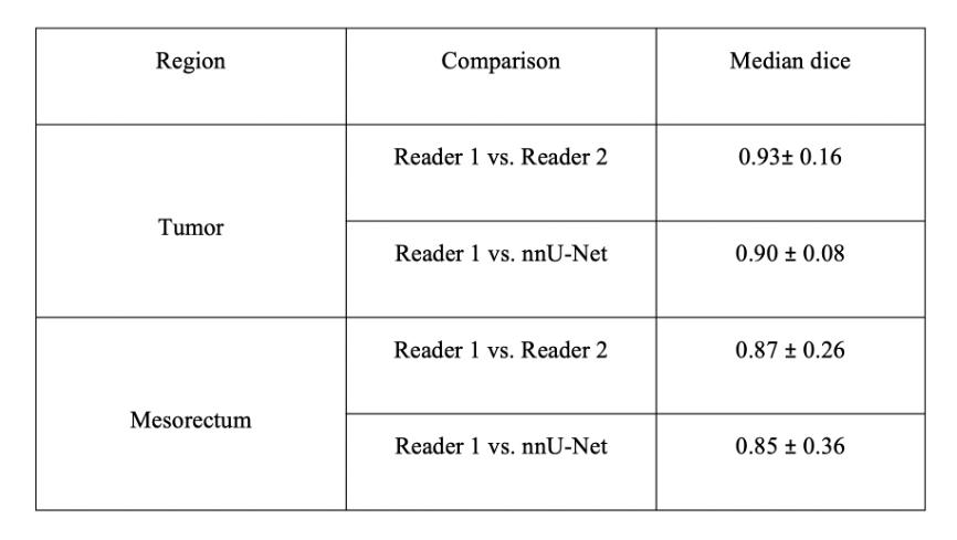
Results: The Dice similarity coefficients for tumor and mesorectum in both the training and testing sets were as follows: 0.91 (training) and 0.88(testing) for tumor, and 0.93 (training) and 0.89 (testing) for mesorectum.
Impact: This study proposes an automatic segmentation scheme for rectal tumor and mesentery using deep learning. It can be used to guide the annotation of new medical images, potentially improving the accuracy of rectal cancer treatment response predictions.
Introduction
Rectal cancer is a common digestive tract tumor, accounting for the third most common tumor worldwide [1,2]. Studies have shown that whole-volume tumor segmentations provide more accurate estimates of true tumor volumes than single slice or sample measurements. However, manual segmentation approaches are time-consuming and unlikely to be implemented into daily clinical practice. Song et al. found that mesorectal fascia has good predictive value for rectal cancer treatment response, but it requires segmentation of both lesions and mesorectal fascia, increasing workload. Therefore, there is a need for smarter algorithms that can automatically localize and accurately segment rectal tumours and mesorectum, reducing the need for expert input.Methods
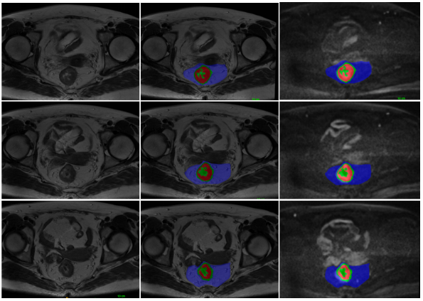
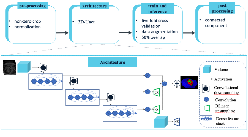
We developed segmentation models using T2WI sequences. Two radiologists independently annotated the outer tumor, rectal wall, and mesorectum wall in a blinded fashion (Figure 1). The dataset was divided into a training set (n=85) and testing set (n=38). We employed the nnU-Net deep learning network to train an automated segmentation model [4]. By utilizing nnU-Net, we streamlined the key decisions involved in designing an effective segmentation pipeline for our specific dataset. This process is illustrated in Figure 2. Prior to training, several pre-processing steps were conducted, which are outlined below. To minimize computational burden and reduce matrix size, all images were cropped to the region with non-zero values. Then, the datasets were resampled to median voxel spacing by using third-order spline interpolation for images and neighbor interpolation for masks. This resampling approach enabled the neural networks to better learn the spatial semantics of the scans. Finally, the entire dataset was normalized by clipping to the [0.50, 99.5] percentile of intensity values and then z-score normalized according to the mean and standard deviation of all collected intensity values. In the training procedure, we used the 3d_fullres model and combined dice and cross-entropy loss, as shown in Formula 1. We also performed 5-fold cross validation during training. Connected component analysis was used as a postprocessing technique. After inference, tumors and mesorectum could be automatically segmented. And segmentation performance was evaluated using the Dice similarity coefficient (DSC).Results
We developed an nnU-Net based network using independent discovery and validation datasets to validate its performance. For each patient, a total of 2400 patches were created by combining T2WI and DWI images for both tumor and rectal mesorectum areas. The Dice similarity coefficients for tumor and mesorectum in both the training and testing sets were as follows: 0.91 (training) and 0.88 (testing) for tumor, and 0.93 (training) and 0.89 (testing) for mesorectum.Discussion
We employed an innovative deep learning approach (nnU-Net model) for rapid and accurate detection and segmentation of tumor and mesorectum on T2WI MR examinations in the context of rectal cancer. Our findings indicate that tumor size is a significant factor affecting the performance of the model for tumor detection, with the sensitivity of the nnU-Net model increasing as the size of the tumor increases in both internal and external testing datasets. The model demonstrated higher efficacy in identifying tumors than rectal mesentery, which may be attributed to the unclear boundary between the two. To improve the accuracy of recognizing mesentery, future studies could consider increasing the number of samples or adjusting the network structure.Conclusion
This paper proposes an automatic segmentation scheme for rectal tumor and mesentery using deep learning, which greatly reduces the annotation time for clinicians and minimizes the adverse effects of individual differences in labeling. In addition, it can be used to guide the annotation of new medical images.Acknowledgements
No acknowledgement found.References
[1]Siegel RL, Miller KD, Fuchs HE, Jemal A. Cancer Statistics, 2021 [published correction appears in CA Cancer J Clin. 2021 Jul;71(4):359]. CA Cancer J Clin. 2021;71(1):7-33. doi:10.3322/caac.21654[2]Sung H, Ferlay J, Siegel RL, et al. Global Cancer Statistics 2020: GLOBOCAN Estimates of Incidence and Mortality Worldwide for 36 Cancers in 185 Countries. CA Cancer J Clin. 2021;71(3):209-249. doi:10.3322/caac.21660[3]Song G, Li P, Wu R, et al. Development and validation of a high-resolution T2WI-based radiomic signature for the diagnosis of lymph node status within the mesorectum in rectal cancer. Front Oncol. [4]2022;12:945559. Published 2022 Sep 16. doi:10.3389/fonc.2022.945559Isensee, F., Jaeger, P. F., Kohl, S. A., Petersen, J., & Maier-Hein, K. H. (2021). nnU-Net: a self-configuring method for deep learning-based biomedical image segmentation. Nature methods, 18(2), 203-211.Figures