2102
Deep learning-based comprehensive multi-sequence liver lesion segmentation for accurate tumor burden assessment1Shukun Technology Co.,Ltd, Beijing, China
Synopsis
Keywords: Analysis/Processing, Liver
Motivation: Accurate lesion segmentation is crucial for tumor burden assessment and subsequent patient-specific treatment prediction.
Goal(s): To develop and validate a deep learning-based automated segmentation model for accurate hepatocellular carcinoma (HCC) lesion detection across various imaging sequences.
Approach: A total of 2800 patients with focal liver lesions (FLLs) were included for developing automated segmentation models.
The automated segmentation models were trained involving preprocessing, lesion detection using mask R-CNN, and lesion segmentation using a 3D-UNet framework.
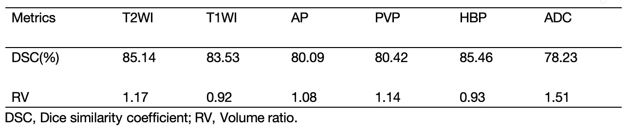
Results: The 3D-UNet framework is used for lesion segmentation, achieving a DSC accuracy ranging from 78.23% to 85.14% and volume ratios from 0.92 to 1.51 across different sequences.
Impact: The model demonstrates promising potential in accurately segmenting HCC lesions.
Introduction
Accurate lesion segmentation is crucial for tumor burden assessment and subsequent patient-specific treatment prediction. Automated and standardized image processing can save time and costs, offering vital clinical evidence for precision diagnosis and treatment. Moreover, previous tumor burden assessments in Hepatocellular Carcinoma have lacked a unified standard. Most measurements have been based on one-dimensional diameter assessments, which may not accurately represent the true tumor size. Moreover, variations in tumor size measurements across different imaging sequences raise concerns about which sequence should be prioritized for accurate assessments. The lack of extensive comparative data further complicates the selection of the most appropriate imaging sequence for reliable tumor burden evaluations. To our knowledge, there is currently no research on the comprehensive segmentation of hepatocellular carcinoma lesions using multiple liver MR sequences. Therefore, this study aimed to develop a deep learning-based comprehensive multi-sequence liver lesion fully automated segmentation model. External validation was performed on six sequences of contrast-enhanced MR images from 911 patients with hepatocellular carcinoma, and differences in lesion burden measurements across different sequences were compared.Method
A total of 2800 patients with focal liver lesions (FLLs) from seven tertiary hospitals in China were included for developing automated deep learning (DL) segmentation models between December 2013 and February 2021. Among them, 1889 patients were allocated into training (n=1511), validation (n=189), and test (n=189) sets at an 8:1:1 ratio. Additionally, 911 cases of hepatocellular carcinoma (HCC) were identified as an independent test set, confirmed by pathology. All magnetic resonance (MR) images in DICOM format were acquired from the picture archiving and communication system. Manual segmentations of FLLs were independently conducted by two abdominal radiologists, carefully avoiding intrahepatic vasculatures. Various imaging sequences were utilized for segmentations. To ensure quality control, a senior radiologist reviewed all regions of interest (ROIs). The resulting sketched images were used to train the automated segmentation models in a sequential modular approach. The independent HCC test set exclusively involved outlining HCC lesions.Preprocessing
Using a 3D-CNN model[1], we isolate the liver region within MRI scans by obtaining a liver segmentation mask. Rigid image registration aligns multiple MRI sequences onto a unified spatial reference frame, ensuring accurate analysis.
Lesion Detection
The core component, Unified Multi-Sequence Lesion Detector (MSLD), is a 3D CNN model[2,3]. The MSLD consists of four Single Lesion Detectors (SLDs) and a False Positive Reduction (FPR) module. (a) Tailored for each MRI sequence, four SLDs accommodate diverse tissue appearances across different sequence groups, including pre/post-contrast T1WI, T2WI, and DWI. Each SLD framework includes four modules: RPN, ROI alignment, lesion identification, and lesion segmentation. (b) The FPR module distinguishes artifacts from genuine lesions using a 3D-CNN, enhancing the algorithm's performance by extracting image features from each ROI.
Lesion Segmentation
We employed a 3D-UNet framework with an encoder-decoder architecture, 3D convolutions, and pooling layers. The Adam optimizer with an initial learning rate of 0.001 was gradually reduced every 30 epochs by a factor of 0.1, culminating after 60 training epochs. Tumor volume was calculated based on the DL-generated lesion segmentation and physical spacing.
Results
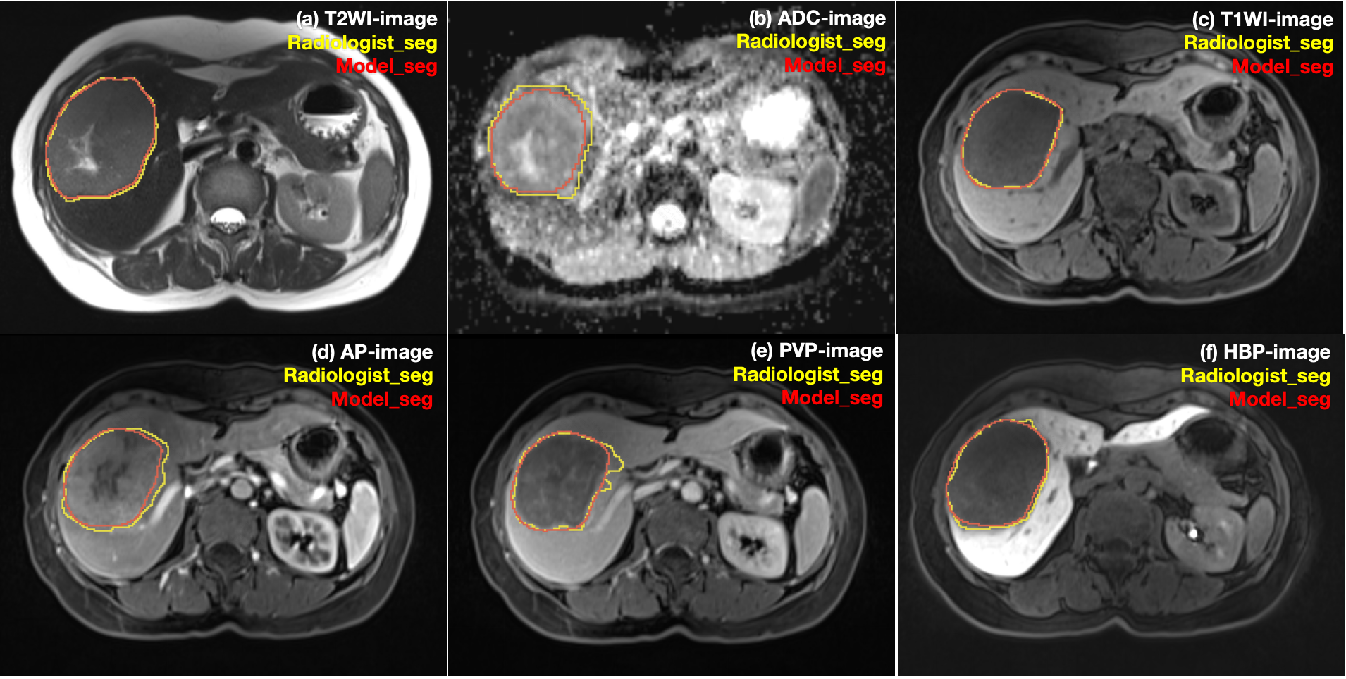
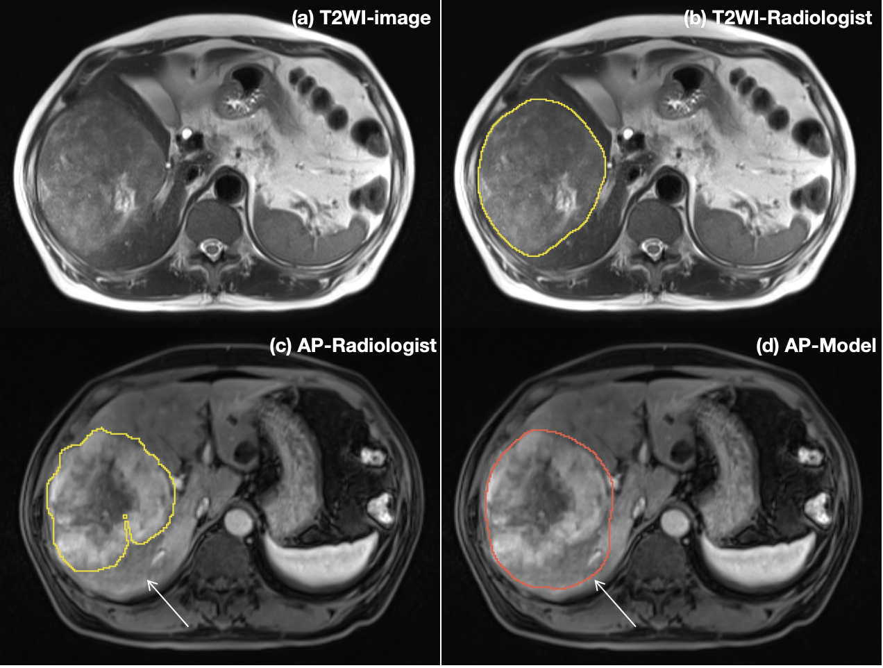
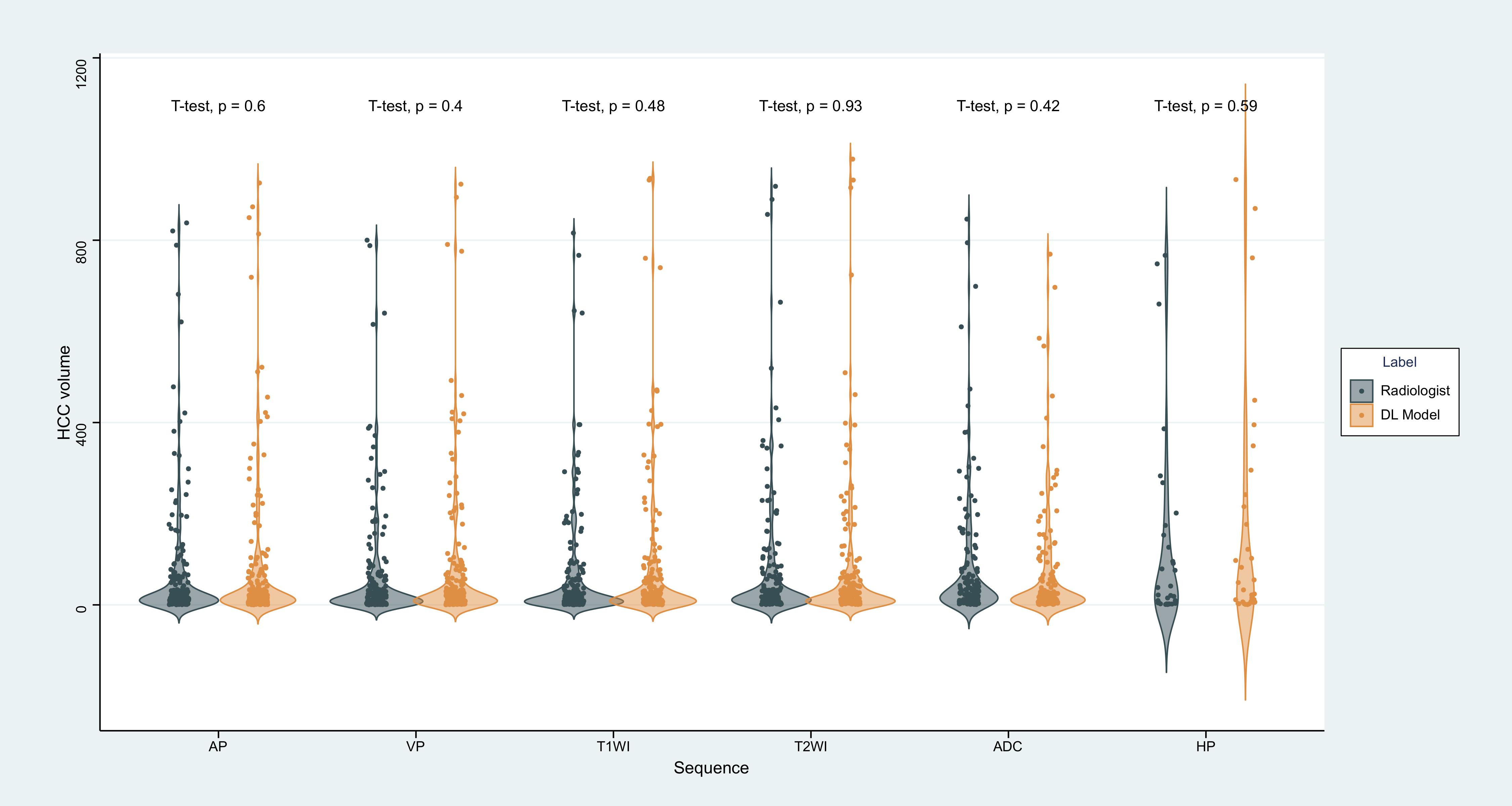
Our model's dice loss and segmentation accuracy are shown in Table 1. The DSC (Dice Similarity Coefficient) accuracy for lesion segmentation across different sequences ranged from 78.23% to 85.14%, with volume ratios ranging from 0.92 to 1.51. Considering the relatively poor signal-to-noise ratio of ADC images, the segmentation accuracy was correspondingly lower. Representative results of automated HCC lesion segmentation are illustrated in Figures 1 and 2. The distribution and difference of lesion volume measurements across different sequences is depicted in the violin plot in Figure 3. On a GPU-accelerated computing platform using Xeon(R) Silver 4210R (CPU) and GeForce RTX 2070 (GPU), the model exhibited an average runtime of 16.8±2.4 s.Conclusion
Our study presents a novel deep learning-based automated segmentation model for accurate and efficient HCC lesion detection. Despite variations in segmentation accuracy across different imaging sequences, our model demonstrates promising performance in delineating HCC lesions. The robust performance of the model in accurately segmenting HCC lesions provides valuable support for its potential clinical application in precision medicine and individualized treatment planning for HCC patients.Acknowledgements
We thank all radiologists and patients who participated in this study.References
1. Han X, Wu X, Wang S, et al. Automated segmentation of liver segment on portal venous phase MR images using a 3D convolutional neural network. Insights Imaging 2022;13(1):26.
2. He, K., et al., Mask R-CNN, in 2017 IEEE International Conference on Computer Vision (ICCV). 2017. p. 2980-2988.
3. Lin, T.-Y., et al., Feature Pyramid Networks for Object Detection, in 2017 IEEE Conference on Computer Vision and Pattern Recognition (CVPR). 2017. p. 936-944.
Figures